Intramedullary solitary fibrous tumor of cervicothoracic spinal cord
- PMID: 25368773
- PMCID: PMC4217067
- DOI: 10.3340/jkns.2014.56.3.265
Intramedullary solitary fibrous tumor of cervicothoracic spinal cord
Abstract
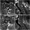
Solitary fibrous tumor is rare benign mesenchymal neoplasm. The spinal solitary fibrous tumor is extremely rare. The authors experienced a case of intramedullary solitary fibrous tumor of cervicothoracic spinal cord in a 48-year-old man with right lower extremity sensory disturbance. Spinal MRI showed intradural mass lesion in the level of C7-T1, the margin between the spinal cord and tumor was not clear on MRI. A Left unilateral laminectomy and mass removal was performed. Intra operative finding, the tumor boundary was unclear from spinal cord and it had intramedullary and extramedullary portion. After surgery, patient had good recovery and had uneventful prognosis. Follow up spinal MRI showed no recurrence of tumor.
Keywords: Benign; Intramedulla; Solitary fibrous tumor; Spine.
Figures


References
-
- Bohinski RJ, Mendel E, Aldape KD, Rhines LD. Intramedullary and extramedullary solitary fibrous tumor of the cervical spine. Case report and review of the literature. J Neurosurg. 2004;100(4 Suppl Spine):358–363. - PubMed
-
- Bouyer B, Guedj N, Lonjon G, Guigui P. Recurrent solitary fibrous tumour of the thoracic spine. A case-report and literature review. Orthop Traumatol Surg Res. 2012;98:850–853. - PubMed
-
- Brigui M, Aldea S, Bernier M, Bennis S, Mireau E, Gaillard S. Two patients with a solitary fibrous tumor of the thoracic spinal cord. J Clin Neurosci. 2013;20:317–319. - PubMed
-
- Briselli M, Mark EJ, Dickersin GR. Solitary fibrous tumors of the pleura : eight new cases and review of 360 cases in the literature. Cancer. 1981;47:2678–2689. - PubMed
-
- Brozzetti S, D'Andrea N, Limiti MR, Pisanelli MC, De Angelis R, Cavallaro A. Clinical behavior of solitary fibrous tumors of the pleura. An immunohistochemical study. Anticancer Res. 2000;20:4701–4706. - PubMed
Publication types
LinkOut - more resources
Full Text Sources
Other Literature Sources
Miscellaneous

