. 2014 Nov 4;12(11):e1001987.
doi: 10.1371/journal.pbio.1001987. eCollection 2014 Nov.

Structural and mechanistic basis of zinc regulation across the E. coli Zur regulon

Affiliations

Structural and mechanistic basis of zinc regulation across the E. coli Zur regulon

Benjamin A Gilston et al. PLoS Biol. .

Abstract

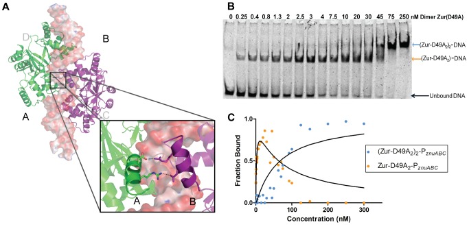
Commensal microbes, whether they are beneficial or pathogenic, are sensitive to host processes that starve or swamp the prokaryote with large fluctuations in local zinc concentration. To understand how microorganisms coordinate a dynamic response to changes in zinc availability at the molecular level, we evaluated the molecular mechanism of the zinc-sensing zinc uptake regulator (Zur) protein at each of the known Zur-regulated genes in Escherichia coli. We solved the structure of zinc-loaded Zur bound to the P(znuABC) promoter and show that this metalloregulatory protein represses gene expression by a highly cooperative binding of two adjacent dimers to essentially encircle the core element of each of the Zur-regulated promoters. Cooperativity in these protein-DNA interactions requires a pair of asymmetric salt bridges between Arg52 and Asp49' that connect otherwise independent dimers. Analysis of the protein-DNA interface led to the discovery of a new member of the Zur-regulon: pliG. We demonstrate this gene is directly regulated by Zur in a zinc responsive manner. The pliG promoter forms stable complexes with either one or two Zur dimers with significantly less protein-DNA cooperativity than observed at other Zur regulon promoters. Comparison of the in vitro Zur-DNA binding affinity at each of four Zur-regulon promoters reveals ca. 10,000-fold variation Zur-DNA binding constants. The degree of Zur repression observed in vivo by comparison of transcript copy number in wild-type and Δzur strains parallels this trend spanning a 100-fold difference. We conclude that the number of ferric uptake regulator (Fur)-family dimers that bind within any given promoter varies significantly and that the thermodynamic profile of the Zur-DNA interactions directly correlates with the physiological response at different promoters.


Conflict of interest statement

The authors have declared that no competing interests exist.

Figures

Figure 1
Figure 1. Structure of (Zur2)2-DNA complex and specific interactions with DNA.
(A) Overall structure of Zn-Zur-33mer DNA complex. The four protein subunits are labeled A–D: dimer 1 contains monomers A and D (green); dimer 2 consists of chains B and C (purple). The DNA axis was generated by Curves+ and is shown in grey. (B) 2D representation of Zur-PznuABC promoter contacts. Amino acid residues of Zur contacting the DNA are colored by dimer, green for dimer 1 and purple for dimer 2. The subscript of each amino acid refers to the monomer chain involved in binding. The extended −10 RNA polymerase binding site is portrayed in a grey outline. The 2-fold axis is shown between bases 15 and 16. Hydrogen bonds between protein and DNA are shown in red. Hydrophobic interactions are shown in blue and lastly electrostatic interactions are shown in orange. Interactions were obtained with the program Monster . (C) Structure based alignment of EcZur and the known structures of the Fur family. The secondary structure elements of the Zur crystal structure are shown above the corresponding sequence of the Fur proteins. Highlighted in yellow are conserved DNA-binding residues. Highlighted in blue are the conserved cooperativity linker salt bridge residues. Highlighted in red are Tyr45 and the conserved Arg65, which make hydrogen bonds to the DNA bases.
Figure 2
Figure 2. Zinc coordination environments used by E. coli Zur.
(A) Site A ligand coordination for the sulfur-rich zinc site (Zn shown in red). (B) Site B ligand coordination for the nitrogen/oxygen-rich zinc site (Zn shown in blue). (C) Structure based alignment highlighting the zinc-coordinating amino acid side chains in known Zur and Fur structures. The sulfur-rich, nitrogen/oxygen-rich, and so-called “third zinc” binding site ligands are shown in red, blue, and yellow, respectively. Note the third zinc binding site is observed in less than half of the structurally characterized family members. This alignment shows a high degree of conservation of the tight-binding sulfur-rich zinc site, while the other two zinc binding sites vary significantly amongst known Fur family structures.
Figure 3
Figure 3. Characterization of WTZur Zn-binding with A-site (C103S) and B-site (C88S) mutant.
(A) Metal contents of Zur measured by inductively coupled plasma mass spectrometry (ICP-MS). Analysis of both purified and EDTA-treated proteins were measured in triplicate. (B) DNA binding activity of WT and mutant Zur proteins analyzed by EMSA gel shifts of the znuABC operator. Using these qualitative gel shift experiments it is apparent that a single site-directed mutation in site A or site B have a dramatic effect on DNA-binding affinity. (C) Analytical gel filtration chromatograms of WT Zur, site A mutant C103S, and site B mutant C88S. The dotted lines in (B) and (C) indicate the position of the elution volume (Ve) of WT protein as a reference (10.9 ml). These experiments demonstrate that site A residues are critical for Zur dimerization. (D) In vivo complementation assay measurement of L31p and zinT expression demonstrate that mutating either site A or site B removes the ability of Zur to repress transcription. See Data S1 for the raw data used to generate each panel.
Figure 4
Figure 4. Affinity determination of WT Zur titrations of PznuABC by EMSA.
(A) Representative gel of the Zur affinity for the znuABC promoter. A Cy5 labeled DNA fluorescent probe was used to monitor the formation of a DNA-protein complex. Each lane represents a different reaction between protein and DNA, where the DNA and Zn2+ concentrations are kept constant (≤45 pM and 50 µM, respectively) as increasing concentrations of protein are added to the sample. The mobility of the shifted species corresponds to an apparent molecular weight of 110 kDa, which corresponds to (Zur2)2-DNA (see Figure S3). Note the absence of any bands corresponding to the single dimer Zur2-DNA intermediate species. (B) Graphical representation of the percentage of bound DNA versus the concentration of Zur protein. The data points presented in this graph are representative of three separate gel shift experiments. A binding isotherm fit to Equation 2b gives a protein-DNA dissociation constant of Kd-app = 8.2 (±0.7)×10−18 M2. Hill plots identify a Hill coefficient of αH≥2.0 indicating that protein-DNA binding is highly cooperative (see Figure S4 and Data S2 for the raw data used to generate each plot.).
Figure 5
Figure 5. Identification of cooperativity linker and effect on protein-DNA binding.
(A) Salt bridge formation between monomers A and B. The image illustrates the communication between the A and B monomers across the dimer-dimer interface. The equivalent interaction is not formed in the other dimer-dimer interface (not shown) (B) Native gel shifts demonstrate the isolation of a single dimer-DNA intermediate in mutant protein D49A unseen in the WT Zur gel shifts. Shown here is a representative gel-shift for Zur(D49A)2 titration of PznuABC. (C) Two-site binding isotherms modeled for the equilibrium for Zur(D49A)2 binding corresponding to Kd1 = 2.1 nM (orange) and Kd2 = 65 nM (blue). See Data S3 for the raw data used to generate each plot.
Figure 6
Figure 6. Affinity determinations of znuABC purine mutations by EMSA.
(A) Sequence of the znuABC with corresponding purines of Dimer 1 recognition (green) and Dimer 2 (purple) recognition sites. (B) Representative binding isotherms between the WT znu promoter and a single mutation, in this case A15T in the center of the DNA sequence, highlighting the difference in binding affinity. (C) Table summarizes the effect of mutating each purine individually and the relative weakening on the Zur-DNA affinity. In all cases Hill plots indicate that DNA-binding occurred in a cooperative manner αH≥1 (See Data S4 for the raw data used to generate each panel.).
Figure 7
Figure 7. Structure-based pattern for DNA recognition by E. coli Zur dimers.
(A) Sequence logo representation of the template strand in Zur-DNA recognition based on the four known Zur operators (see text for details). Each number corresponds to the base number in the Zur-DNA crystal structure. The bases in the motif recognized by dimer 1 are highlighted in green, while the dimer 2 recognized bases are highlighted in purple. The purine-N-N-N-pyrimidine (i.e., R-N-N-N-Y) motif is conserved within all four of the operators regulated by Zur. Overlap of the green and purple recognition motifs at positions 15 and 16 highlight the importance of the central AT bases. (B) Purine and pyrimidine pattern of the two dimer DNA recognition of E. coli Zur. The sequence dyad is shown with dotted lines. (C) Representative gel for the Zur titration of PpliG. The presence of the single dimer intermediate provides the ability to calculate individual macroscopic binding constants. For Zur-PliG binding isotherms see Figure S6.

References

    1. Rink L, Haase H (2007) Zinc homeostasis and immunity. Trends Immunol 28: 1–4. - PubMed
    1. Marvin RG, Wolford JL, Kidd MJ, Murphy S, Ward J, et al. (2012) Fluxes in “free” and total zinc are essential for progression of intraerythrocytic stages of Plasmodium falciparum. Chem Biol 19: 731–741. - PMC - PubMed
    1. Kim AM, Bernhardt ML, Kong BY, Ahn RW, Vogt S, et al. (2011) Zinc sparks are triggered by fertilization and facilitate cell cycle resumption in mammalian eggs. ACS Chem Biol 6: 716–723. - PMC - PubMed
    1. Kim AM, Vogt S, O'Halloran TV, Woodruff TK (2010) Zinc availability regulates exit from meiosis in maturing mammalian oocytes. Nat Chem Biol 6: 674–681. - PMC - PubMed
    1. Gee KR, Zhou ZL, Qian WJ, Kennedy R (2002) Detection and imaging of zinc secretion from pancreatic beta-cells using a new fluorescent zinc indicator. J Am Chem Soc 124: 776–778. - PubMed

Publication types

MeSH terms