. 2014 Nov 4;9(11):e109956.
doi: 10.1371/journal.pone.0109956. eCollection 2014.

"Glowing head" mice: a genetic tool enabling reliable preclinical image-based evaluation of cancers in immunocompetent allografts

Affiliations

"Glowing head" mice: a genetic tool enabling reliable preclinical image-based evaluation of cancers in immunocompetent allografts

Chi-Ping Day et al. PLoS One. .

Abstract

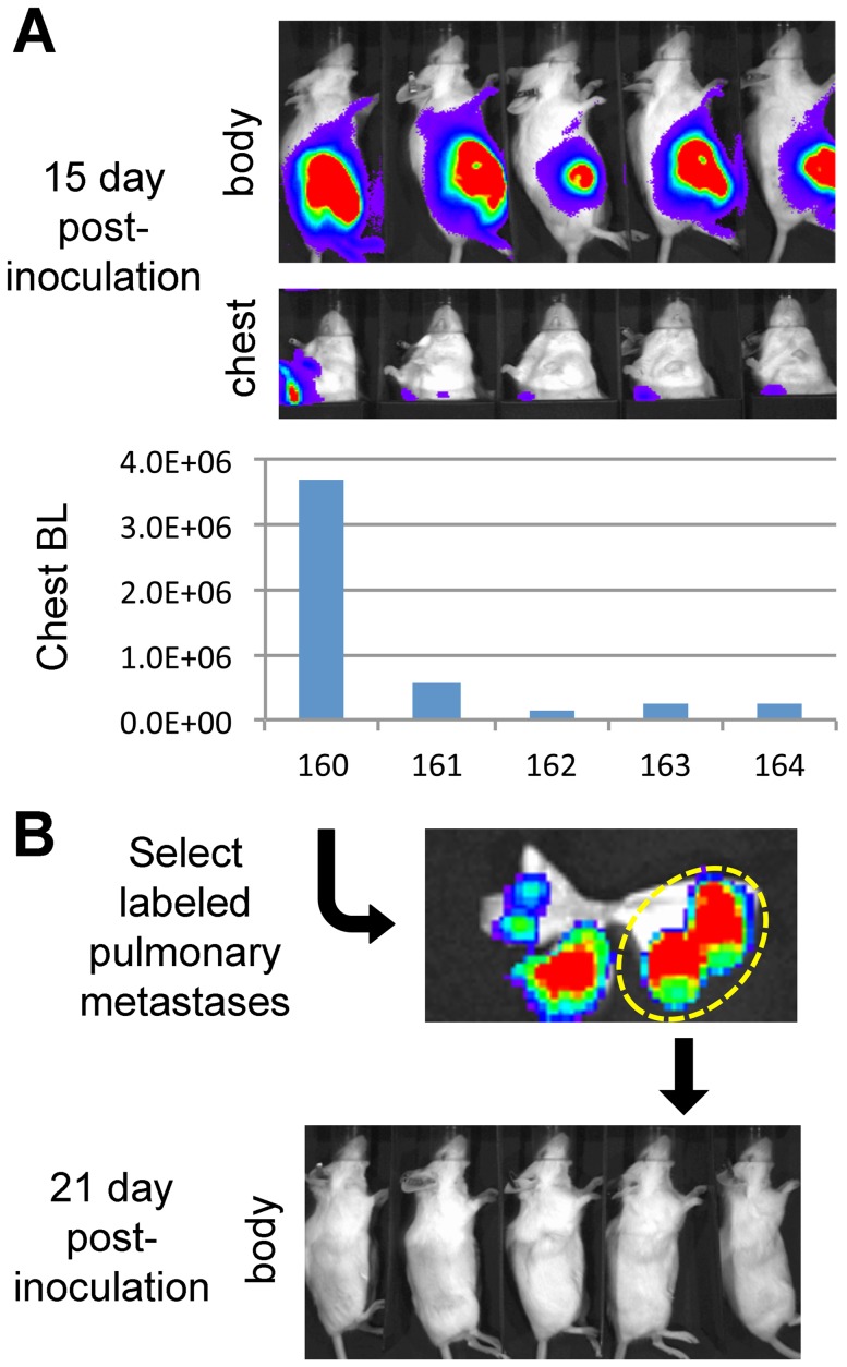
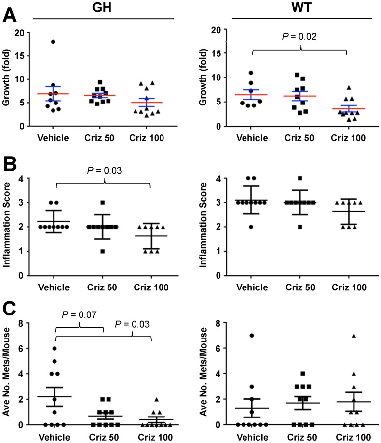
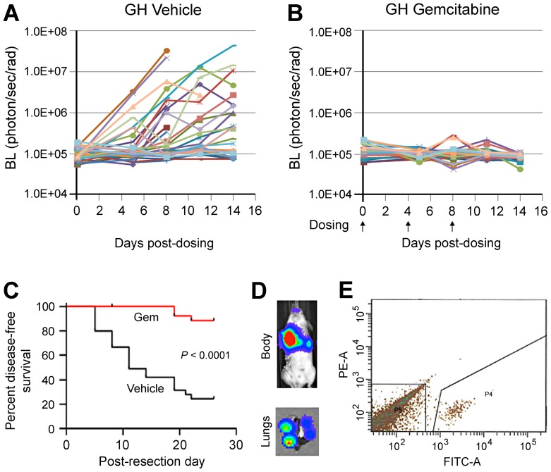
Preclinical therapeutic assessment currently relies on the growth response of established human cell lines xenografted into immunocompromised mice, a strategy that is generally not predictive of clinical outcomes. Immunocompetent genetically engineered mouse (GEM)-derived tumor allograft models offer highly tractable preclinical alternatives and facilitate analysis of clinically promising immunomodulatory agents. Imageable reporters are essential for accurately tracking tumor growth and response, particularly for metastases. Unfortunately, reporters such as luciferase and GFP are foreign antigens in immunocompetent mice, potentially hindering tumor growth and confounding therapeutic responses. Here we assessed the value of reporter-tolerized GEMs as allograft recipients by targeting minimal expression of a luciferase-GFP fusion reporter to the anterior pituitary gland (dubbed the "Glowing Head" or GH mouse). The luciferase-GFP reporter expressed in tumor cells induced adverse immune responses in wildtype mouse, but not in GH mouse, as transplantation hosts. The antigenicity of optical reporters resulted in a decrease in both the growth and metastatic potential of the labeled tumor in wildtype mice as compared to the GH mice. Moreover, reporter expression can also alter the tumor response to chemotherapy or targeted therapy in a context-dependent manner. Thus the GH mice and experimental approaches vetted herein provide concept validation and a strategy for effective, reproducible preclinical evaluation of growth and response kinetics for traceable tumors.


Conflict of interest statement

Competing Interests: Co-authors John Carter, Zoe Weaver-Ohler, Carrie Bonomi, Rajaa El Meskini, Philip Martin, Cari Graff-Cherry and Lionel Feigenbaum are employed by Leidos Biomedical Research Inc. Leidos Biomedical Research Inc., is dedicated to a single contract to operate the Frederick National Laboratory for Cancer Research (FNLCR), a Federally Funded Research and Development Center. It is not involved in any employment, consultancy, patents, products in development or marketed products etc. related to this study. This does not alter the authors' adherence to PLOS ONE policies on sharing data and materials.

Figures

Figure 1
Figure 1. Inconsistency of ffLuc-eGFP reporter activity in labeled tumors during passages in syngeneic immunocompetent mice.
A, Murine Lewis Lung Carcinoma (LLC) cells were infected with ffLuc-eGFP-expressing lentivirus ex vivo, and subcutaneously transplanted into five syngeneic albino C57BL/6 (c-Brd) mice (#160–164). Reporter activity was monitored by BL imaging of subcutaneous tumor growth (body) and pulmonary metastasis (chest). At day 15 after inoculation, a metastatic BL signal was found in one of the mice (#160 in the lower panel). B, The lung was harvested from #160, and a single glowing metastatic nodule selected using ex vivo imaging (upper panel) was transplanted into five c-Brd mice in the second passage. Imaging results showed that the reporter activity could not be consistently maintained in the resulting palpable tumors (lower panel).
Figure 2
Figure 2. Generation of the rGH-ffLuc-eGFP (“Glowing Head”) genetically engineered mouse.
A, Structure of the expression vector for generation of Glowing Head (GH) transgenic mice. Expression of a firefly luciferase-eGFP fusion gene (ffLuc-eGFP) was targeted to the mouse anterior pituitary gland by using the rat growth hormone promoter (rGH) and human growth hormone gene sequences, which include a polyadenylylation site (hGHpA)20. B, Optical expression pattern of transgene in GH mice as visualized by BL imaging. Reporter activity was detected in the anterior pituitary gland of both genders and the testes of male mice. C, Serum levels of growth hormone from age-matched GH mice and wildtype (WT) c-Brd mice was assessed by ELISA (mean ± SE). Blood was withdrawn at the same time of day. No significant differences in circulating growth hormone levels between the GH and WT mice were found. D, ffLuc-eGFP-labeled LLC tumors were subcutaneously transplanted into WT, GH, and NOD-SCID mice. Blood was withdrawn to prepare sera when tumors reached 500 mm3, and the serum levels of anti-GFP antibody were analyzed by ELISA. The levels of anti-GFP antibody in WT mice are significantly higher than those in GH and NOD-SCID mice (p<0.005), but no difference was found between those in GH and NOD-SCID mice (p = 0.19). The sera from healthy mice without tumor transplantation served as controls to define zero point.
Figure 3
Figure 3. Reporter activity and metastasis of ffLuc-eGFP-labeled cancer cells are consistent in GH mice but suppressed in immunocompetent wildtype mice.
A–D, Functional comparison of GH and WT mice as transplantation hosts using a breast cancer model. The GFP+ population from ffLuc-eGFP-transduced Mvt1 mouse breast cancer cells was isolated and expanded in culture. 1×10⁵ cells were injected into the mammary fat pads (m.f.p.) of WT and GH syngeneic FVB/N mice, followed by BL imaging to monitor tumor growth. Though tumors grew in the fat pads of both groups, the BL intensity (mean ± SE) of those in WT mice was highly suppressed relative to GH mice (A). *, P = 0.083; **, P<0.001. (B–C) Upon reaching 500 mm3 m.f.p. tumors were resected, and BL imaging was used to monitor metastatic progression, which is visualized by body BL signal in each mouse. Metastatic disease progressed consistently in GH mice (B), while being suppressed in WT mice (C); the sign and number at side refer to individual mice in each figure. Kaplan-Meier survival analysis showed that GH mice exhibited significantly shorter survival times than WT mice (P = 0.0025). Median survival times in GH and WT groups were 16.5 and 41.5 days, respectively (D). E–H, Behavioral inconsistency of labeled tumors in WT and immunocompromised mice as compared to GH mice. ffLuc-eGFP-labeled LLC tumors were transplanted subcutaneously into syngeneic GH mice, strain-unmatched immunocompromised NOD/SCID (BALB/c) mice, and syngeneic c-Brd (WT) mice. Upon reaching 500 mm3 subcutaneous tumors were resected, and mice were subjected to periodic BL imaging to monitor metastasis. The growth curves representing metastatic growth in GH (E), NOD/SCID (F), and c-Brd WT mice (G) are shown; the sign and number at side refer to individual mice in each figure. Compared to those in GH mice, the metastatic growth in the other two groups exhibited heterogeneous and delayed patterns. In accordance with their more efficient metastatic progression, Kaplan-Meyer survival analysis showed that GH mice exhibited significantly shorter survival time than the other two strains of mice (P = 0.0037). Median survival times in WT, NOD/SCID, and GH groups were 18 days, 16.5 days and 11 days, respectively (H).
Figure 4
Figure 4. Immunogenicity of ffLuc-eGFP alters the response of tumors to chemotherapeutic agents in wildtype mice compared to GH mice.
Labeled LLC tumors were inoculated subcutaneously into WT and GH c-Brd mice. When the average tumor size reached 125 mm3, each strain of mice was randomized into two groups to receive either control vehicle (Cremophor EL + saline) or paclitaxel. Tumor size was measured periodically. A and B, Tumor growth (fold-increase relative to day 1) in WT and GH c-Brd mice (mean ± SE). Paclitaxel treatment was inefficacious in GH mice (A), but delayed tumor growth in WT mice (*, P<0.05 in a two-tailed T-test) (B). Ctrl, control vehicle; Tx, paclitaxel treatment. C, Spleen size in each group (mean ± SE). Spleens in paclitaxel-treated WT c-Brd mice were marginally bigger than those in vehicle-treated c-Brd mice but significantly bigger than those in both groups of GH mice. No significant difference was found between the two GH mouse groups. D and E, Enlarged spleens in paclitaxel-treated WT mice correspond to higher CD8/CD4 ratios. Splenocytes were prepared from spleens harvested from mice from each treatment group. These were stained with anti-mouse CD4 or CD8 antibodies, and analyzed by flow cytometry and Cellometer to obtain the ratio of the CD8+ to CD4+ subpopulation (CD8/CD4) in WT and GH c-Brd host mice (mean ± SE) (D). E, Regressional analysis demonstrated a significant correlation between CD8/CD4 ratio and spleen size (P<0.01).
Figure 5
Figure 5. Immunogenicity of ffLuc-eGFP alters the response of tumors, including metastases, to targeted therapy in wildtype mice compared to GH mice.
Labeled melanoma tissues from the HGF/CDK4R24C-transgenic mouse were subcutaneously inoculated into WT and GH c-Brd mice. When the average tumor size reached 125 mm3, mice from each strain were randomized into two groups to receive either control vehicle (saline) or the MET inhibitor crizotinib (Criz). Tumor size was measured periodically. A, Fold-tumor growth in WT and GH c-Brd mice (mean ± SE). 100 mg/kg Criz treatment delayed tumor growth in WT mice (right panel, P<0.02 in two-tailed T-test), but no efficacy was found in GH mice (left panel). B, When primary tumors reached 2000 mm3, the mice were euthanized to harvest tumors and lungs. Tumors were subjected to pathological analysis of inflammatory infiltrates according to the scoring system: minimal  = 1, mild  = 2, moderate  = 3, severe  = 4. Criz at 100 mg/kg significantly reduced inflammatory infiltration of the primary tumors in GH mice (left panel), but not in WT mice (right panel). C, The fixed lungs were subjected to the quantitation of metastatic foci. Criz reduced the number of pulmonary metastases in GH mice in a dose-dependent manner (left panel), but had no effect in WT mice (right panel).
Figure 6
Figure 6. The GH mouse allows for consistent tracking of the progression of labeled metastases, their therapeutic responses and isolation in a preclinical adjuvant study.
ffLuc-eGFP-labeled LLC tumors were transplanted subcutaneously into GH mice. Upon reaching 500 mm3, the primary tumors were resected. A and B, GH mice were randomized into control and treated groups to receive vehicle and gemcitabine at 25 mg/kg, respectively. Metastatic progression in mice was periodically monitored by BL imaging. Metastatic growth was efficient in untreated GH mice (A) but suppressed by gemcitabine (B). C, Kaplan-Meyer analysis showed that survival was significantly shorter in control compared to treated GH mice. D and E, GFP+ cancer cells can be readily isolated from whole lungs of GH mice. The lungs were harvested from control GH mice, made into a single-cell suspension, and subjected to sorting by FACS to isolate GFP+ cells. The representative result from mouse #806 is shown. The in vivo image of mice and ex vivo image of lung (D) showed BL signal from pulmonary metastases and individual lung nodules, respectively. The GFP+ cancer cells were successfully isolated from whole lung by FACS sorting (P4 subpopulation in E).

References

    1. Herper M (2012) The Truly Staggering Cost Of Inventing New Drugs. Forbes. Available: http://www.forbes.com/sites/matthewherper/2012/02/10/the-truly-staggerin....
    1. Sharpless NE, Depinho RA (2006) The mighty mouse: genetically engineered mouse models in cancer drug development. Nat Rev Drug Discov 5: 741–754. - PubMed
    1. Suggitt M, Bibby MC (2005) 50 years of preclinical anticancer drug screening: empirical to target-driven approaches. Clin Cancer Res 11: 971–981. - PubMed
    1. Administration USFaD (2004) Innovation or Stagnation: Challenge and Opportunity on the Critical Path to New Medical Products.
    1. Zitvogel L, Apetoh L, Ghiringhelli F, Kroemer G (2008) Immunological aspects of cancer chemotherapy. Nat Rev Immunol 8: 59–73. - PubMed

Publication types

MeSH terms