. 2014 Nov 4;9(11):e109575.
doi: 10.1371/journal.pone.0109575. eCollection 2014.

YAP/TEAD co-activator regulated pluripotency and chemoresistance in ovarian cancer initiated cells

Affiliations

YAP/TEAD co-activator regulated pluripotency and chemoresistance in ovarian cancer initiated cells

Yan Xia et al. PLoS One. .

Abstract

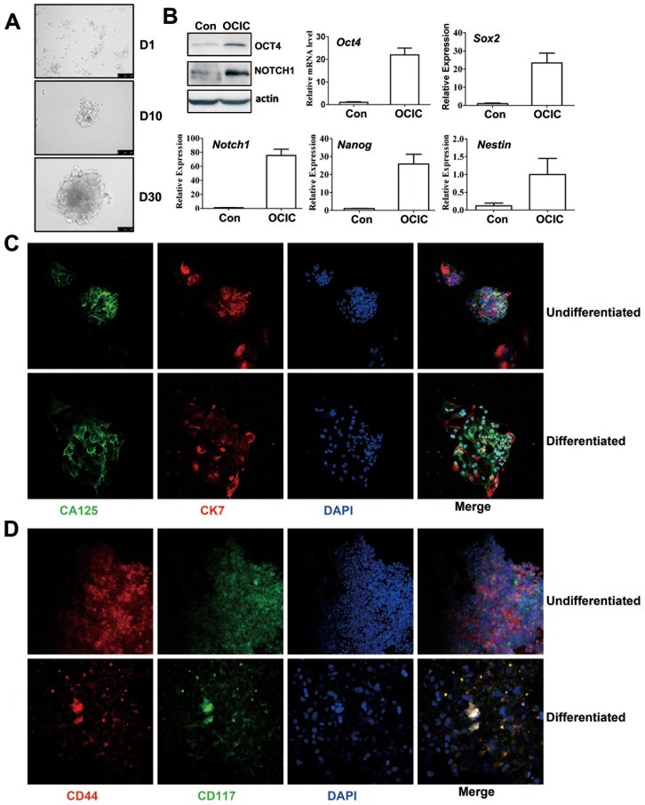
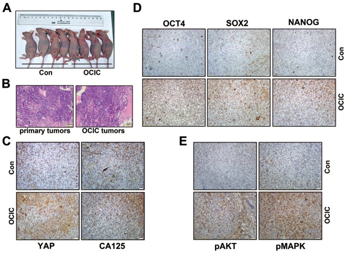
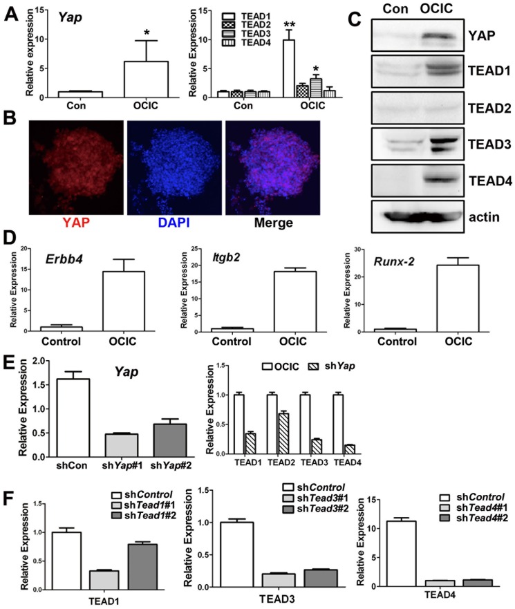
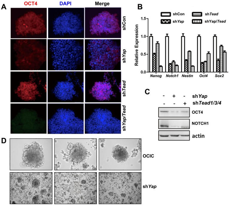
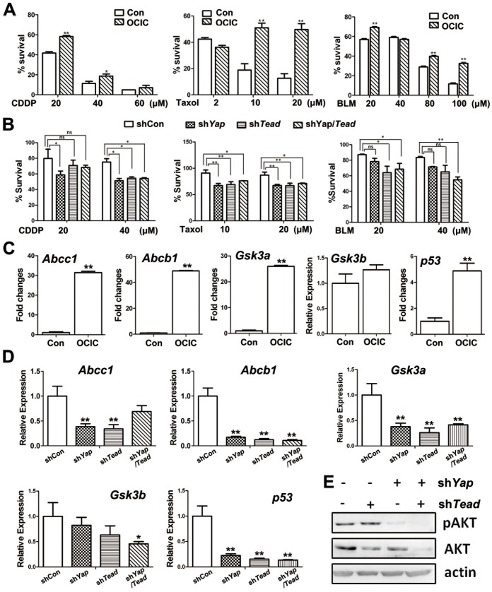
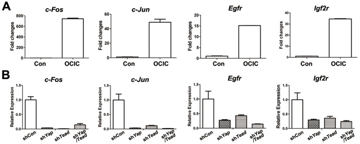
Recent evidence suggests that some solid tumors, including ovarian cancer, contain distinct populations of stem cells that are responsible for tumor initiation, growth, chemo-resistance, and recurrence. The Hippo pathway has attracted considerable attention and some investigators have focused on YAP functions for maintaining stemness and cell differentiation. In this study, we successfully isolated the ovarian cancer initiating cells (OCICs) and demonstrated YAP promoted self-renewal of ovarian cancer initiated cell (OCIC) through its downstream co-activator TEAD. YAP and TEAD families were required for maintaining the expression of specific genes that may be involved in OCICs' stemness and chemoresistance. Taken together, our data first indicate that YAP/TEAD co-activator regulated ovarian cancer initiated cell pluripotency and chemo-resistance. It proposed a new mechanism on the drug resistance in cancer stem cell that Hippo-YAP signal pathway might serve as therapeutic targets for ovarian cancer treatment in clinical.


Conflict of interest statement

Competing Interests: The authors have declared that no competing interests exist.

Figures

Figure 1
Figure 1. Isolation and culture of ovarian cancer-initiating cell (OCIC) spheroids with characteristics of self-renewal and anchorage-independent growth.
A: Images of non-adherent spherical cell clusters derived from cultured primary ovarian cancer cells. Scale bars  = 100 µM. B: Western blotting and real-time RT-PCR results showing enhanced expression of the indicated genes in OCICs. Relative mRNA levels were determined by normalizing to endogenous β-actin mRNA levels (used as an internal control) using Microsoft EXCEL. For each indicated gene, the relative transcript level of the control sample (left-hand bar of each graph) was set at 1. The relative transcript levels of other samples were compared to the control, and fold-changes are shown in the graph. C–D: Representative immunofluorescence staining results for CA125, CK7, CD44, and CD117 expression in undifferentiated and differentiated OCICs. Nuclei were stained with DAPI. Scale bar  = 100 µM for all panels.
Figure 2
Figure 2. OCICs have stronger tumorigenic capability than primary ovarian cancer cells.
A: Images of nude mice showing xenograft ovarian tumor formation after injecting OCIC spheres. Cell concentrations used 104 in each group. B: H&E staining results showing the histology of tumors derived from subcutaneously transplanted OCIC spheroids. C: Representative IHC results for CA125 and YAP expression in human xenograft tumors derived from OCICs. Specific protein expression is indicated by the brown color and nuclei (blue) were counterstained with DAPI. D: Representative IHC results for the expression of the pluripotency markers OCT4, SOX2, and NANOG in xenograft ovarian tumors derived from OCIC spheroids. E: Representative IHC results for phosphorylated AKT and pMAPK (ERK1/2) expression in the xenograft tumors derived from OCICs. Scale bar  = 200 µM for B-E panels.
Figure 3
Figure 3. YAP and TEAD are required for maintaining OCIC pluripotency.
A-C: Real-time RT-PCR (A), immunofluorescence staining (B), and Western blotting (C) results for YAP and TEAD1-4 expression levels in primary ovarian cancer cells (control) and OCICs. Nuclei were stained with DAPI. D: Real-time RT-PCR results for mRNA levels of known YAP/TEAD target genes, including Runx2, Itgb2, and Erbb4, in primary ovarian cancer cells (control) and OCIC cells. E-F: Real-time RT-PCR results for the RNAi depletion efficiencies of YAP, TEAD1, TEAD3, and TEAD4, after using two different shRNAs for each gene.
Figure 4
Figure 4. Pluripotency markers' expression in YAP- and TEAD1/3/4-silenced OCICs.
A: Representative immunofluorescence staining results for OCT-4 expression in Yap- and Tead1/3/4-silenced OCICs. B-C: Real-time RT-PCR (B) and Western blotting (C) results showing that the indicated genes were down-regulated in OCICs after RNAi depletion of Yap and Tead1/3/4. D: Images of OCIC spherical clusters without (upper panels) and with (lower panels) shYap-treatment by 200 times magnification.
Figure 5
Figure 5. YAP-TEAD confers chemotherapeutic drug resistance to OCICs by regulating specific target genes' expression.
A. Survival rates of primary ovarian cancer cells (control) and OCICs after treatment with cisplatin (CDDP), taxol, or bleomycin at the indicated concentrations. OCICs and control cells were treated with drugs for 48 h. *, P<0.01, compared with the corresponding control group. **, P<0.001, compared with the corresponding control group. B. Survival rates of OCICs with or without Yap and Tead1/3/4 knockdown after treatment with CDDP, taxol, or bleomycin at the indicated concentrations for 48 h. ns, not significant; *, P<0.01; **, P<0.001. C-D: Real-time RT-PCR results showing that the indicated genes were expressed in OCICs at higher levels than in primary ovarian cancer cells (C). Indicated genes' expression levels in OCICs were significantly down-regulated with Yap and Tead1/3/4 RNAi (D). *, P<0.01; **, P<0.001. E. Western blotting results for AKT and pAKT levels in OCICs with or without Yap/Tead1/3/4 shRNA treatment.
Figure 6
Figure 6. MAPK pathway genes regulated by YAP in OCICs.
A. Real-time RT-PCR results showing that the indicated genes were expressed in OCICs at higher levels than in primary ovarian cancer cells (control). B. Real-time RT-PCR results showing that the indicated genes' expression levels were significantly down-regulated in OCICs with Yap/Tead1/3/4 RNAi.

Similar articles

Cited by

References

    1. Jemal A, Siegel R, Ward E, Hao Y, Xu J, et al. (2008) Cancer statistics, 2008. CA Cancer J Clin 58: 71–96. - PubMed
    1. Auersperg N, Wong AS, Choi KC, Kang SK, Leung PC (2001) Ovarian surface epithelium: biology, endocrinology, and pathology. Endocr Rev 22: 255–288. - PubMed
    1. Steg AD, Bevis KS, Katre AA, Ziebarth A, Dobbin ZC, et al. (2012) Stem cell pathways contribute to clinical chemoresistance in ovarian cancer. Clin Cancer Res 18: 869–881. - PMC - PubMed
    1. Kulkarni-Datar K, Orsulic S, Foster R, Rueda BR (2013) Ovarian tumor initiating cell populations persist following paclitaxel and carboplatin chemotherapy treatment in vivo. Cancer Lett 339: 237–246. - PubMed
    1. Wang L, Mezencev R, Bowen NJ, Matyunina LV, McDonald JF (2012) Isolation and characterization of stem-like cells from a human ovarian cancer cell line. Mol Cell Biochem 363: 257–268. - PubMed

Publication types

MeSH terms