Review
. 2015 Jan;88(1045):20140599.
doi: 10.1259/bjr.20140599.

Cardiac MR assessment of cardiac myxomas

Affiliations
Review

Cardiac MR assessment of cardiac myxomas

A Abbas et al. Br J Radiol. 2015 Jan.

Abstract

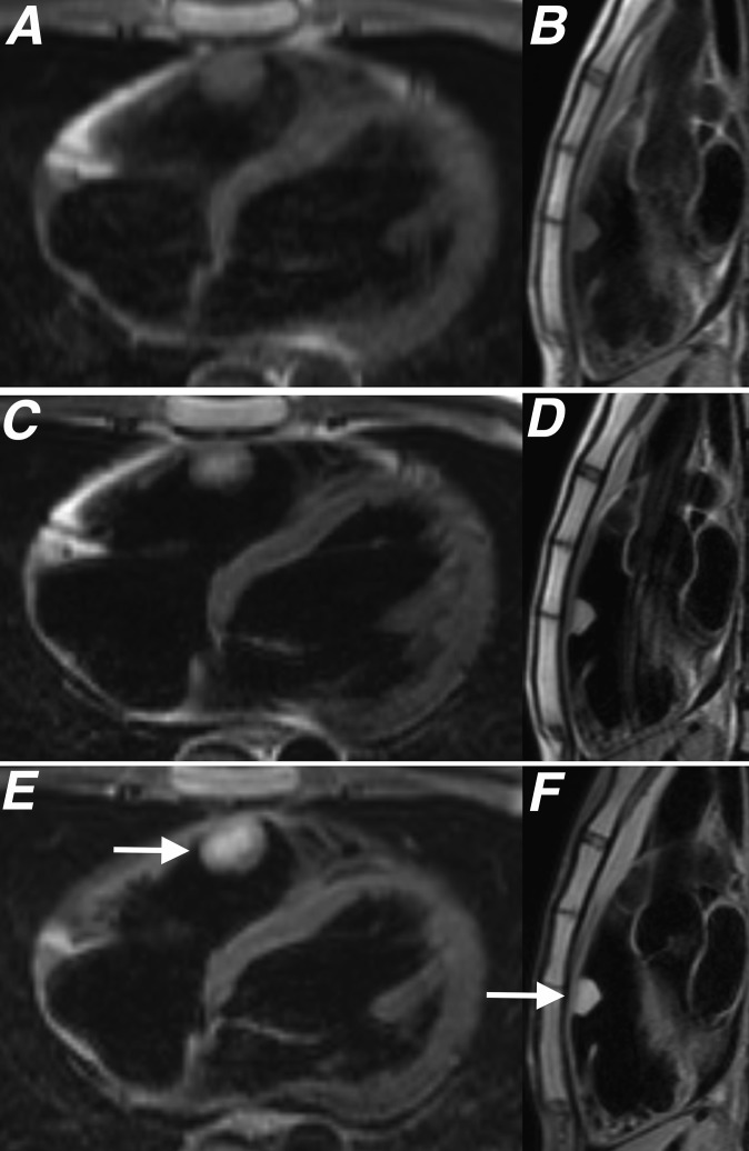
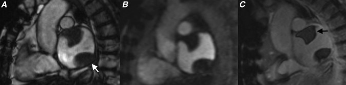
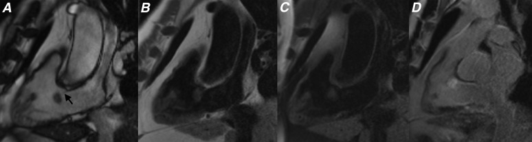
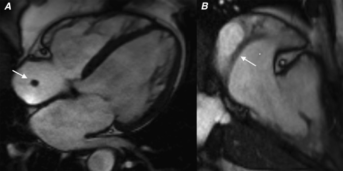
Cardiac myxomas are the most common benign primary cardiac tumour to present in adulthood. While most patients present with symptoms of cardiac obstruction, embolic phenomena or constitutional impairment, up to a fifth of patients remain asymptomatic and are incidentally diagnosed on imaging. Although echocardiography is usually the initial imaging modality used to evaluate these patients, cardiac MRI (CMR) has emerged over the past decade as the primary imaging modality in the assessment of patients with cardiac tumours. The superior tissue characterization capability of CMR means that it is able to determine the nature of some tumours pre-operatively and performs well in differentiating myxomas from thrombus. We present a pictorial review highlighting the key CMR features of myxomas and show how these lesions can be differentiated from thrombus and other cardiac masses.


Figures

Figure 1.
Figure 1.
Schematic showing typical cardiac MRI protocol used for patients being assessed for cardiac myxomas. FS, fat saturated; LGE, late gadolinium enhancement; LV, left ventricle; SSFP, steady-state free procession.
Figure 2.
Figure 2.
(a) T1 weighted, (b) T2 weighted, (c) T2 weighted with fat saturation, (d) cine steady-state free procession, (e) first pass perfusion and (f) late gadolinium enhancement four-chamber sequences showing the typical appearance of a left atrial myxoma (black and white arrows). A diagnosis of cardiac myxoma was histologically confirmed following excision.
Figure 3.
Figure 3.
(a) Cine steady-state free procession, (b) first pass perfusion and (c) late gadolinium enhancement (LGE) four-chamber sequences showing a left atrial myxoma. Note how the lesion shows limited enhancement on first pass perfusion sequences (black arrow) and heterogeneous enhancement on LGE sequences (white arrow).
Figure 4.
Figure 4.
(a) Four-chamber steady-state free procession (SSFP) during systole, (b) four-chamber SSFP during diastole, (c) four-chamber first pass perfusion and (d) two-chamber right ventricle long axis first pass perfusion sequences showing a right atrial myxoma. The lesion shows no enhancement on first pass perfusion, which is an imaging feature demonstrated by some myxomas. Also note how the lesion prolapses through the tricuspid valve during diastole (white arrows). Following excision, this lesion demonstrated the typical histological features of a cardiac myxoma with a predominant myxoid tissue composition.
Figure 5.
Figure 5.
(a, b) T1 weighted, (c, d) T2 weighted and (e, f) T2 fat-saturated sequences acquired in two planes orientated perpendicular to a myxoma (white arrows) located in the right ventricular outflow tract. Histological assessment of this lesion after surgical excision showed the lesion to be composed of spindled and stellate cells in a myxoid matrix with associated haemosiderin typical for a cardiac myxoma.
Figure 6.
Figure 6.
(a) Parasagittal cine steady-state free procession, (b) parasagittal first pass perfusion and (c) parasagittal late gadolinium enhancement (LGE) sequences through the left atrium showing two separate left atrial thrombi (white and black arrows). Note how these thrombi show no enhancement on first pass perfusion or LGE sequences (black arrow).
Figure 7.
Figure 7.
(a) Cine steady-state free procession (SSFP), (b) T1 weighted, (c) T2 weighted with fat saturation and (d) late gadolinium enhancement (LGE) sequences showing a small lesion with a thin attachment to the tricuspid valve leaflets (black arrow). This lesion was very mobile on cine SSFP sequences and the relatively homogenous increased signal seen on the LGE sequences suggest focal gadolinium accumulation. These appearances are typical for a fibroelastoma.
Figure 8.
Figure 8.
(a) Four-chamber cine steady-state free procession (SSFP) and (b) parasagittal cine SSFP showing typical appearances of a prominent rather tubular crista terminalis.

References

    1. Lam KY, Dickens P, Chan AC. Tumors of the heart. A 20-year experience with a review of 12,485 consecutive autopsies. Arch Pathol Lab Med 1993; 117: 1027–31. - PubMed
    1. Motwani M, Kidambi A, Herzog BA, Uddin A, Greenwood JP, Plein S. MR imaging of cardiac tumors and masses: a review of methods and clinical applications. Radiology 2013; 268: 26–43. doi: 10.1148/radiol.13121239 - DOI - PubMed
    1. Grebenc ML, Rosado-de-Christenson ML, Green CE, Burke AP, Galvin JR. Cardiac myxoma: imaging features in 83 patients. Radiographics 2002; 22: 673–89. - PubMed
    1. O'Donnell DH, Abbara S, Chaithiraphan V, Yared K, Killeen RP, Cury RC, et al. . Cardiac tumors: optimal cardiac MR sequences and spectrum of imaging appearances. AJR Am J Roentgenol 2009; 193: 377–87. doi: 10.2214/AJR.08.1895 - DOI - PubMed
    1. Buckley O, Mada R, Kwong R, Rybicki FJ, Hunsaker A. Cardiac masses, part 1: imaging strategies and technical considerations. AJR Am J Roentgenol 2013; 197: W837–41. doi: 10.2214/AJR.10.7260 - DOI - PMC - PubMed

MeSH terms