. 2014 Nov 18;111(46):16496-501.
doi: 10.1073/pnas.1418087111. Epub 2014 Nov 4.

Cerebral small vessel disease-related protease HtrA1 processes latent TGF-β binding protein 1 and facilitates TGF-β signaling

Affiliations

Cerebral small vessel disease-related protease HtrA1 processes latent TGF-β binding protein 1 and facilitates TGF-β signaling

Nathalie Beaufort et al. Proc Natl Acad Sci U S A. .

Abstract

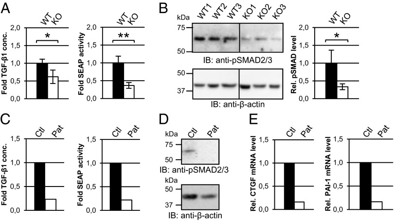
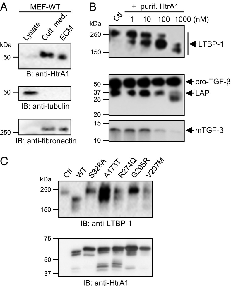
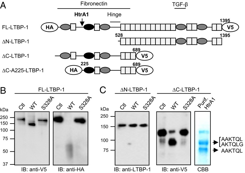
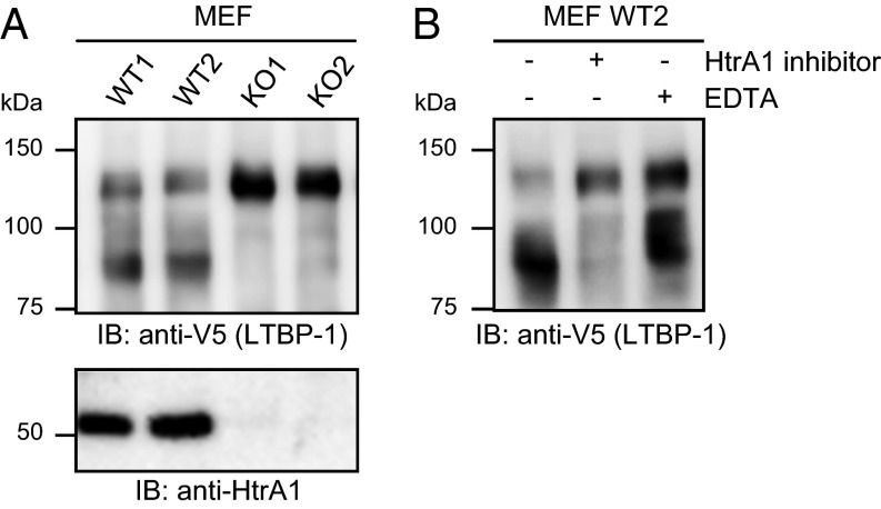
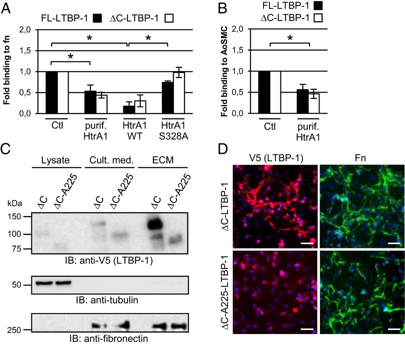
High temperature requirement protein A1 (HtrA1) is a primarily secreted serine protease involved in a variety of cellular processes including transforming growth factor β (TGF-β) signaling. Loss of its activity causes cerebral autosomal recessive arteriopathy with subcortical infarcts and leukoencephalopathy (CARASIL), an inherited form of cerebral small vessel disease leading to early-onset stroke and premature dementia. Dysregulated TGF-β signaling is considered to promote CARASIL pathogenesis, but the underlying molecular mechanisms are incompletely understood. Here we present evidence from mouse brain tissue and embryonic fibroblasts as well as patient skin fibroblasts for a facilitating role of HtrA1 in TGF-β pathway activation. We identify latent TGF-β binding protein 1 (LTBP-1), an extracellular matrix protein and key regulator of TGF-β bioavailability, as a novel HtrA1 target. Cleavage occurs at physiological protease concentrations, is prevented under HtrA1-deficient conditions as well as by CARASIL mutations and disrupts both LTBP-1 binding to fibronectin and its incorporation into the extracellular matrix. Hence, our data suggest an attenuation of TGF-β signaling caused by a lack of HtrA1-mediated LTBP-1 processing as mechanism underlying CARASIL pathogenesis.

Keywords: LTBP-1; extracellular matrix; proteolysis; small vessel disease.


Conflict of interest statement

The authors declare no conflict of interest.

Figures

Fig. 1.
Fig. 1.
TGF-β signaling is impaired in HtrA1-deficient mouse brains. (A) Relative HtrA1 mRNA levels in mouse brain, kidney and lung were determined by real-time PCR using β-actin for normalization (WT: wild-type; HET: heterozygous; KO: HTRA1 knockout). The mean value measured for WT mouse tissues was set to 1. n = 3. (B) Mouse brain lysates were subjected to HtrA1 immunoprecipitation (IP) using rabbit anti-HtrA1 antiserum, followed by HtrA1 immunoblotting (IB) using anti-HtrA1 (23E1) antibody. (C) Brain sections of 20- to 26-mo-old mice were stained with phosphoSMAD2/3 antibody (red) and nuclei were visualized with DAPI (blue). (Scale bar: 50 µm.) Quantification of the immunopositive area in corresponding brain regions of HTRA1-KO and HTRA1-WT mice is shown as percentage of the total image area. n = 6. (D) Relative CTGF mRNA levels from 2 and 18- to 26-mo-old mouse brains were determined by real-time PCR. 2 mo: n = 4–7; 18–26 mo: 7–10. Results are expressed as mean ± SEM; *P < 0.05, **P < 0.01.
Fig. 2.
Fig. 2.
Decreased TGF-β signaling in HtrA1-deficient and patient fibroblasts. (A) Culture medium from MEF cell lines was used to measure TGF-β1 protein concentration by ELISA (Left) and bioactive TGF-β levels by a cellular bioassay using secreted alkaline phosphatase (SEAP) as readout (Right). After normalization to total protein amounts in the medium HtrA1-WT values were set to 1. (B) MEF lysates were analyzed by immunoblotting using an anti-phosphoSMAD2/3 or an anti-β-actin antibody. PhosphoSMAD signals were quantified densitometrically and normalized to β-actin. (C) Culture medium from skin fibroblasts derived from a control individual (Ctl) or from a CARASIL patient (Pat) was collected. Total (Left) and bioactive TGF-β1 levels (Right) were measured. (D) Human fibroblast lysates were analyzed by immunoblotting using an anti-phosphoSMAD2/3 or an anti-β-actin antibody. (E) CTGF and PAI-1 mRNA levels were measured by real-time PCR and normalized to β-actin. Results are representative of at least two independent experiments.
Fig. 3.
Fig. 3.
HtrA1 efficiently processes LTBP-1. (A) Upon sequential extraction, HtrA1 expression was analyzed by immunoblotting (IB) in MEF lysate, culture medium (Cult. med.) and extracellular matrix (ECM). All fractions were also probed with anti-tubulin and anti-fibronectin antibodies. (B) Medium from LTBP-1- (Upper) or TGF-β1- (Lower) expressing HEK293 cells was treated with the indicated concentrations of purified HtrA1 or with PBS (Ctl) followed by immunoblotting. pro-TGF-β: TGF-β proform; LAP: latency-associated peptide; mTGF-β: mature TGF-β. (C) LTBP-1-containing medium was exposed to medium from vector-transfected cells (Ctl) or cells expressing either wild-type HtrA1 (WT) or one of four different CARASIL mutants. LTBP-1 (Upper) and HtrA1 (Lower) were detected by immunoblotting.
Fig. 4.
Fig. 4.
Mapping of the HtrA1 cleavage site within LTBP-1. (A) Domain organization of LTBP-1 including cysteine-rich domains (gray ovals), a hybrid domain (black oval) and EGF-like domains (rectangles). The protease-sensitive hinge region as well as binding regions for fibronectin and TGF-β are shown. The HtrA1 cleavage site is indicated by an arrow. Numbers represent amino acids. The various LTBP-1 constructs used in this study are depicted. HA: Hemagglutinin tag; V5: V5 tag. (B) Medium from HEK293 cells expressing full-length (FL-) LTBP-1 was exposed to medium from vector- (Ctl), HtrA1 WT-, or HtrA1 S328A-transfected cells and LTBP-1 analyzed by immunoblotting. (C) HEK293 cell-derived ΔN-LTBP-1 (Left) or ΔC-LTBP-1 (Center) was treated with control, HtrA1 WT-, or HtrA1 S328A-containing medium and immunoblotting was performed. Purified ΔC-LTBP-1 (Right) was treated with purified HtrA1 and upon SDS/PAGE and Coomassie brilliant blue (CBB) staining two proteolytic fragments were isolated and subjected to Edman degradation. The resulting amino-terminal sequences are indicated. Note that the smaller fragment (∼75 kDa) could not be detected by an anti-V5 antibody indicating carboxyl-terminal processing.
Fig. 5.
Fig. 5.
LTBP-1 processing by endogenous HtrA1. (A) Concentrated culture medium from wild-type (WT) or HTRA11 knockout (KO) MEF lines was incubated with ∆C-LTBP-1-containing medium. LTBP-1 and HtrA1 were analyzed by immunoblotting using anti-V5 and anti-HtrA1 (16C8) antibodies. (B) ∆C-LTBP-1 was exposed to medium from the HtrA1-WT2 line in the absence or presence of a selective HtrA1 inhibitor or EDTA, before LTBP-1 was immunodetected.
Fig. 6.
Fig. 6.
Regulation of the LTBP-1 interaction with ECM by HtrA1-mediated proteolysis. (A) HEK293 cell-derived full-length LTBP-1 (FL-LTBP-1) or ΔC-LTBP-1 was incubated with PBS or medium from vector-transfected cells (Ctl), with 250 nM purified HtrA1 or with medium containing HtrA1-WT or -S328A, before LTBP-1 binding to immobilized fibronectin was measured and values obtained by control treatment were set to 1. (B) HEK293 cell-derived FL-LTBP-1 or ΔC-LTBP-1 was incubated with PBS (Ctl) or 250 nM purified HtrA1 before binding to fixed AoSMC cultures was measured. Results are expressed as mean ± SEM of 4–6 experiments; *P < 0.05. (C) Cell culture medium (Cult. med.) as well as cellular (Lysate) and matrix (ECM) fractions from MEF cells transfected with plasmids encoding ΔC-LTBP-1 or ΔC-A225-LTBP-1 were analyzed by anti-V5 (LTBP-1), anti-tubulin or anti-fibronectin immunoblotting. (D) Transfected MEF cells were subjected to immunofluorescence microscopy using anti-V5 (LTBP-1) or anti-fibronectin antibodies. (Scale bar: 50 µm.)

Comment in

References

    1. Wardlaw JM, Smith C, Dichgans M. Mechanisms of sporadic cerebral small vessel disease: Insights from neuroimaging. Lancet Neurol. 2013;12(5):483–497. - PMC - PubMed
    1. Dichgans M. Genetics of ischaemic stroke. Lancet Neurol. 2007;6(2):149–161. - PubMed
    1. Federico A, et al. Hereditary cerebral small vessel diseases: A review. J Neurol Sci. 2012;322(1-2):25–30. - PubMed
    1. Chabriat H, Joutel A, Dichgans M, Tournier-Lasserve E, Bousser MG. Cadasil. Lancet Neurol. 2009;8(7):643–653. - PubMed
    1. Fukutake T. Cerebral autosomal recessive arteriopathy with subcortical infarcts and leukoencephalopathy (CARASIL): From discovery to gene identification. J Stroke Cerebrovasc Dis. 2011;20(2):85–93. - PubMed

Publication types

MeSH terms

Supplementary concepts