

Passive transfer of antibodies to the linear epitope 60 kD Ro 273-289 induces features of Sjögren's syndrome in naive mice
- PMID: 25370295
- PMCID: PMC4367090
- DOI: 10.1111/cei.12480
Passive transfer of antibodies to the linear epitope 60 kD Ro 273-289 induces features of Sjögren's syndrome in naive mice
Abstract
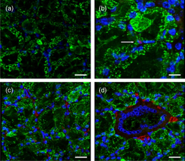
Sjögren's syndrome (SS) is an autoimmune inflammatory disease that primarily affects the lacrimal and salivary glands causing dry eyes and mouth. Antibodies to Ro60 are observed frequently in patients with SS; however, the role of these antibodies in SS initiation and progression remains unclear. The sequence Ro60 273-289 (Ro274) is a known B cell epitope of Ro60 and antibodies to this epitope have been observed in a subset of SS patients and in animals immunized with Ro60 protein. Animals immunized with Ro274 linear peptide develop a Sjögren's-like illness. We hypothesized that passive transfer of anti-Ro274-specific immunoglobulin (Ig)G would induce a Sjögren's-like phenotype. To evaluate this hypothesis, we adoptively transferred affinity-purified Ro274 antibodies into naive BALB/c animals, then evaluated salivary gland histology, function and IgG localization 4 days post-transfer. At this time-point, there was no demonstrable mononuclear cell infiltration and salivary glands were histologically normal, but we observed a functional deficit in stimulated salivary flow of animals receiving Ro274 antibodies compared to animals receiving control IgG. Cellular fractionation and enzyme-linked immunosorbent assay revealed Ro274-specific antibodies in the nucleus and cytoplasmic fractions of isolated parotid salivary gland cells that was confirmed by immunohistochemistry. These data support the hypothesis that antibodies to Ro274 deposit in salivary glands can enter intact salivary gland cells and are involved in the dysregulation of salivary flow in SS.
Keywords: antigens/peptides/epitopes; autoantibodies; autoimmunity; rodent; systemic lupus erythematosus.
© 2014 British Society for Immunology.
Figures


Similar articles
-
Experimental induction of anti-muscarinic type-3-receptor extracellular loop antibodies by immunization with 4-hydroxy-2-nonenal modified Ro60 and unmodified Ro60.Clin Exp Immunol. 2025 Jan 21;219(1):uxae114. doi: 10.1093/cei/uxae114. Clin Exp Immunol. 2025. PMID: 39658078
-
Immunization with 60 kD Ro peptide produces different stages of preclinical autoimmunity in a Sjögren's syndrome model among multiple strains of inbred mice.Clin Exp Immunol. 2013 Jul;173(1):67-75. doi: 10.1111/cei.12094. Clin Exp Immunol. 2013. PMID: 23607771 Free PMC article.
-
Immunization with short peptides from the 60-kDa Ro antigen recapitulates the serological and pathological findings as well as the salivary gland dysfunction of Sjogren's syndrome.J Immunol. 2005 Dec 15;175(12):8409-14. doi: 10.4049/jimmunol.175.12.8409. J Immunol. 2005. PMID: 16339583
-
The complexity of Sjögren's syndrome: novel aspects on pathogenesis.Immunol Lett. 2011 Dec 30;141(1):1-9. doi: 10.1016/j.imlet.2011.06.007. Epub 2011 Jul 12. Immunol Lett. 2011. PMID: 21777618 Review.
-
Sjögren's syndrome: autoantibodies to cellular antigens. Clinical and molecular aspects.Int Arch Allergy Immunol. 2000 Sep;123(1):46-57. doi: 10.1159/000024423. Int Arch Allergy Immunol. 2000. PMID: 11014971 Review.
Cited by
-
Polygenic autoimmune disease risk alleles impacting B cell tolerance act in concert across shared molecular networks in mouse and in humans.Front Immunol. 2022 Aug 24;13:953439. doi: 10.3389/fimmu.2022.953439. eCollection 2022. Front Immunol. 2022. PMID: 36090990 Free PMC article. Review.
-
Sjögren's syndrome and systemic lupus erythematosus: links and risks.Open Access Rheumatol. 2019 Jan 29;11:33-45. doi: 10.2147/OARRR.S167783. eCollection 2019. Open Access Rheumatol. 2019. PMID: 30774485 Free PMC article. Review.
-
Experimental induction of anti-muscarinic type-3-receptor extracellular loop antibodies by immunization with 4-hydroxy-2-nonenal modified Ro60 and unmodified Ro60.Clin Exp Immunol. 2025 Jan 21;219(1):uxae114. doi: 10.1093/cei/uxae114. Clin Exp Immunol. 2025. PMID: 39658078
-
Serological Influences on Dry Eye: Insights from the Sjögren's International Collaborative Clinical Alliance.Ophthalmol Sci. 2025 Jun 2;5(6):100843. doi: 10.1016/j.xops.2025.100843. eCollection 2025 Nov-Dec. Ophthalmol Sci. 2025. PMID: 40661175 Free PMC article.
References
-
- Scofield RH, Henry WE, Kurien BT, James JA, Harley JB. Immunization with short peptides from the sequence of the systemic lupus erythematosus-associated 60-kDa Ro autoantigen results in anti-Ro ribonucleoprotein autoimmunity. J Immunol. 1996;156:4059–4066. - PubMed
-
- Scofield RH, Kaufman KM, Baber U, James JA, Harley JB, Kurien BT. Immunization of mice with human 60-kd Ro peptides results in epitope spreading if the peptides are highly homologous between human and mouse. Arthritis Rheum. 1999;42:1017–1024. - PubMed
-
- Scofield RH, Asfa S, Obeso D, Jonsson R, Kurien BT. Immunization with short peptides from the 60-kDa Ro antigen recapitulates the serological and pathological findings as well as the salivary gland dysfunction of Sjogren's syndrome. J Immunol. 2005;175:8409–8414. - PubMed
-
- Reynolds J, Albouainain A, Duda MA, Evans DJ, Pusey CD. Strain susceptibility to active induction and passive transfer of experimental autoimmune glomerulonephritis in the rat. Nephrol Dial Transplant. 2006;21:3398–3408. - PubMed
MeSH terms
Substances
LinkOut - more resources
Full Text Sources
Other Literature Sources
Medical
Molecular Biology Databases
Research Materials
