. 2015 Jan;35(1):121-30.
doi: 10.1038/jcbfm.2014.185. Epub 2014 Nov 5.

Increased BDNF protein expression after ischemic or PKC epsilon preconditioning promotes electrophysiologic changes that lead to neuroprotection

Affiliations

Increased BDNF protein expression after ischemic or PKC epsilon preconditioning promotes electrophysiologic changes that lead to neuroprotection

Jake T Neumann et al. J Cereb Blood Flow Metab. 2015 Jan.

Abstract

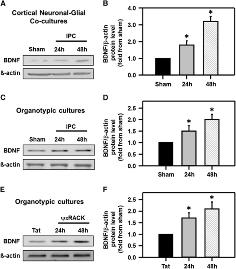
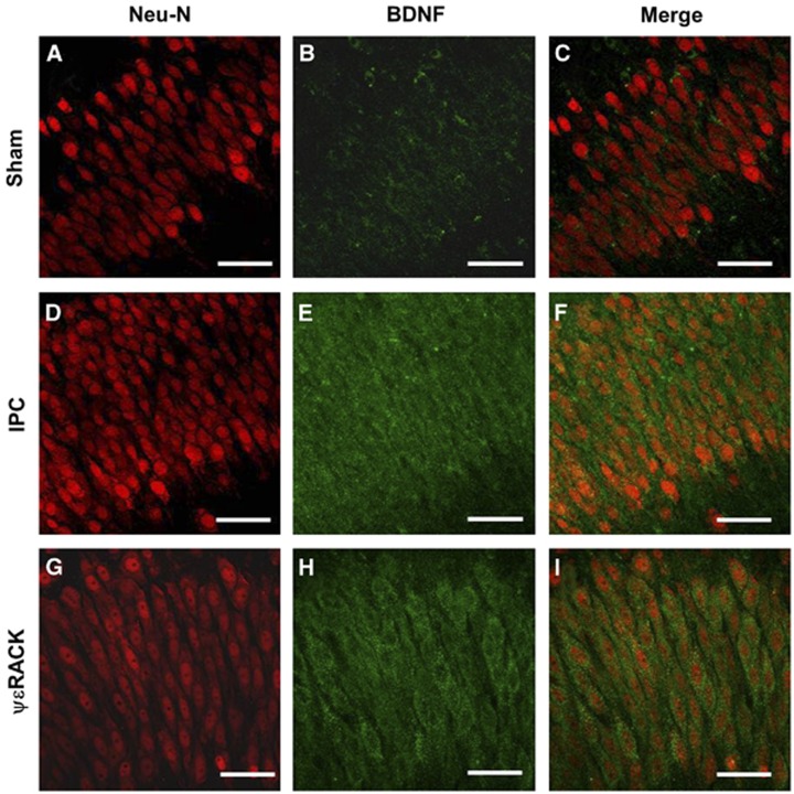
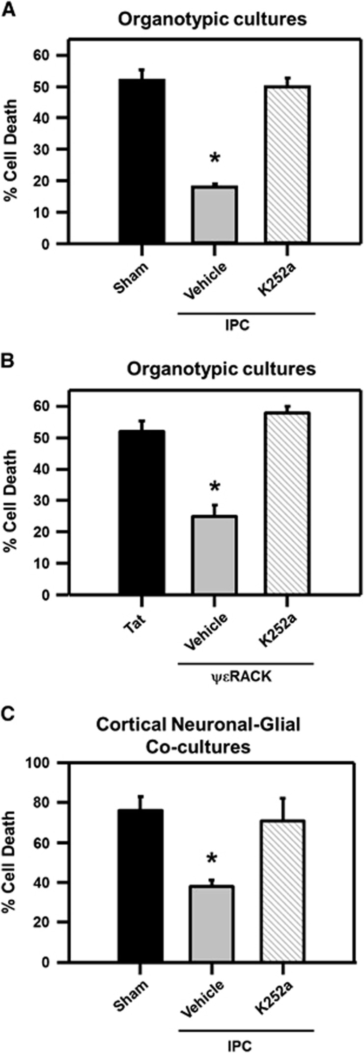
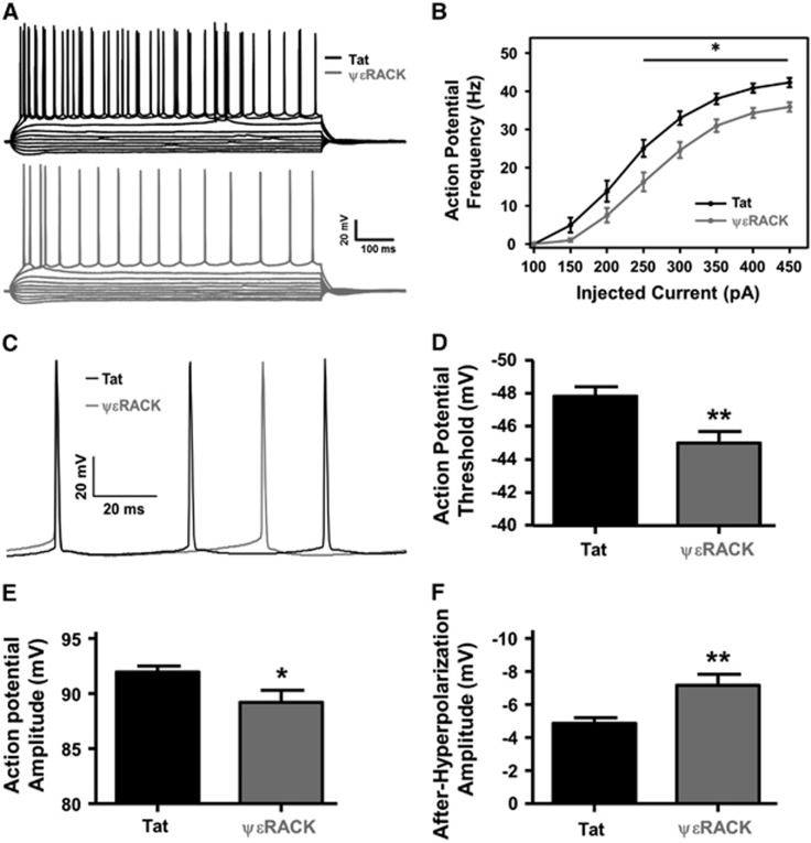
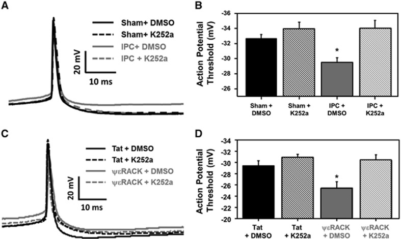
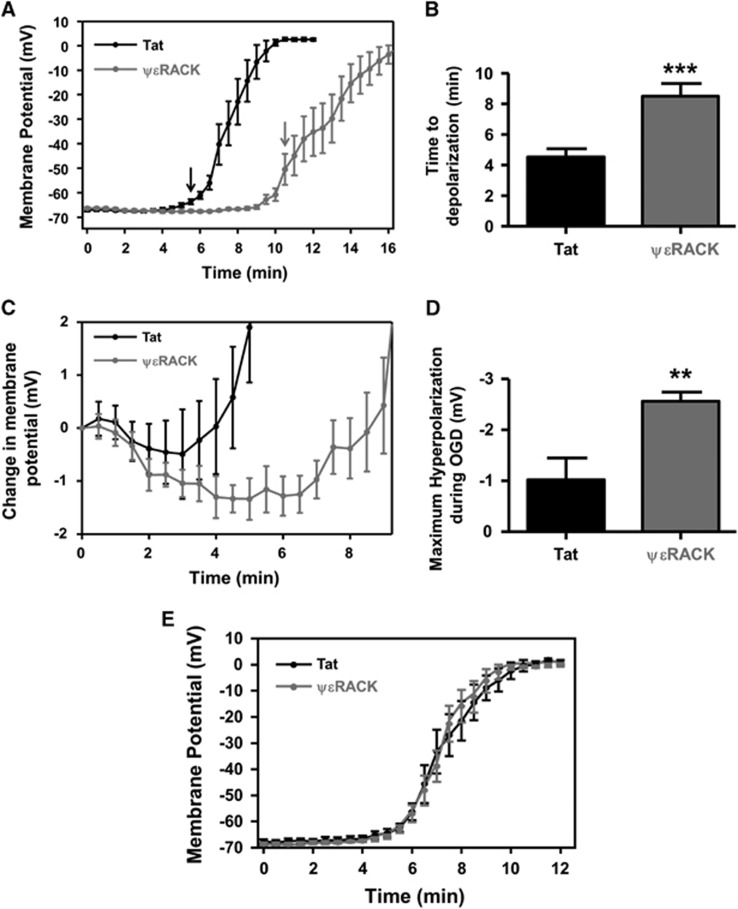
Ischemic preconditioning (IPC) via protein kinase C epsilon (PKCɛ) activation induces neuroprotection against lethal ischemia. Brain-derived neurotrophic factor (BDNF) is a pro-survival signaling molecule that modulates synaptic plasticity and neurogenesis. Interestingly, BDNF mRNA expression increases after IPC. In this study, we investigated whether IPC or pharmacological preconditioning (PKCɛ activation) promoted BDNF-induced neuroprotection, if neuroprotection by IPC or PKCɛ activation altered neuronal excitability, and whether these changes were BDNF-mediated. We used both in vitro (hippocampal organotypic cultures and cortical neuronal-glial cocultures) and in vivo (acute hippocampal slices 48 hours after preconditioning) models of IPC or PKCɛ activation. BDNF protein expression increased 24 to 48 hours after preconditioning, where inhibition of the BDNF Trk receptors abolished neuroprotection against oxygen and glucose deprivation (OGD) in vitro. In addition, there was a significant decrease in neuronal firing frequency and increase in threshold potential 48 hours after preconditioning in vivo, where this threshold modulation was dependent on BDNF activation of Trk receptors in excitatory cortical neurons. In addition, 48 hours after PKCɛ activation in vivo, the onset of anoxic depolarization during OGD was significantly delayed in hippocampal slices. Overall, these results suggest that after IPC or PKCɛ activation, there are BDNF-dependent electrophysiologic modifications that lead to neuroprotection.


Figures

Figure 1
Figure 1
Increased brain-derived neurotrophic factor (BDNF) protein expression after preconditioning. Western blot images depicting increased BDNF protein expression from (A) cortical neuronal-glial cocultures and (C) organotypic hippocampal slices after the induction of ischemic preconditioning (IPC), or (E) ΨɛRACK treatment, 24 and 48 hours as compared with sham treatment. Quantification of western blot images (BDNF/β-actin) are depicted in (B, D, and F) to their respective blots (*P<0.05; n=4 to 6).
Figure 2
Figure 2
Preconditioning increases brain-derived neurotrophic factor (BDNF) protein expression in the cornu ammonis 1 (CA1) region of hippocampus. Typical micrographs from the CA1 region of organotypic hippocampal slices with (A, D, and G) NeuN (red, neuronal marker) and (B, E, and H) BDNF (green) depicting increased (C, F, and I) colocalization 48 hours after ischemic preconditioning (IPC) or ΨɛRACK treatment versus sham treatment. Scale bar, 50 μm; n=4.
Figure 3
Figure 3
Brain-derived neurotrophic factor (BDNF) receptor activation is required for ischemic preconditioning (IPC)- and protein kinase C epsilon (PKCɛ) activation-induced ischemic tolerance. (A) Bar graph representing the percentage of cell death after lethal ischemia in the cornu ammonis 1 (CA1) region of organotypic hippocampal slice cultures that were treated with sham or IPC procedures (with or without K252a) or (B) Tat or ΨɛRACK treatment with or without K252a. (C) Bar graph representing cortical neuronal-glial cocultures cell death after lethal oxygen and glucose deprivation (OGD) in sham- or IPC-treated neurons with or without K252a. *P<0.05; n=4.
Figure 4
Figure 4
Protein kinase C epsilon (PKCɛ) activation decreases cornu ammonis 1 (CA1) neuronal firing frequency and excitability. (A) Representative tracings of action potential recordings made 48 hours after Tat (black) or ΨɛRACK (gray) treatment (0.2 mg/kg) with current injections (−250 to 200 pA in 50 pA increments). (B) Action potential firing frequency (1 Hz) recorded 48 hours after the intraperitoneal injection of Tat or ΨɛRACK. Firing frequency in the ΨɛRACK-treated neurons was significantly reduced at current injections at and above 250 pA. (C) Representative tracing of individual action potentials 48 hours after Tat- or ΨɛRACK-treated neurons with significant alterations in (D) action potential threshold, (E) action potential amplitude, and (F) action potential after-hyperpolarization (*P<0.05; **P<0.01; n=18 to 19).
Figure 5
Figure 5
Preconditioning modulation of neuronal threshold potential is mediated by brain-derived neurotrophic factor (BDNF). (A) Typical tracing of a neuronal action potential from cortical neuronal-gial cocultures recorded 48 hours after sham or ischemic preconditioning (IPC) treatment (with or without the Trk inhibitor K252a). (B) The average action potential threshold value for each treatment group (*P<0.05; n=9 to 10). (C) Typical tracing of a neuronal action potential from cortical neuronal-gial cocultures recorded 48 hours after Tat or ΨɛRACK treatment (with or without the Trk inhibitor K252a). (D) The average action potential threshold value for each treatment group (*P<0.05; n=7 to 8).
Figure 6
Figure 6
Protein kinase C epsilon (PKCɛ) activation delays the anoxic depolarization of hippocampal cornu ammonis 1 (CA1) neurons. (A) Graphical representation of CA1 neuronal membrane potential from acute slices measured every 30 seconds using whole-cell patch-clamp from Tat- or ΨɛRACK-treated neurons during continuous oxygen and glucose deprivation (OGD). Arrows indicate significant depolarization of the membrane potential compared to the resting membrane potential (time 0). (B) Bar graph representing the average time neurons maintained their resting membrane potential during OGD before depolarization. (C) Graphical representation of the normalized membrane potential during OGD measured every 30 seconds, 48 hours after Tat or ΨɛRACK treatment. (D) Bar graph representing the maximal hyperpolarization amplitude recorded during OGD after Tat or ΨɛRACK treatment. (E) Graphical representation of CA1 neuronal membrane potential measured every 30 seconds from acute Tat- or ΨɛRACK-treated hippocampal slices (slices from naive rats). (↓P<0.05; **P<0.01; ***P<0.001; n=7 to 9).

References

    1. Go AS, Mozaffarian D, Roger VL, Benjamin EJ, Berry JD, Blaha MJ, et al. Heart disease and stroke statistics—2014 update: a report from the American Heart Association. Circulation. 2014;129:e28–e292. - PMC - PubMed
    1. Nichols-Larsen DS, Clark PC, Zeringue A, Greenspan A, Blanton S. Factors influencing stroke survivors' quality of life during subacute recovery. Stroke. 2005;36:1480–1484. - PubMed
    1. DeFazio RA, Raval AP, Lin HW, Dave KR, Della-Morte D, Perez-Pinzon MA. GABA synapses mediate neuroprotection after ischemic and epsilonPKC preconditioning in rat hippocampal slice cultures. J Cereb Blood Flow Metab. 2009;29:375–384. - PMC - PubMed
    1. Tauskela JS, Fang H, Hewitt M, Brunette E, Ahuja T, Thivierge JP, et al. Elevated synaptic activity preconditions neurons against an in vitro model of ischemia. J Biol Chem. 2008;283:34667–34676. - PMC - PubMed
    1. Dave KR, DeFazio RA, Raval AP, Torraco A, Saul I, Barrientos A, et al. Ischemic preconditioning targets the respiration of synaptic mitochondria via protein kinase C epsilon. J Neurosci. 2008;28:4172–4182. - PMC - PubMed

Publication types

MeSH terms

Substances