

Culture phases, cytotoxicity and protein expressions of agarose hydrogel induced Sp2/0, A549, MCF-7 cell line 3D cultures
- PMID: 25371010
- PMCID: PMC4846635
- DOI: 10.1007/s10616-014-9795-z
Culture phases, cytotoxicity and protein expressions of agarose hydrogel induced Sp2/0, A549, MCF-7 cell line 3D cultures
Abstract
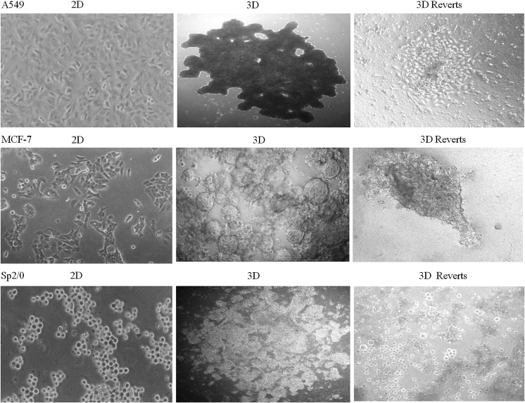
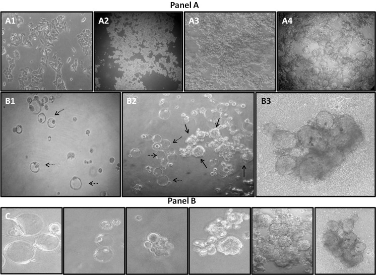
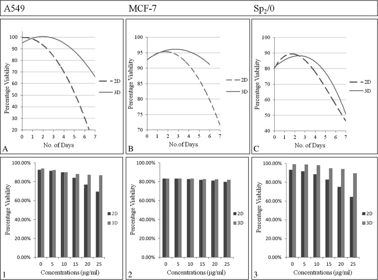
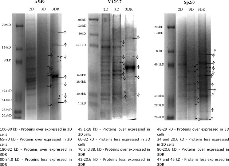
Advancements in cell cultures are occurring at a rapid pace, an important direction is culturing cells in 3D conditions. We demonstrate the usefulness of agarose hydrogels in obtaining 3 dimensional aggregates of three cell lines, A549, MCF-7 and Sp2/0. The differences in culture phases, susceptibility to cisplatin-induced cytotoxicity are studied. Also, the 3D aggregates of the three cell lines were reverted into 2D cultures and the protein profile differences among the 2D, 3D and revert cultures were studied. The analysis of protein profile differences using UniProt data base further augment the usefulness of agarose hydrogels for obtaining 3D cell cultures.
Keywords: 3D aggregates; Agarose hydrogels; Cell culture phases; Cytotoxicity; Protein expressions.
Figures
Similar articles
-
Significance of cell culture phases for utilizing 3D cultures of A549 cells for in vitro studies.Cell Biol Int. 2023 Oct;47(10):1760-1764. doi: 10.1002/cbin.12075. Epub 2023 Jul 20. Cell Biol Int. 2023. PMID: 37471709
-
Agarose hydrogel induced MCF-7 and BMG-1 cell line progressive 3D and 3D revert cultures.J Cell Physiol. 2018 Apr;233(4):2768-2772. doi: 10.1002/jcp.25965. Epub 2017 May 8. J Cell Physiol. 2018. PMID: 28422283
-
Differences of SiHa (human cancer of cervix) and BMG-1 (brain glioma) cell lines as 2D and 3D cultures.J Cell Physiol. 2014 Feb;229(2):127-31. doi: 10.1002/jcp.24433. J Cell Physiol. 2014. PMID: 23881600
-
Human Brain Malignant Glioma (BMG-1) 3D Aggregate Morphology and Screening for Cytotoxicity and Anti-Proliferative Effects.J Cell Physiol. 2017 Apr;232(4):685-690. doi: 10.1002/jcp.25603. Epub 2016 Nov 10. J Cell Physiol. 2017. PMID: 27639069
-
Applications of Three-Dimensional Cell Cultures in the Early Stages of Drug Discovery, Focusing on Gene Expressions, Drug Metabolism, and Susceptibility.Crit Rev Eukaryot Gene Expr. 2017;27(1):53-62. doi: 10.1615/CritRevEukaryotGeneExpr.2017018935. Crit Rev Eukaryot Gene Expr. 2017. PMID: 28436332 Review.
Cited by
-
Polymeric Hydrogels for In Vitro 3D Ovarian Cancer Modeling.Int J Mol Sci. 2022 Mar 17;23(6):3265. doi: 10.3390/ijms23063265. Int J Mol Sci. 2022. PMID: 35328686 Free PMC article. Review.
-
Cytotoxic responses of carnosic acid and doxorubicin on breast cancer cells in butterfly-shaped microchips in comparison to 2D and 3D culture.Cytotechnology. 2017 Apr;69(2):337-347. doi: 10.1007/s10616-016-0062-3. Epub 2017 Feb 13. Cytotechnology. 2017. PMID: 28191587 Free PMC article.
References
-
- Bazou D (2010) Biochemical properties of encapsulated high density 3-D HepG2 aggregates formed in an ultrasound trap for application in hepatotoxicity studies: biochemical responses of encapsulated 3-D HepG2 aggregates. Cell Biol Toxicol 26:127–141 - PubMed
-
- Cella N, Contreras A, Latha K, Rosen JM, Zhang M (2006) Maspin is physically associated with b- integrin regulating cell adhesion in mammary epithelial cells. FASEB J 20:1510–1512 - PubMed
LinkOut - more resources
Full Text Sources
Other Literature Sources
