. 2015 Jan 2;290(1):142-56.
doi: 10.1074/jbc.M114.598110. Epub 2014 Nov 3.

Structure-function analysis of heterodimer formation, oligomerization, and receptor binding of the Staphylococcus aureus bi-component toxin LukGH

Affiliations

Structure-function analysis of heterodimer formation, oligomerization, and receptor binding of the Staphylococcus aureus bi-component toxin LukGH

Adriana Badarau et al. J Biol Chem. .

Abstract

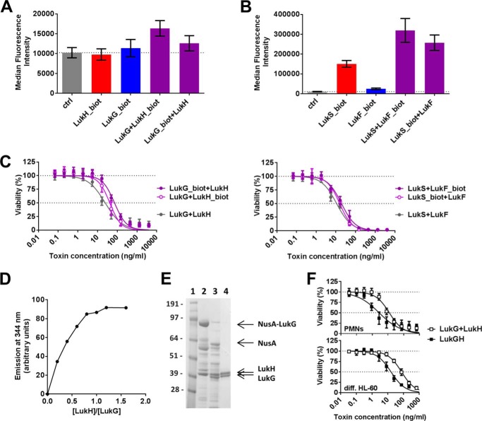
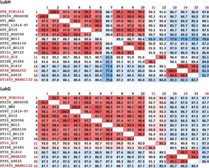
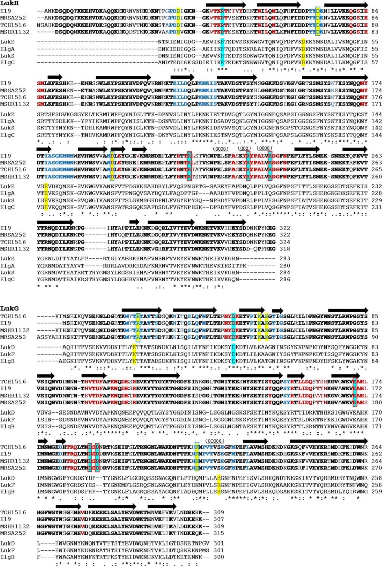
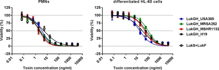
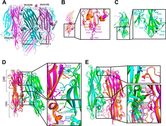
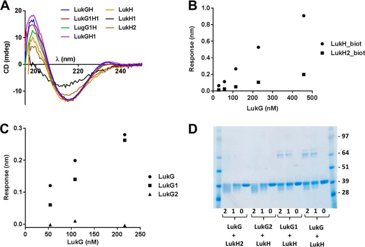
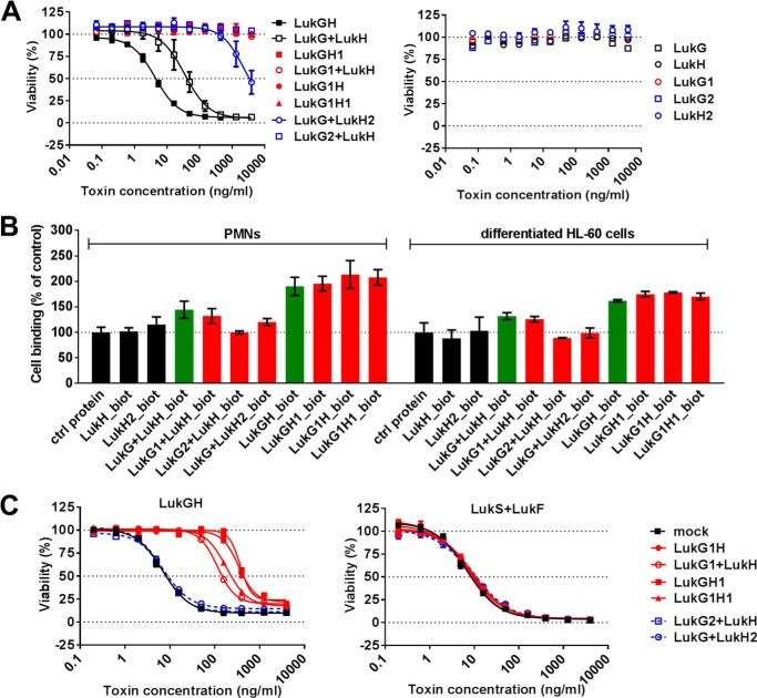
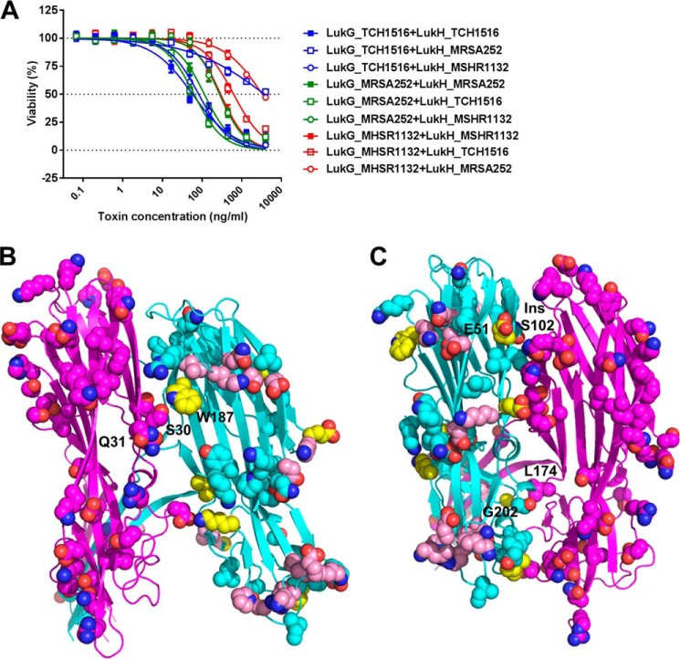
The bi-component leukocidins of Staphylococcus aureus are important virulence factors that lyse human phagocytic cells and contribute to immune evasion. The γ-hemolysins (HlgAB and HlgCB) and Panton-Valentine leukocidin (PVL or LukSF) were shown to assemble from soluble subunits into membrane-bound oligomers on the surface of target cells, creating barrel-like pore structures that lead to cell lysis. LukGH is the most distantly related member of this toxin family, sharing only 30-40% amino acid sequence identity with the others. We observed that, unlike other leukocidin subunits, recombinant LukH and LukG had low solubility and were unable to bind to target cells, unless both components were present. Using biolayer interferometry and intrinsic tryptophan fluorescence we detected binding of LukH to LukG in solution with an affinity in the low nanomolar range and dynamic light scattering measurements confirmed formation of a heterodimer. We elucidated the structure of LukGH by x-ray crystallography at 2.8-Å resolution. This revealed an octameric structure that strongly resembles that reported for HlgAB, but with important structural differences. Structure guided mutagenesis studies demonstrated that three salt bridges, not found in other bi-component leukocidins, are essential for dimer formation in solution and receptor binding. We detected weak binding of LukH, but not LukG, to the cellular receptor CD11b by biolayer interferometry, suggesting that in common with other members of this toxin family, the S-component has the primary contact role with the receptor. These new insights provide the basis for novel strategies to counteract this powerful toxin and Staphylococcus aureus pathogenesis.

Keywords: Bacterial Toxin; Crystal Structure; Crystallography; Microbial Pathogenesis; Mutagenesis; Staphylococcus aureus (S. aureus).


Figures

FIGURE 1.
FIGURE 1.
The LukGH toxin binds to target cells as a dimer. Biotinylated LukH or LukG (A) and LukS or LukF (B) was incubated with human PMNs in the absence or presence of the unlabeled toxin pair. Surface bound, biotinylated proteins were detected with Alexa 488-labeled streptavidin and quantified by flow cytometry. Results of three independent experiments are shown (mean ± S.E.). C, biotin-labeled toxin components were tested for functionality in PMN toxicity assays and compared with unlabeled counterparts. Left panel, LukG and LukH; right panel, LukS and LukF. Results represent the mean from three independent experiments ± S.E. D, complex formation by LukG and LukH was investigated by fluorescence spectroscopy detecting changes in intrinsic tryptophan fluorescence upon the addition of increasing amounts of LukH to a constant amount of LukG (250 nm). E, N-terminal NusA/His-tagged LukG and untagged LukH (derived from the S. aureus TCH1516 strain) were co-expressed in E. coli. Proteins isolated from the soluble fraction of E. coli and eluted from the IMAC column were visualized by SDS-PAGE. Lane 1, marker proteins (molecular masses indicated in kDa); lane 2, NusA-LukG and LukH co-eluted from the IMAC column; lane 3, enterokinase digestion of the NusA-LukG and LukH fractions; lane 4, LukGH fraction eluted from the Sulfo-Propyl column. F, cytotoxic potency of the mixture of individually expressed LukH and LukG was compared with the co-expressed complex (derived from the S. aureus TCH1516 strain) using human PMNs or differentiated HL-60 cells. Viability of cells was determined by luminescent measurement of cellular ATP levels. Results of three independent experiments are shown (mean ± S.E.).
FIGURE 2.
FIGURE 2.
Conservation of LukG and LukH sequences from different S. aureus strains. Percent amino acid sequence identities between LukH and LukG variants derived from different strains for which annotated genome data are available. Those used in this study are highlighted in red.
FIGURE 3.
FIGURE 3.
Amino acid alignment of the bi-component toxins of S. aureus. Amino acid conservations among S- or F-components of all bi-component toxins of the strain TCH1516 are shown. LukH and LukG sequences from MRSA252, MSHR1132, and H19 strains are included for comparison. Multiple sequence alignment was performed with the ClustalW2 program (36). Amino acids conserved between the LukH or LukG variants are shown in bold, and those involved in contacts in interface 1 and 2 are shown in blue and red, respectively. Residues involved in salt bridges in interface 1 and 2 are highlighted in yellow and cyan, respectively, and those mutated in this study are boxed. Secondary structure elements are indicated; arrows for β-strands and coils for α-helices.
FIGURE 4.
FIGURE 4.
All LukGH variants display comparable toxicity toward human granulocytes. Recombinant LukG and LukH proteins derived from the TCH1516, MRSA252, H19, and MSHR1132 S. aureus strains co-expressed in E. coli cells were incubated with freshly isolated human PMNs or differentiated HL-60 cells at the indicated concentration range. Cell viability was determined by luminescent measurement of cellular ATP levels. Results of three independent experiments (mean ± S.E.) are shown.
FIGURE 5.
FIGURE 5.
X-ray structure of the LukG and LukH monomers in the LukGH octamer. A, structure of the LukGH octamer formed by LukG (pink) and LukH (cyan). B, overlay of LukG (pink) and HlgB (orange) (on chains A) with the side chains of the residues involved in membrane binding shown as sticks and MPD and Met178 as orange and pink spheres, respectively. C, overlay of LukH (cyan) and HlgA (green) (on chains B), with LukH residues Thr35-Ile40 and Tyr314-Glu323 colored magenta and red, respectively; the side chains of the residues delineating the loops with a different conformation in LukH are shown as sticks. D, interface 1: LukG and LukH superposed on HlgB and HlgA (chains A and B, respectively, overlay on chains A). E, interface 2: LukH and LukG superposed on HlgA and HlgB (chains B and C, respectively, overlay on chains B). The side chains of the residues involved in electrostatic interactions are shown as sticks and the salt bridges as dotted lines. Electron density map (2FoFc) around residues involved in salt bridges in panel E contoured at 1 σ, is shown as a gray mesh.
FIGURE 6.
FIGURE 6.
Interaction of wild-type and mutated LukG with the LukH proteins. A, CD spectra of wild-type and mutant LukGH complexes and LukH normalized to a concentration of 0.5 mg/ml were acquired in 50 mm sodium phosphate, pH 7.5, in the presence of 200 or 500 mm NaCl, respectively. B and C, binding of LukG to biotinylated LukH and LukH2 (B) or binding of LukG, LukG1, and LukG2 to biotinylated LukH (C) was detected by BLI measurements and expressed as response values (obtained after subtracting the values for nonspecific binding when biotinylated HlgC was immobilized on the sensor). D, cross-linking of LukG and LukH. Mixtures of wild-type and mutant LukG and LukH were cross-linked with glutaraldehyde for 2 or 1 min (lanes 2 and 1 in each group, respectively) in the indicated combinations and resolved by SDS-PAGE. Untreated samples are shown in lanes 0.
FIGURE 7.
FIGURE 7.
Cytolytic activity and binding of LukGH is abolished by mutations affecting the electrostatic interactions of LukG and LukH at both interface 1 and 2. A, mixtures of individually expressed LukG and LukH monomers and co-expressed complexes were used in the indicated concentration range to intoxicate differentiated HL-60 cells. Single components were used as controls. Cell viability was determined by luminescent measurement of cellular ATP content. Data are expressed as mean ± S.E. from two independent experiments. B, cell binding of wild-type and mutant proteins to human PMNs and differentiated HL-60 cells was determined using biotinylated toxin components as indicated and quantified by flow cytometry measurement. Results represent the mean ± S.E. of three and two independent experiments with PMNs and differentiated HL-60 cells, respectively. C, differentiated HL-60 cells were preincubated with 14 nm LukGH mutants as indicated for 30 min, followed by intoxication with wild-type LukGH (left panel) and LukSF (right panel) for 4 h in the indicated concentration range. Results represent the mean ± S.E. of three independent experiments.
FIGURE 8.
FIGURE 8.
Active toxin formation by heterologous pairing by LukG and LukH variants. A, human PMNs were treated with mixtures of LukG and LukH monomers derived from TCH1516, MSHR1132, and MRSA252 S. aureus strains in the indicated concentration ranges. Cell viability was determined by luminescent measurement of cellular ATP content. Data are expressed as mean ± S.E. from three independent experiments. B, structure of LukG_TCH1516 (chain A, magenta) and LukH_TCH1516 (chain B, cyan) at interface 1. The side chains of the amino acids that differ in the MRSA252, MSHR1322, and H19 variants are shown as spheres. The side chains of the amino acids that are different in LukH_MRSA252 compared with both LukH_TCH1516 and LukH_MSHR1132 are shown as spheres in pink or yellow if found at the LukGH interface. C, structure of LukH (chain B, cyan) and LukG (chain C, magenta) at interface 2. Representations as described in B.

References

    1. Spaan A. N., Surewaard B. G., Nijland R., van Strijp J. A. (2013) Neutrophils versus Staphylococcus aureus: a biological tug of war. Annu. Rev. Microbiol. 67, 629–650 - PubMed
    1. Rigby K. M., DeLeo F. R. (2012) Neutrophils in innate host defense against Staphylococcus aureus infections. Semin. Immunopathol. 34, 237–259 - PMC - PubMed
    1. Vandenesch F., Lina G., Henry T. (2012) Staphylococcus aureus hemolysins, bi-component leukocidins, and cytolytic peptides: a redundant arsenal of membrane-damaging virulence factors? Front. Cell Infect. Microbiol. 2, 12. - PMC - PubMed
    1. Alonzo F., 3rd, Torres V. J. (2014) The bicomponent pore-forming leucocidins of Staphylococcus aureus. Microbiol. Mol. Biol. Rev. 78, 199–230 - PMC - PubMed
    1. Aman M. J., Adhikari R. P. (2014) Staphylococcal bicomponent pore-forming toxins: targets for prophylaxis and immunotherapy. Toxins 6, 950–972 - PMC - PubMed

Publication types

MeSH terms

LinkOut - more resources