. 2015 Jan 2;290(1):359-70.
doi: 10.1074/jbc.M114.617597. Epub 2014 Nov 4.

Non-canonical interleukin 23 receptor complex assembly: p40 protein recruits interleukin 12 receptor β1 via site II and induces p19/interleukin 23 receptor interaction via site III

Affiliations

Non-canonical interleukin 23 receptor complex assembly: p40 protein recruits interleukin 12 receptor β1 via site II and induces p19/interleukin 23 receptor interaction via site III

Jutta Schröder et al. J Biol Chem. .

Abstract

IL-23, composed of the cytokine subunit p19 and the soluble α receptor subunit p40, binds to a receptor complex consisting of the IL-23 receptor (IL-23R) and the IL-12 receptor β1 (IL-12Rβ1). Complex formation was hypothesized to follow the "site I-II-III" architectural paradigm, with site I of p19 being required for binding to p40, whereas sites II and III of p19 mediate binding to IL-12Rβ1 and IL-23R, respectively. Here we show that the binding mode of p19 to p40 and of p19 to IL-23R follow the canonical site I and III paradigm but that interaction of IL-23 to IL-12Rβ1 is independent of site II in p19. Instead, binding of IL-23 to the cytokine binding module of IL-12Rβ1 is mediated by domains 1 and 2 of p40 via corresponding site II amino acids of IL-12Rβ1. Moreover, domains 2 and 3 of p40 were sufficient for complex formation with p19 and to induce binding of p19 to IL-23R. The Fc-tagged fusion protein of p40_D2D3/p19 did, however, not act as a competitive IL-23 antagonist but, at higher concentrations, induced proliferation via IL-23R but independent of IL-12Rβ1. On the basis of our experimental validation, we propose a non-canonical topology of the IL-23·IL-23R·IL-12Rβ1 complex. Furthermore, our data help to explain why p40 is an antagonist of IL-23 and IL-12 signaling and show that site II of p19 is dispensable for IL-23 signaling.

Keywords: Cytokine; Cytokine Action; Interleukin; Interleukin 12 Receptor; Interleukin 23; Interleukin 23 Receptor; Molecular Modeling; Mutagenesis; p19; p40.


Figures

FIGURE 1.
FIGURE 1.
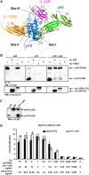
Binding of p19 to IL-12Rβ1 and IL-23R is dependent on p40. A, IL-23_p40 (green, domains D1-D3) and IL-23_p19 (blue) bind to IL-12Rβ1 (orange, domains D1 and D2) and IL-23R (pink, domain D1). B, coimmunoprecipitation of FLAG-tagged p40 or p19 and Fc-tagged sIL-12Rβ1 or sIL-23R using protein A-agarose. One of three independent experiments is shown. L, lysates; +, coimmunoprecipitates; C, controls (without sIL-12Rβ1 or sIL-23R); WB, Western blot. C, Western blotting of secreted p40 and/or p19 from transfected COS-7 cells. D, cell viability assay of Ba/F3-IL-12Rβ1-IL-23R cells stimulated with increasing amounts of the indicated cytokine-conditioned supernatants (CS). Data are mean ± S.D. The concentration of p19/p40 and p40-p19-Fp was 200 ng/ml and 685 ng/ml, respectively, as determined by p40-ELISA.
FIGURE 2.
FIGURE 2.
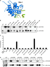
Characterization of site I in p19. A, model of site I of p19 (blue) toward p40 (green). B, Western blotting (WB) of secreted p40 and/or p19_R179E, K66A, I176E, A178W, and R179A from transfected COS-7 cells. C, cell viability assay of Ba/F3-IL-12Rβ1-IL-23R cells stimulated with the indicated cytokines (10% conditioned cell culture supernatant). One of three independent experiments is shown. Data are mean ± S.D. D, co-IP of c-myc-tagged p40 and FLAG-tagged p19 or p19_R179E, K66A, I176E, A178W, and R179A. L, lysates; +, coimmunoprecipitates; C, controls (without mAbs).
FIGURE 3.
FIGURE 3.
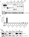
Targeted site I mutations in p40 prevent the formation of IL-23. A, model of p40 (green) toward site I of p19 (blue). B, Western blotting (WB) of secreted p19, p40_Y265K, and p40_Y318K. One of three independent experiments is shown. C, cell viability assay of Ba/F3-IL-12Rβ1-IL-23R cells stimulated with the indicated cytokines (10% conditioned cell culture supernatant). One of three independent experiments is shown. Data are mean ± S.D. D, co-IP of c-myc-tagged p19 and FLAG-tagged p40 or the p40 mutants Y265K and Y318K. L, lysates; +, coimmunoprecipitates; C, controls (without mAbs). One of three independent experiments is shown.
FIGURE 4.
FIGURE 4.
Binding of deletion variants of p40 to p19. A, Western blotting (WB) of secreted p19 and p40, p40_D1, p40_D2, p40_D3, p40_D1D2, and p40_D2D3. One of three independent experiments is shown. B, cell viability assay of Ba/F3-IL-12Rβ1-IL-23R cells stimulated with the indicated cytokines (10% conditioned cell culture supernatant). One of three independent experiments is shown. Data are mean ±S.D. C, co-IP of FLAG-tagged p40 variants and c-myc-tagged p19. L, lysates; +, coimmunoprecipitates; C, controls (without mAbs). One of three independent experiments is shown.
FIGURE 5.
FIGURE 5.
Characterization of p19 site III. A, model of the predicted site III of p19 (blue) toward IL-23R (pink). B, Western blotting (WB) of secreted p40 and p19 or p19_W157A, p40, p40-p19-Fp, or p40-p19_W157A-Fp. One of three independent experiments is shown. C, cell viability assay of Ba/F3-IL-12Rβ1-IL-23R cells stimulated with the indicated cytokines (10% conditioned cell culture supernatant). One of three independent experiments is shown. Data are mean ± S.D. D, co-IP of FLAG-tagged p40 and p19 or p19_W157A and Fc-tagged sIL-12Rβ1 or sIL-23R. One of three independent experiments is shown. E, co-IP of FLAG-tagged p40-p19_wt and _W157A fusion proteins and Fc-tagged sIL-12Rβ1 or sIL-23R. L, lysates; +, coimmunoprecipitates; C, controls (without sIL-12Rβ1-Fc or sIL-23R-Fc). One of three independent experiments is shown.
FIGURE 6.
FIGURE 6.
Absence of a classical site II in p19. A, model of the predicted site II of p19 (blue) toward IL-12Rβ1 (orange). B, Western blotting (WB) of secreted p40 and p19_L36E, p19_L135E, or p19_R38E. One of three independent experiments is shown. C, cell viability assay of Ba/F3-IL-12Rβ1-IL-23R cells stimulated with the indicated cytokines (10% conditioned cell culture supernatant). One of three independent experiments is shown. Data are mean ± S.D. D, co-IP of FLAG-tagged p40 and p19, p19_L36E, p19_L135E, or p19_R38E with Fc-tagged sIL-12Rβ1 or sIL-23R. L, lysates; +, coimmunoprecipitates; C, controls (without sIL-12Rβ1-Fc or sIL-23R-Fc). One of three independent experiments is shown. E, Western blotting of secreted p40-p19-Fp and p40-p19_L36E_R38E_L135E-Fp. F, cell viability assay of Ba/F3-IL-12Rβ1-IL-23R cells stimulated with the indicated cytokines (10% conditioned cell culture supernatant). One of three independent experiments is shown. Data are mean ± S.D. G, co-IP of FLAG-tagged p40-p19-Fp or p40-p19_L36E_L135E_R38E-Fp with Fc-tagged sIL-12Rβ1 or sIL-23R. One of three independent experiments is shown.
FIGURE 7.
FIGURE 7.
p40 substitutes for the site II interaction of p19 toward IL-12Rβ1. A, co-IP of FLAG-tagged p40 variants and Fc-tagged sIL-12Rβ1. One of three independent experiments is shown. WB, Western blot. B, co-IP of FLAG-tagged p40 and Fc-tagged sIL-12Rβ1 deletion variants. One of three independent experiments is shown. C, model of the predicted site II of p19 (blue) toward IL-12Rβ1 (orange). D, co-IP of FLAG-tagged p40 and Fc-tagged sIL-12Rβ1 point mutation variants. One of three independent experiments is shown. L, p40 lysate; +, coimmunoprecipitates; C, control (without sIL-12Rβ1 variant).
FIGURE 8.
FIGURE 8.
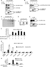
p40_D2D3-p19 specifically interacts with IL-23R but not with IL-12Rβ1 and acts as an agonist independently of IL-12Rβ1. A, co-IP of FLAG-tagged p40_D2D3 and p19 and Fc-tagged sIL-12Rβ1 or sIL-23R. L, lysates; +, coimmunoprecipitates; C, controls (without sIL-12Rβ1-Fc or sIL-23R-Fc); WB, Western blot. One of three independent experiments is shown. B, co-IP of the fusion protein p40_D2D3-p19-Fp with Fc-tagged sIL-12Rβ1 or sIL-23R. One of three independent experiments is shown. C and D, Coomassie staining (C) and Western blotting (D) of purified p40_D2D3-p19Fc. E, co-IP of myc-tagged sIL-23R and recombinant p40_D2D3-p19Fc. L, lysates; +, coimmunoprecipitates; C, controls (without sIL-23R). One of three independent experiments is shown. F–H, cell viability assay of Ba/F3-IL-12Rβ1-IL-23R cells (F and G), Ba/F3-IL-12Rβ1 cells (H), and Ba/F3-IL-23R cells (H) stimulated with the indicated recombinant cytokines. One of three independent experiments is shown. Data are mean ± S.D.
FIGURE 9.
FIGURE 9.
Predicted and experimentally validated model of the IL-23 receptor complex. A, predicted model with canonical sites I, II, and III of p19. B, experimentally validated model with sites I and III in p19 and major interaction of p40 with IL-12Rβ1. The proposed binding site II of p19 did not contribute to IL-12Rβ1 binding. Instead, the interaction of p40_D1D2 with IL-12Rβ1 was sufficient for receptor complex formation. Shown are p40 (green, D1-D3), p19 (blue, site I), IL-12Rβ1 (orange, domains D1 and D2), and IL-23R (pink, D1).

References

    1. Vignali D. A., Kuchroo V. (2012) IL-12 family cytokines: immunological playmakers. Nat. Immunol. 13, 722–728 - PMC - PubMed
    1. Garbers C., Hermanns H. M., Schaper F., Müller-Newen G., Grötzinger J., Rose-John S., Scheller J. (2012) Plasticity and cross-talk of interleukin 6-type cytokines. Cytokine Growth Factor Rev. 23, 85–97 - PubMed
    1. Park H., Li Z., Yang X. O., Chang S. H., Nurieva R., Wang Y. H., Wang Y., Hood L., Zhu Z., Tian Q., Dong C. (2005) A distinct lineage of CD4 T cells regulates tissue inflammation by producing interleukin 17. Nat. Immunol. 6, 1133–1141 - PMC - PubMed
    1. Ouyang W., Kolls J. K., Zheng Y. (2008) The biological functions of T helper 17 cell effector cytokines in inflammation. Immunity 28, 454–467 - PMC - PubMed
    1. Korn T., Bettelli E., Oukka M., Kuchroo V. K. (2009) IL-17 and Th17 Cells. Annu. Rev. Immunol. 27, 485–517 - PubMed

Publication types

MeSH terms

LinkOut - more resources