Case Reports
. 2015 Sep;9(3):405-11.
doi: 10.1007/s12105-014-0577-x. Epub 2014 Nov 5.

Angiosarcoma Developing in a Vagal Schwannoma: A Rare Case Report

Affiliations
Case Reports

Angiosarcoma Developing in a Vagal Schwannoma: A Rare Case Report

Vrushali Mahajan et al. Head Neck Pathol. 2015 Sep.

Abstract

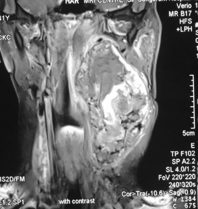
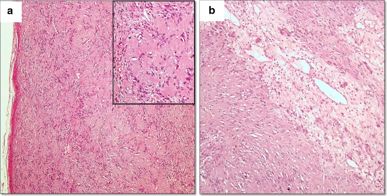
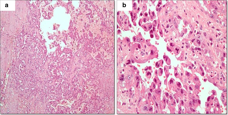
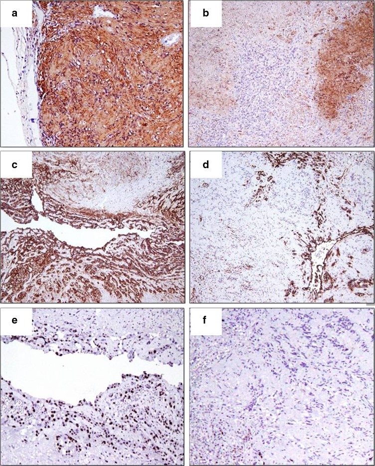
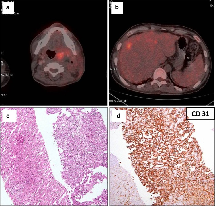
Angiosarcoma arising in a schwannoma is extremely rare with only fourteen cases having been reported in the literature to the best of our knowledge. Amongst these fourteen, only five cases developed from vagal schwannoma. We describe a case of epithelioid angiosarcoma arising in a long standing vagal schwannoma in a 41 years male patient. Grossly the tumor was well encapsulated with variegated cut surface. On microscopy the tumor had two distinct components composed of benign schwannoma and malignant angiosarcoma which were further confirmed by immunohistochemistry. On further work up, he was found to have multiple distant metastases. This is the sixth reported case of angiosarcoma arising in a vagal schwannoma. The proposed histogenesis of this rare transformation, its prognostic factors and a review of literature regarding this entity is discussed.


Figures

Fig. 1
Fig. 1
MRI with contrast: image showing a well defined heterogeneously enhancing soft tissue mass in left parapharyngeal space with hemorrhage
Fig. 2
Fig. 2
Gross photographs showing a well encapsulated mass (a), cut surface showing grey white gelatinous areas at the periphery, and rest of the mass showing hemorrhage, necrosis and cystic change (b)
Fig. 3
Fig. 3
Well encapsulated spindle cell tumor with hypercellular Antoni A areas (inset Verocay bodies) (a), and hypocellular Antoni B areas (b) (H&E ×10)
Fig. 4
Fig. 4
Anastomosing vascular spaces lined with atypical endothelial cells (a ×10), epithelioid morphology and increased mitotic activity with some atypical mitoses (b ×40) (H&E)
Fig. 5
Fig. 5
IHC (af) S100—schwann cells show positivity (a) while atypical endothelial cells are negative for S100 (b). CD 31—atypical endothelial cells show positivity (c) while schwann cells are negative for CD 31 (d). Ki 67—atypical endothelial cells show high proliferative activity—40–50 % (e) while schwann cells show low proliferative activity—1–2 % (f)
Fig. 6
Fig. 6
PET CT scan showing residual disease in left lateral neck (a), FDG avid lesion in the liver (b), metastatic angiosarcoma in the liver (c H&E ×10), and tumor cells showing positive staining for CD 31 (d)

References

    1. McMenamin ME, Fletcher CD. Expanding the spectrum of malignant change in Schwannomas: epithelioid malignant change, epithelioid malignant peripheral nerve sheath tumor, and epithelioid angiosarcoma: a study of 17 cases. Am J Surg Pathol. 2001;25(1):13–25. doi: 10.1097/00000478-200101000-00002. - DOI - PubMed
    1. Mentzel T, Katenkamp D. Intraneural angiosarcoma and angiosarcoma arising in benign and malignant peripheral nerve sheath tumours: clinicopathological and immunohistochemical analysis of four cases. Histopathology. 1999;35(2):114–120. doi: 10.1046/j.1365-2559.1999.00714.x. - DOI - PubMed
    1. Scheithauer B, Louis D, Hunter S, Woodruff JM, et al. Schwannoma. In: Louis D, Ohgaki H, Wiestler O, Cavenee W, et al., editors. WHO classification of tumours of the central nervous system. 4. Lyon: International Agency for Research on Cancer (IARC); 2007. pp. 152–155.
    1. Woodruff JM, Selig AM, Crowley K, Allen PW. Schwannoma (neurilemoma) with malignant transformation, A rare, distinctive peripheral nerve tumor. Am J Surg Pathol. 1994;18(9):882–895. doi: 10.1097/00000478-199409000-00003. - DOI - PubMed
    1. Trassard M, Le Doussal V, Bui BN, Coindre JM. Angiosarcoma arising in a solitary schwannoma (neurilemoma) of the sciatic nerve. Am J Surg Pathol. 1996;20(11):1412–1417. doi: 10.1097/00000478-199611000-00014. - DOI - PubMed

Publication types

Substances

LinkOut - more resources