Review
. 2014 Nov;141(22):4206-18.
doi: 10.1242/dev.107086.

Stem cells and the impact of ROS signaling

Affiliations
Review

Stem cells and the impact of ROS signaling

Carolina L Bigarella et al. Development. 2014 Nov.

Abstract

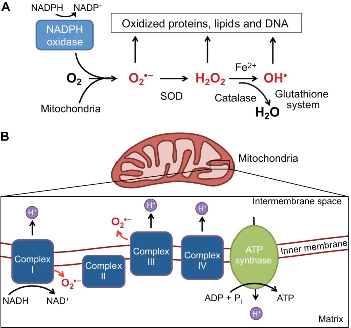
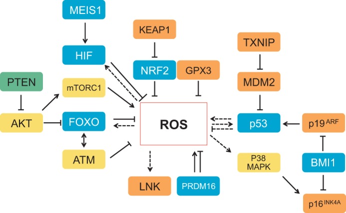
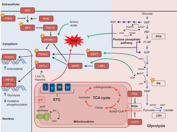
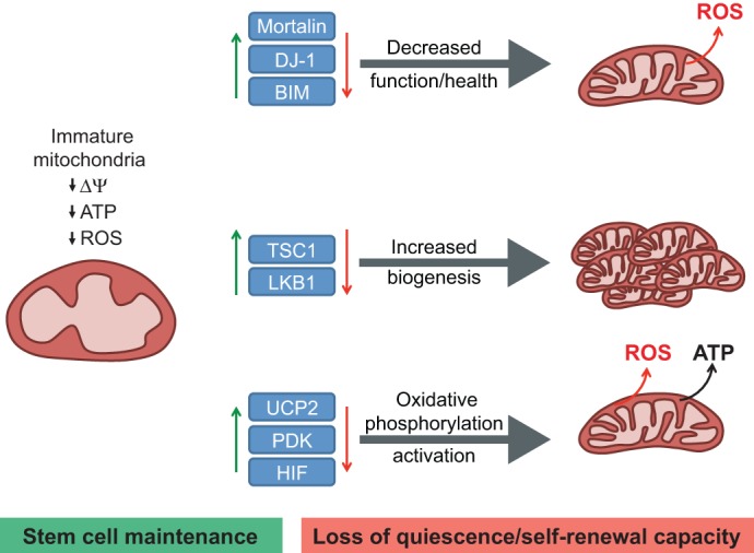
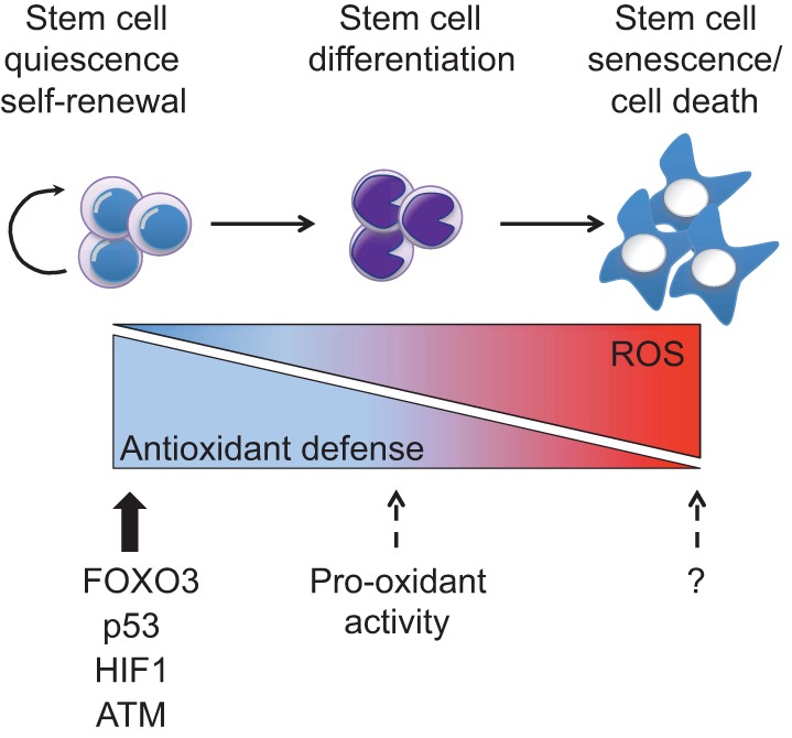
An appropriate balance between self-renewal and differentiation is crucial for stem cell function during both early development and tissue homeostasis throughout life. Recent evidence from both pluripotent embryonic and adult stem cell studies suggests that this balance is partly regulated by reactive oxygen species (ROS), which, in synchrony with metabolism, mediate the cellular redox state. In this Primer, we summarize what ROS are and how they are generated in the cell, as well as their downstream molecular targets. We then review recent findings that provide molecular insights into how ROS signaling can influence stem cell homeostasis and lineage commitment, and discuss the implications of this for reprogramming and stem cell ageing. We conclude that ROS signaling is an emerging key regulator of multiple stem cell populations.

Keywords: Embryonic stem cells; Hematopoietic stem cells; Metabolism; Mitochondria; ROS.


Figures

Fig. 1.
Fig. 1.
ROS generation and scavenging. (A) Reactive oxygen species (ROS) include superoxide (O2.−), hydrogen peroxide (H2O2) and the highly reactive hydroxyl radical (OH.) (shown in red). O2.− can be generated from complexes I and III (shown in B) or through the oxidation of NADPH by NADPH oxidases. Subsequent reduction to H2O2 is catalyzed by superoxide dismutase (SOD). H2O2 can be further reduced to water (H2O) by catalase or can spontaneously oxidize iron (Fe2+) to form the highly reactive OH.. Under conditions of oxidative stress, when ROS generation outpaces the ROS scavenging system, accumulating levels of ROS oxidize and damage various cellular components. (B) The electron transport chain complexes I-IV harness electrons from NADH in a series of redox reactions, which are coupled to pumping protons (H+) into the mitochondrial intermembrane space. The proton motive force, a combination of the membrane potential (charge) and the concentration gradient (pH), powers ATP synthase (complex V). Normally, O2 acts as the final electron acceptor at complex IV, but aberrant reduction of O2 can occur at complexes I and III (red arrows), leading to the generation of O2.− (red).
Fig. 2.
Fig. 2.
Redox sensor molecules. Intricate control of reactive oxygen species (ROS) can be either directly or indirectly mediated by several transcription factors (blue), as well as by kinases (yellow) and phosphatases (green). Other regulators, such as the cytokine signaling inhibitor LNK, the modulator KEAP1, the E3 ubiquitin ligase MDM2, the cell cycle inhibitors p16INK4A and p19ARF (which are negatively modulated by the polycomb group member BMI1), the complex mTORC1, TXNIP, and the antioxidant enzyme GPX3 (all shown in orange) can also control ROS levels. Dashed arrows and lines indicate regulations that have not been explicitly shown to occur in stem cells; unbroken lines represent interactions that have been shown in stem cells. p53 has both antioxidant and pro-oxidant functions (shown in somatic cells). AKT, protein kinase B; FOXO, forkhead box O protein; GPX3, glutathione peroxidase 3; HIF, hypoxia-inducible factor; KEAP1, kelch-like ECH-associated protein 1; MDM2, transformed mouse 3T3 cell double minute 2; MEIS1, Meis homeobox 1; mTORC1, mammalian target of rapamycin complex 1; NRF2, nuclear factor erythroid 2-related factor 2; PTEN, phosphate and tensin homolog; TXNIP, thioredoxin-interacting protein;
Fig. 3.
Fig. 3.
Metabolic crosstalk between key signaling pathways in stem cells via ROS and other metabolic co-factors. Glycolysis (depicted by light-blue arrows) is a catabolic process, creating energy via the conversion of glucose to pyruvate. The glycolytic intermediate glucose 6-phosphate (G6P) can also be shunted into the pentose phosphate pathway (dark-blue arrows) to produce precursors for nucleotide biosynthesis and also to regenerate NADPH, a co-factor that replenishes the reduced glutathione pools. In turn, the antioxidant glutathione (GSH) mitigates oxidative stress. The pentose phosphate pathway is especially important for the anabolic (energy consuming) demands of stem cells and for the regeneration of glutathione. Pyruvate can be catalyzed to lactate to regenerate NAD+, a required co-factor for continued flux through glycolysis and is the preferred path for stem cells. Pyruvate can also be further metabolized inside the mitochondria, beginning with the conversion to acetyl CoA, which feeds into the tricarboxylic acid cycle (TCA) cycle. The TCA cycle generates reducing co-factors that power the electron transport chain (ETC) and subsequent production of ATP, a process known as oxidative phosphorylation. Some key metabolic enzymes that are described in the text are outlined in black. The metabolic processes described can affect concentrations of mitochondrial membrane potential (ΔΨ), metabolic intermediates (acetyl CoA, α-ketoglutarate), co-factors (NADPH, NAD+, AMP/ADP) and ROS, which in turn affect the function of nutrient-sensing (depicted by green arrows) and redox-sensitive proteins (depicted by gold stars). Many of these proteins can then alter cellular processes and ultimately regulate stem cell fate. Additionally, some proteins, such as the transcription factors FOXO3 and HIF1α, can modulate the expression of metabolic genes to fit the needs of stem cells (pink background depicts the metabolic reactions and blue background indicates signaling pathways. AKT, protein kinase B; AMPK, 5′ adenosine monophosphate-activated protein kinase; BMI1, Bmi1 polycomb ring finger oncogene; F6P, fructose 6-phosphate; F1,6P, fructose 1,6-bisphosphate; FOXO, forkhead box O protein; G3P, glyceraldehyde 3-phosphate; G6P, glucose 6-phosphate; GSSH, oxidized glutathione; HIF1α, hypoxia-inducible factor 1α; LDH, lactate dehydrogenase; LKB1 (STK11), serine/threonine kinase 11; LNK, SH2B adaptor protein 3 (SH2B3; mTORC1, mammalian target of rapamycin complex 1; p16INK4A, cyclin-dependent kinase inhibitor 2A (CDKN2A); p19ARF, cyclin-dependent kinase inhibitor 2A (CDKN2A); p38 MAPK, p38 mitogen-activated protein kinase; p53, transformation related protein 53 (TRP53); PDH, pyruvate dehydrogenase complex; PDK, pyruvate dehydrogenase kinase; PEP, phosphoenolpyruvate; PFK, phosphofructokinase; PI3K, phosphatidylinositol 3-kinase; PK, pyruvate kinase; PRDM16, PR domain containing 16; PTEN, phosphate and tensin homolog; R5P, ribulose 5-phosphate; RTK, receptor tyrosine kinase; SIRT1, sirtuin 1; TSC1/2, tuberous sclerosis 1/2; UCP2, uncoupling protein 2.
Fig. 4.
Fig. 4.
Effects of mitochondrial function on HSC maintenance and ROS production. Mitochondria in hematopoietic stem cells (HSCs) are characterized as having an immature morphology with lower metabolic activity, as determined by lower ATP output, O2 consumption, total mass and membrane potential (ΔΨ) when compared with more differentiated cells. These attributes appear to be required for stem cell and especially for HSC maintenance. The balance between stem cell maintenance (green) and the loss of quiescence and self-renewal capacity (red) can be influenced by the abundance and activity of certain proteins (blue boxes). Knock out of the corresponding genes leads to disruption of normal mitochondrial status in stem cells and changes in ROS either by decreasing the function and health of the mitochondria (top), increasing mitochondria biogenesis (middle) or activating the mitochondria oxidative phosphorylation pathway (bottom). BIM, BCL2-like 11; DJ-1, Parkinson disease (autosomal recessive, early onset) 7 (PARK7); HIF, hypoxia-inducible factor; LKB1 (STK11), serine/threonine kinase 11; PDK, pyruvate dehydrogenase kinase; TSC1, tuberous sclerosis 1; UCP2, uncoupling protein 2.
Fig. 5.
Fig. 5.
ROS effects on stem cells. Quiescent and/or self-renewing stem cells display low reactive oxygen species (ROS) levels due to their strong antioxidant machinery, which is maintained by proteins such as FOXO3 (forkhead box O3), p53 [transformation related protein 53 (TRP53)], HIF1 (hypoxia-inducible factor 1) and ATM (ataxia telangiectasia mutated kinase). Intermediate ROS levels prime stem cells for differentiation and under this context some proteins (such as p53) might act as pro-oxidant factors (dashed arrows). High ROS levels cause stem cell senescence and death. Question marks indicate unidentified proteins responsible for senescence and cell death under high ROS conditions in stem cells.

References

    1. Abbas H. A., MacCio D. R., Coskun S., Jackson J. G., Hazen A. L., Sills T. M., You M. J., Hirschi K. K. and Lozano G. (2010). Mdm2 is required for survival of hematopoietic stem cells/progenitors via dampening of ROS-induced p53 activity. Cell Stem Cell 7, 606-617 10.1016/j.stem.2010.09.013 - DOI - PMC - PubMed
    1. Anastasiou D., Poulogiannis G., Asara J. M., Boxer M. B., Jiang J.-k., Shen M., Bellinger G., Sasaki A. T., Locasale J. W., Auld D. S. et al. (2011). Inhibition of pyruvate kinase M2 by reactive oxygen species contributes to cellular antioxidant responses. Science 334, 1278-1283 10.1126/science.1211485 - DOI - PMC - PubMed
    1. Angkeow P., Deshpande S. S., Qi B., Liu Y.-X., Park Y. C., Jeon B. H., Ozaki M. and Irani K. (2002). Redox factor-1: an extra-nuclear role in the regulation of endothelial oxidative stress and apoptosis. Cell Death Differ. 9, 717-725 10.1038/sj.cdd.4401025 - DOI - PubMed
    1. Armstrong L., Tilgner K., Saretzki G., Atkinson S. P., Stojkovic M., Moreno R., Przyborski S. and Lako M. (2010). Human induced pluripotent stem cell lines show stress defense mechanisms and mitochondrial regulation similar to those of human embryonic stem cells. Stem Cells 28, 661-673 10.1002/stem.307 - DOI - PubMed
    1. Baccelli I. and Trumpp A. (2012). The evolving concept of cancer and metastasis stem cells. J. Cell Biol. 198, 281-293 10.1083/jcb.201202014 - DOI - PMC - PubMed

Publication types

Substances