. 2015 Jan;167(1):251-61.
doi: 10.1104/pp.114.251892. Epub 2014 Nov 4.

Involvement of the electrophilic isothiocyanate sulforaphane in Arabidopsis local defense responses

Affiliations

Involvement of the electrophilic isothiocyanate sulforaphane in Arabidopsis local defense responses

Mats X Andersson et al. Plant Physiol. 2015 Jan.

Abstract

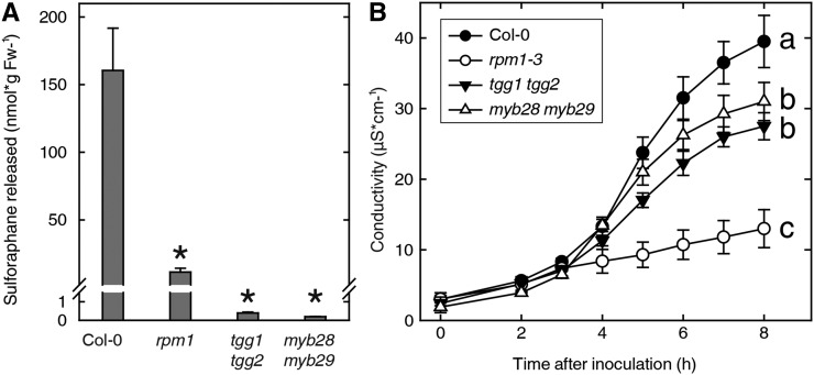
Plants defend themselves against microbial pathogens through a range of highly sophisticated and integrated molecular systems. Recognition of pathogen-secreted effector proteins often triggers the hypersensitive response (HR), a complex multicellular defense reaction where programmed cell death of cells surrounding the primary site of infection is a prominent feature. Even though the HR was described almost a century ago, cell-to-cell factors acting at the local level generating the full defense reaction have remained obscure. In this study, we sought to identify diffusible molecules produced during the HR that could induce cell death in naive tissue. We found that 4-methylsulfinylbutyl isothiocyanate (sulforaphane) is released by Arabidopsis (Arabidopsis thaliana) leaf tissue undergoing the HR and that this compound induces cell death as well as primes defense in naive tissue. Two different mutants impaired in the pathogen-induced accumulation of sulforaphane displayed attenuated programmed cell death upon bacterial and oomycete effector recognition as well as decreased resistance to several isolates of the plant pathogen Hyaloperonospora arabidopsidis. Treatment with sulforaphane provided protection against a virulent H. arabidopsidis isolate. Glucosinolate breakdown products are recognized as antifeeding compounds toward insects and recently also as intracellular signaling and bacteriostatic molecules in Arabidopsis. The data presented here indicate that these compounds also trigger local defense responses in Arabidopsis tissue.


Figures

Figure 1.
Figure 1.
Induction of cell death by an aqueous extract from Arabidopsis tissue undergoing the HR and identification of sulforaphane. A and B, Transgenic Arabidopsis plants expressing the bacterial P. syringae effector AvrRpm1 (DEX:AvrRpm1/Col-0 and DEX:AvrRpm1/rpm1-3) were incubated in water with the inducer DEX. Small molecules recovered from the bathing solution of the Col-0 (A) and rpm1-3 (B) plants were infiltrated into wild-type (nontransgenic) plants at the indicated dilutions. C, The material obtained was further analyzed by HPLC. Fractions were collected, dried, dissolved in water, and infiltrated into wild-type leaves. Visible effects and Trypan Blue staining of leaves receiving the fractions from the DEX:AvrRpm1/Col-0 extract are shown at bottom. D, The HPLC-purified fraction from the DEX:AvrRpm1/Col-0 fraction was subjected to GC-MS with electron-impact ionization. E, Mass spectrum for the major peak. F, The fraction was also dissolved in methanol, and the UV absorption spectrum was recorded. G, Structure of the identified compound, sulforaphane. The experiments depicted in A to C were performed twice with identical results.
Figure 2.
Figure 2.
Release of sulforaphane during effector-induced HR. A, Transgenic Arabidopsis plants expressing the bacterial P. syringae effector AvrRpm1 (DEX:AvrRpm1/Col-0 and DEX:AvrRpm1/rpm1-3) were incubated in water with DEX. B and C, Wild type Col-0 Arabidopsis leaf discs were infiltrated with wild-type P. syringae pv tomato DC3000 (B) or the Δsax mutant (C) expressing the effector AvrRpm1 (OD600 = 0.1) and incubated in water. At the indicated times, the discs were removed and the amount of sulforaphane in the bathing solution was analyzed. Average and range values for duplicate samples are shown. The experiments in A and B were performed three times with similar results, and the experiment in C was repeated twice with similar results. Fw, Fresh weight.
Figure 3.
Figure 3.
Infiltration of pure sulforaphane causes cell death in leaf. A to C, Sulforaphane was suspended in water to the indicated concentrations (A) or 1 mm (B and C) and syringe infiltrated into wild-type Col-0 Arabidopsis (A), broad bean (B), and sunflower (C) leaves. The left side of the leaf was infiltrated with sulforaphane, and the right side was mock infiltrated with deionized water. The leaves were detached and photographed after 24 h (A). D, Leaves were syringe infiltrated with the indicated isothiocyanates (ITC); leaf discs were punched out, washed, and incubated in water for the indicated times; and the conductance of the bathing solution was measured. Average and range values for duplicate samples are shown. Asterisks indicate statistical significance compared with mock treatment at the indicated times (one-way ANOVA, P < 0.05). The experiments were performed twice with similar results.
Figure 4.
Figure 4.
Infiltration of pure sulforaphane causes oxidation of the cellular glutathione pool. Leaf discs from wild-type Arabidopsis were vacuum infiltrated with sulforaphane suspended in water at the indicated concentrations. Reduced and oxidized glutathione contents were measured 30 min after infiltration, and the redox potential was calculated. Average and range values for triplicate samples are shown. The experiment was performed twice with similar results. Fw, Fresh weight.
Figure 5.
Figure 5.
Compromised PCD in sulforaphane-deficient mutants. A, Leaf discs from Arabidopsis wild-type and mutant lines were infiltrated with P. syringae DC3000:AvrRpm1, and the amount of sulforaphane released into the bathing solution was determined at 6 hpi. Mean and range values for duplicate samples are shown. Asterisks indicate statistical significance compared with Col-0 (one-way ANOVA, P < 0.05). Fw, Fresh weight. B, Electrolyte leakage was measured at the indicated time points for the wild type (black circles), double mutant myb28 myb29 (white triangles), double mutant tgg1 tgg2 (black triangles), and rpm1-3 (white circles). Mean ± sd of six replicate samples are shown. Letters a to c indicate statistically significant groups (one-way ANOVA with Tukey’s posthoc test, P < 0.05). The experiments were performed twice with similar results.
Figure 6.
Figure 6.
Reduced resistance to a biotrophic oomycete in sulforaphane-deficient mutants. The indicated lines were inoculated at the cotyledon stage with Hpa conidia of the isolates Cala2 (A–D) or Emwa1 (E). The cotyledons were stained with Trypan Blue 7 (A–C) or 2 (D) dpi, and the extent of cell death was determined or sporophores were counted at 7 dpi (E). Rapid cell death in the wild type is shown in B, and trailing necrosis is shown in C. Average and range values for three replicate experiments, each including 200 interaction sites, are shown in A and D. Average and sd for 15 cotyledons are shown in E. Letters a to c indicate statistically significant groups (one-way ANOVA with Tukey’s posthoc test, P < 0.05). The experiments were performed three times with similar results. Bars in B and C = 0.5 mm.
Figure 7.
Figure 7.
Sulforaphane treatment of plants provides increased resistance against pathogens. Seedlings of wild-type Landsberg erecta (Ler) or eds1 in the Wassilewskija (Ws) background were sprayed with 200 µm sulforaphane 24 h before or after, as indicated, inoculation with Hpa isolate Cala2, and the resulting sporulation was counted at 4 dpi. Average numbers of spores and range values for three replicate samples are shown. Letters a to c indicate statistically significant groups (one-way ANOVA with Tukey’s posthoc test, P < 0.05). The experiment was repeated twice with identical results.

References

    1. Agerbirk N, Olsen CE (2012) Glucosinolate structures in evolution. Phytochemistry 77: 16–45 - PubMed
    1. Ahmad S, Veyrat N, Gordon-Weeks R, Zhang Y, Martin J, Smart L, Glauser G, Erb M, Flors V, Frey M, et al. (2011) Benzoxazinoid metabolites regulate innate immunity against aphids and fungi in maize. Plant Physiol 157: 317–327 - PMC - PubMed
    1. Andersson MX, Kourtchenko O, Dangl JL, Mackey D, Ellerström M (2006) Phospholipase-dependent signalling during the AvrRpm1- and AvrRpt2-induced disease resistance responses in Arabidopsis thaliana. Plant J 47: 947–959 - PubMed
    1. Bailey K, Cevik V, Holton N, Byrne-Richardson J, Sohn KH, Coates M, Woods-Tör A, Aksoy HM, Hughes L, Baxter L, et al. (2011) Molecular cloning of ATR5(Emoy2) from Hyaloperonospora arabidopsidis, an avirulence determinant that triggers RPP5-mediated defense in Arabidopsis. Mol Plant Microbe Interact 24: 827–838 - PubMed
    1. Barth C, Jander G (2006) Arabidopsis myrosinases TGG1 and TGG2 have redundant function in glucosinolate breakdown and insect defense. Plant J 46: 549–562 - PubMed

Publication types