

Reusing cadaveric humeri for fracture testing after testing simulated rotator cuff tendon repairs
- PMID: 25371862
- PMCID: PMC4215328
- DOI: 10.1089/biores.2014.0020
Reusing cadaveric humeri for fracture testing after testing simulated rotator cuff tendon repairs
Abstract
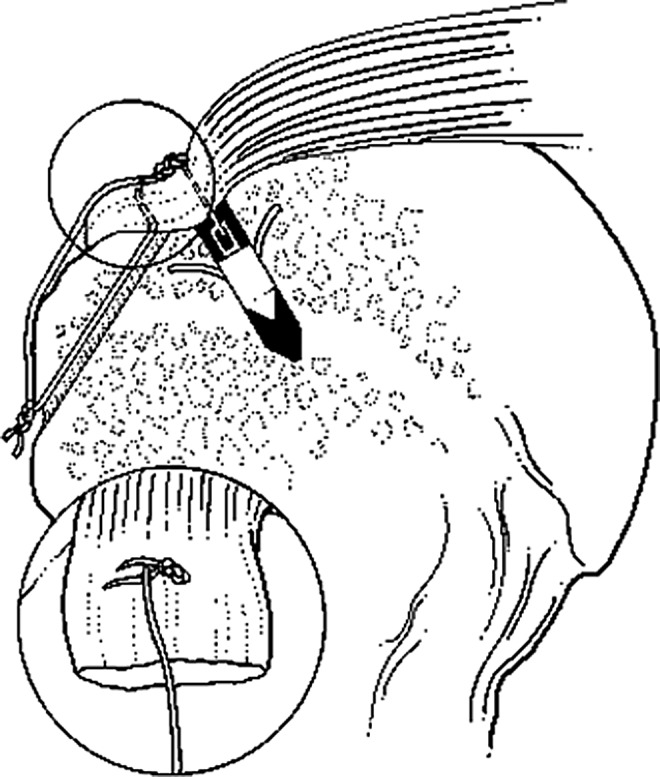
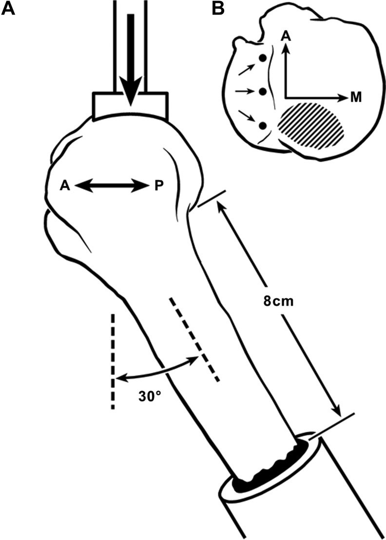
The financial cost of using human tissues in biomedical testing and surgical reconstruction is predicted to increase at a rate that is disproportionately greater than other materials used in biomechanical testing. Our first hypothesis is that cadaveric proximal humeri that had undergone monotonic failure testing of simulated rotator cuff repairs would not differ in ultimate fracture loads or in energy absorbed to fracture when compared to controls (i.e., bones without cuff repairs). Our second hypothesis is that there can be substantial cost savings if these cadaveric proximal humeri, with simulated cuff repairs, can be re-used for fracture testing. Results of fracture tests (conducted in a backwards fall configuration) and cost analysis support both hypotheses. Hence, the bones that had undergone monotonic failure tests of various rotator cuff repair techniques can be re-used in fracture tests because their load-carrying capacity is not significantly reduced.
Keywords: bone; cost analysis; fracture; humerus; in vitro; rotator cuff repair.
Figures
References
-
- Nandi SK, Roy S, Mukherjee P, Kundu B, De DK, Basu D. Orthopaedic applications of bone graft & graft substitutes: a review. Indian J Med Res. 2010;132:15–30 - PubMed
-
- Urabe K, Naruse K, Uchino M, et al. . The expense for one implantation of a banked bone allograft from a cadaveric donor and the issues affecting current advanced medical treatment in the Japanese orthopaedic field. Cell Tissue Bank. 2009;10:259–265 - PubMed
-
- Bloebaum RD, Lauritzen RS, Skedros JG, et al. . Roentgenographic procedure for selecting proximal femur allograft for use in revision arthroplasty. J Arthroplasty. 1993;8:347–360 - PubMed
LinkOut - more resources
Full Text Sources
Other Literature Sources