Clinical Trial
. 2014 Nov 5;9(11):e112325.
doi: 10.1371/journal.pone.0112325. eCollection 2014.

Spinal fMRI reveals decreased descending inhibition during secondary mechanical hyperalgesia

Affiliations
Clinical Trial

Spinal fMRI reveals decreased descending inhibition during secondary mechanical hyperalgesia

Torge Rempe et al. PLoS One. .

Abstract

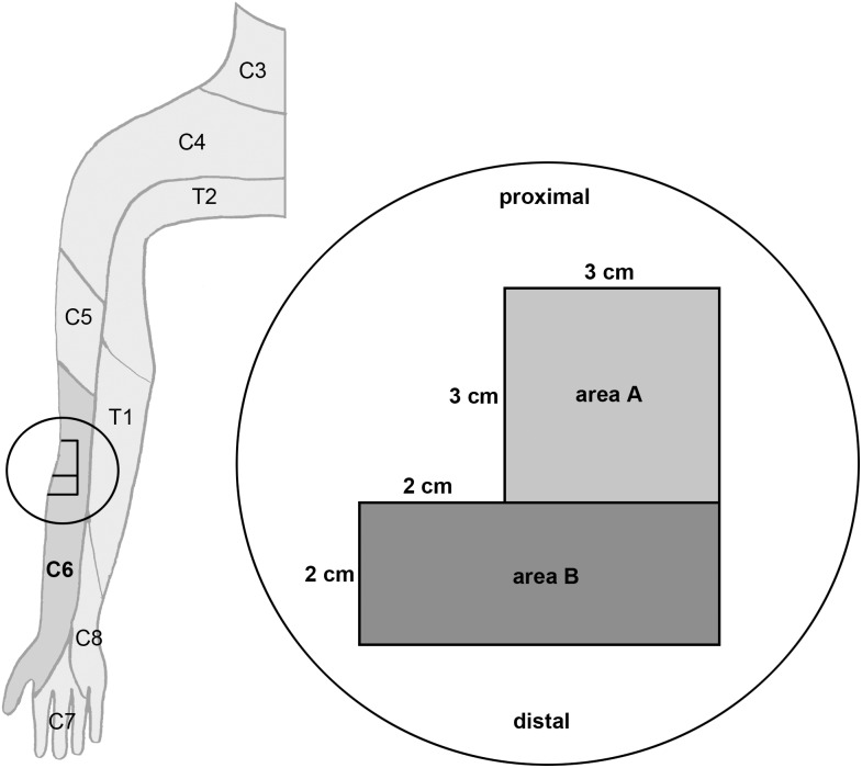
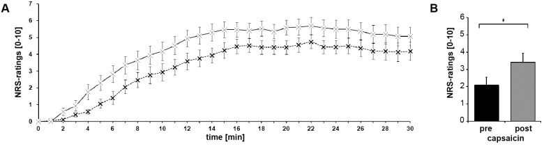
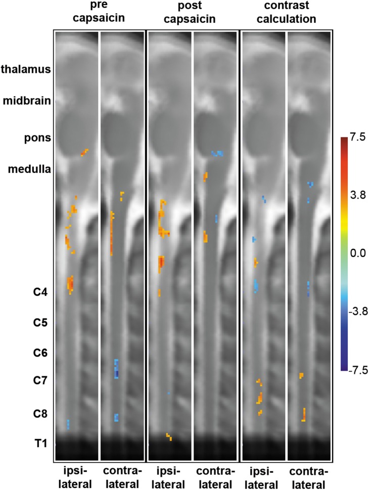
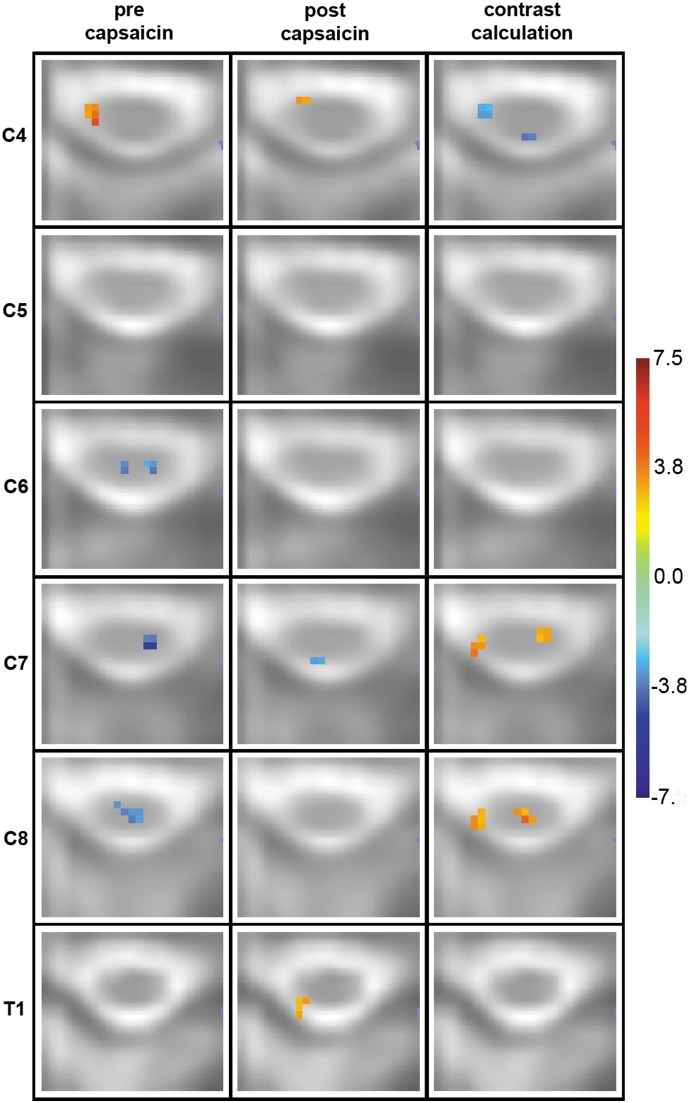
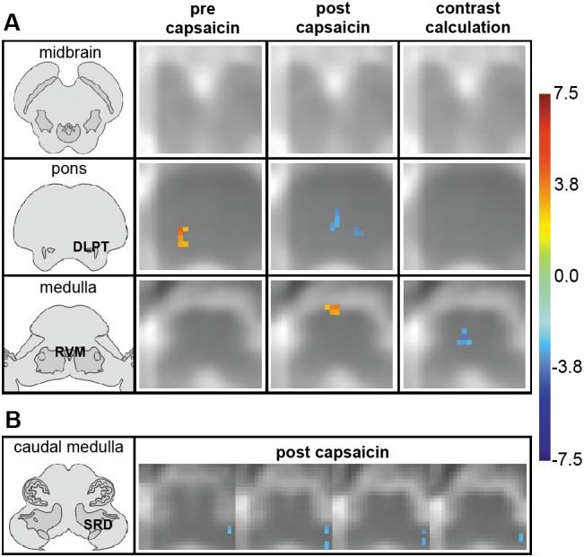
Mechanical hyperalgesia is one distressing symptom of neuropathic pain which is explained by central sensitization of the nociceptive system. This sensitization can be induced experimentally with the heat/capsaicin sensitization model. The aim was to investigate and compare spinal and supraspinal activation patterns of identical mechanical stimulation before and after sensitization using functional spinal magnetic resonance imaging (spinal fMRI). Sixteen healthy subjects (6 female, 10 male, mean age 27.2 ± 4.0 years) were investigated with mechanical stimulation of the C6 dermatome of the right forearm during spinal fMRI. Testing was always performed in the area outside of capsaicin application (i.e. area of secondary mechanical hyperalgesia). During slightly noxious mechanical stimulation before sensitization, activity was observed in ipsilateral dorsolateral pontine tegmentum (DLPT) which correlated with activity in ipsilateral spinal cord dorsal gray matter (dGM) suggesting activation of descending nociceptive inhibition. During secondary mechanical hyperalgesia, decreased activity was observed in bilateral DLPT, ipsilateral/midline rostral ventromedial medulla (RVM), and contralateral subnucleus reticularis dorsalis, which correlated with activity in ipsilateral dGM. Comparison of voxel-based activation patterns during mechanical stimulation before/after sensitization showed deactivations in RVM and activations in superficial ipsilateral dGM. This study revealed increased spinal activity and decreased activity in supraspinal centers involved in pain modulation (SRD, RVM, DLPT) during secondary mechanical hyperalgesia suggesting facilitation of nociception via decreased endogenous inhibition. Results should help prioritize approaches for further in vivo studies on pain processing and modulation in humans.


Conflict of interest statement

Competing Interests: Ralf Baron reported grant/research support by: Pfizer, Genzyme, Grünenthal. Member of the IMI „Europain“ collaboration and industry members of this are: Astra Zeneca, Pfizer, Esteve, UCB-Pharma, Sanofi Aventis, Grünenthal, Eli Lilly and Boehringer Ingelheim. German Federal Ministry of Education and Research (BMBF): German Research Network on Neuropathic Pain, Modelling Pain Switches. German Research Foundation (DFG). He received honoraria as a speaker from Pfizer, Genzyme, Grünenthal, Mundipharma, Sanofi Pasteur, Medtronic, Eisai, UCB BioSciences, Lilly, Boehringer Ingelheim, Astellas, Desitin and as a consultant from Pfizer, Genzyme, Grünenthal, Mundipharma, Allergan, Sanofi Pasteur, Medtronic, Eisai, UCB BioSciences, Lilly, Boehringer Ingelheim, Astellas, Novartis, Bristol-Myers Squibb, Biogenidec, AstraZeneca. Olav Jansen received honoraria as a speaker from Penumbra and as a consultant from Strykker, Philips and Boehringer Ingelheim. Janne Gierthmühlen received honoraria as a speaker from Pfizer and travel grants from Grünenthal. There are no conflicts of interests relevant to this article and none of the competing interests alter the authors’ adherence to PLOS ONE policies on sharing data and materials.

Figures

Figure 1
Figure 1. Areas of sensitization and stimulation on the right lateral volar forearm.
For orientation, the schematic drawing on the left shows a dermatome map of the right upper extremity (redrawn and modified from [20]). The area of testing (marked by the circle) is situated on the right lateral volar forearm in the C6 dermatome. Area A: Site of sensitization with the heat/capsaicin model (3×3 cm square area 3 cm distal to the elbow in the C6 dermatome). Area B: Site of mechanical stimulation corresponding to the area of secondary mechanical hyperalgesia (2×5 cm area distal to area A).
Figure 2
Figure 2. Psychophysical data.
(A) Mean ratings of pain intensity (dashed line) and temperature perception (solid line) during capsaicin application. Capsaicin was applied at time 0. Mean ± standard error of the mean (SEM). (B) Mean pain ratings for the mechanical stimulus before and after application of capsaicin. Capsaicin induced secondary mechanical hyperalgesia. Mean ± SEM. *: p<0.05.
Figure 3
Figure 3. Sagittal slices of group activation patterns and contrast maps.
Columns 1 to 4 show areas of activity across brain stem and cervical spinal cord before (1st and 2nd column) and after (3rd and 4th column) sensitization with the heat capsaicin model representing the significance (T-value) of each active voxel across the 16 subjects. Columns 5 and 6 show partial-least squares (PLS) results of contrast calculations on a voxel-by-voxel basis. The left column of each 2 columns (e.g. 1st, 3rd and 5th) corresponds to the ipsilateral side of the stimulus, the right column to the contralateral side. The color bar on the right indicates the corresponding significance, i.e. T-value (columns 1–4) or bootstrap-ratio (columns 5–6) for each color.
Figure 4
Figure 4. Spinal group activation patterns before (left column) and after (middle column) sensitization and contrast maps (right column).
The transverse slices are in radiological orientation with the left side corresponding to the right body side and approximate the corresponding spinal cord segment for a rostral-caudal span from C4 to T1. They show spinal regions of signal intensity change before (left column) and after (middle column) sensitization with the heat/capsaicin model representing the significance (T-value) of each active voxel across the 16 subjects. The right column shows partial-least squares (PLS) results of contrast calculations on a voxel-by-voxel basis. The color bar in figure 3 indicates the corresponding significance, i.e. T-value (left and middle column) or bootstrap-ratio (right column) for each color. Left Column: Activations in ipsilateral vGM of C4. Deactivations in bilateral deep dGM of C6 and C8 and contralateral deep dGM of C7. Middle column: Ipsilateral activations in superficial dGM of T1 and vGM of C4. Deactivations in ipsilateral superficial dGM of C7. Right column: Activations in ipsilateral superficial dGM (C7, C8) and in contralateral vGM (C7) and deep dGM (C8). Deactivations in ipsilateral vGM and contralateral dGM of C4.
Figure 5
Figure 5. Supraspinal group activation patterns and contrast maps.
Slices are in radiological orientation with the left side corresponding to the right body side. The color bar in figure 3 indicates the corresponding significance, i.e. T-value (combined group results pre/post capsaicin) or bootstrap-ratio (contrast calculation) for each color. Anatomical transverse sections on the left were modified from . (A) The transverse slices approximate the corresponding brainstem region (midbrain, pons, medulla) for a rostral-caudal span. They show supraspinal regions of signal intensity change before (left column) and after (middle column) sensitization with the heat/capsaicin model representing the significance (T-value) of each active voxel across the 16 subjects. The right column shows partial-least squares (PLS) results of contrast calculations on a voxel-by-voxel basis. Left column: Activations in the ipsilateral DLPT (dorsolateral pontine tegmentum) during mechanical stimulation prior to application of capsaicin. Middle column: Deactivations in the contralateral DLPT and in the RVM (rostral ventromedial medulla) during secondary mechanical hyperalgesia (note that visible medial pontine deactivations are situated in the caudal pons/rostral medulla and therefore most likely correspond to the location of the RVM). Right column: Deactivations in the RVM. (B) The adjacent 1 mm thick transverse slices in consecutive arrangement located in the medulla show signal intensity changes during secondary mechanical hyperalgesia. Deactivations are observed in contralateral subnucleus reticularis dorsalis (SRD).

Similar articles

Cited by

References

    1. Baron R, Binder A, Wasner G (2010) Neuropathic pain: diagnosis, pathophysiological mechanisms, and treatment. Lancet Neurol 9: 807–819. - PubMed
    1. Simone DA, Sorkin LS, Oh U, Chung JM, Owens C, et al. (1991) Neurogenic hyperalgesia: central neural correlates in responses of spinothalamic tract neurons. J Neurophysiol 66: 228–246. - PubMed
    1. Westlund KN (2006) The dorsal horn and hyperalgesia. In: Cervero F, Jensen TS, editors. Handbook of Clinical Neurology, Vol 81 (3rd series): Pain. Amsterdam: Elsevier B.V. pp. 178–186. - PMC - PubMed
    1. Petersen KL, Rowbotham MC (1999) A new human experimental pain model: the heat/capsaicin sensitization model. Neuroreport 10: 1511–1516. - PubMed
    1. Villanueva L, Lopez-Avila A, Le Bars D (2006) Ascending nociceptive pathways. In: Cervero F, Jensen TS, editors. Handbook of Clinical Neurology, Vol 81 (3rd series): Pain. Amsterdam: Elsevier B.V. pp. 93–102. - PubMed

Publication types