. 2014 Nov 5;9(11):e111539.
doi: 10.1371/journal.pone.0111539. eCollection 2014.

Caspase-1/ASC inflammasome-mediated activation of IL-1β-ROS-NF-κB pathway for control of Trypanosoma cruzi replication and survival is dispensable in NLRP3-/- macrophages

Affiliations

Caspase-1/ASC inflammasome-mediated activation of IL-1β-ROS-NF-κB pathway for control of Trypanosoma cruzi replication and survival is dispensable in NLRP3-/- macrophages

Nilay Dey et al. PLoS One. .

Abstract

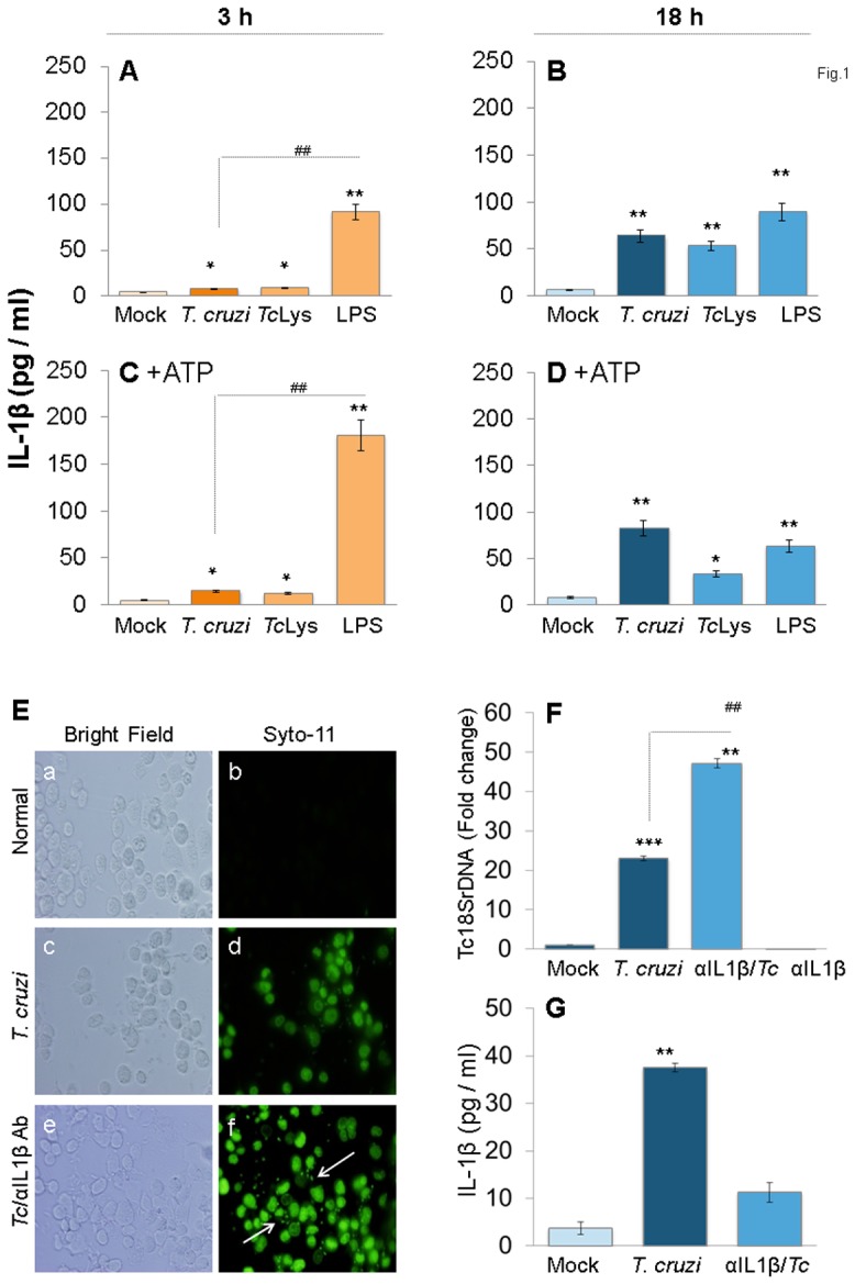
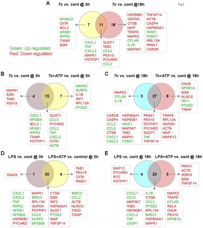
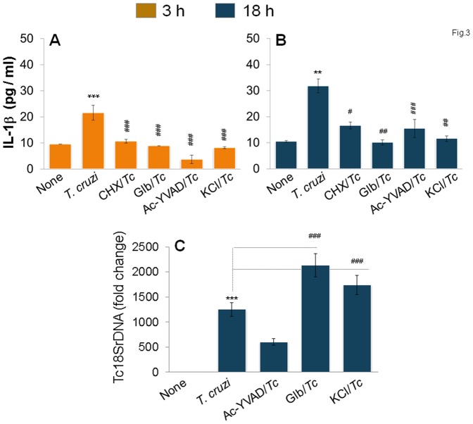
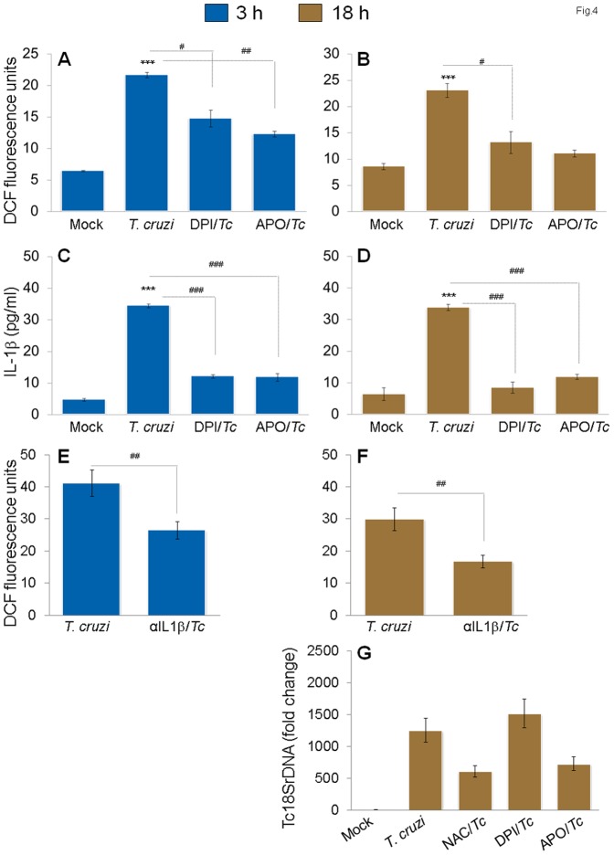
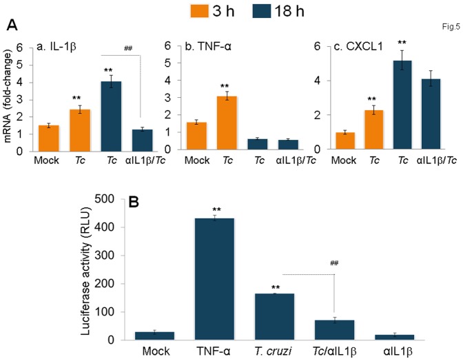
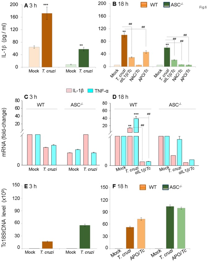
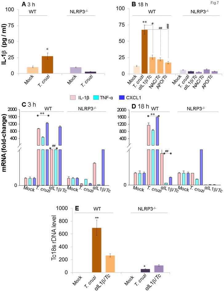
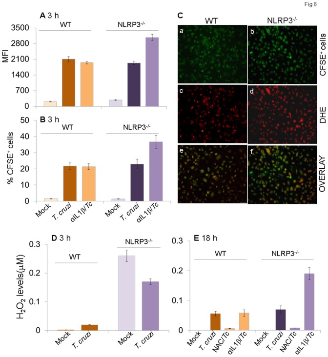
In this study, we have utilized wild-type (WT), ASC-/-, and NLRP3-/- macrophages and inhibition approaches to investigate the mechanisms of inflammasome activation and their role in Trypanosoma cruzi infection. We also probed human macrophages and analyzed published microarray datasets from human fibroblasts, and endothelial and smooth muscle cells for T. cruzi-induced changes in the expression genes included in the RT Profiler Human Inflammasome arrays. T. cruzi infection elicited a subdued and delayed activation of inflammasome-related gene expression and IL-1β production in mφs in comparison to LPS-treated controls. When WT and ASC-/- macrophages were treated with inhibitors of caspase-1, IL-1β, or NADPH oxidase, we found that IL-1β production by caspase-1/ASC inflammasome required reactive oxygen species (ROS) as a secondary signal. Moreover, IL-1β regulated NF-κB signaling of inflammatory cytokine gene expression and, subsequently, intracellular parasite replication in macrophages. NLRP3-/- macrophages, despite an inability to elicit IL-1β activation and inflammatory cytokine gene expression, exhibited a 4-fold decline in intracellular parasites in comparison to that noted in matched WT controls. NLRP3-/- macrophages were not refractory to T. cruzi, and instead exhibited a very high basal level of ROS (>100-fold higher than WT controls) that was maintained after infection in an IL-1β-independent manner and contributed to efficient parasite killing. We conclude that caspase-1/ASC inflammasomes play a significant role in the activation of IL-1β/ROS and NF-κB signaling of cytokine gene expression for T. cruzi control in human and mouse macrophages. However, NLRP3-mediated IL-1β/NFκB activation is dispensable and compensated for by ROS-mediated control of T. cruzi replication and survival in macrophages.


Conflict of interest statement

Competing Interests: The authors have declared that no competing interests exist.

Figures

Figure 1
Figure 1. IL-1β production in macrophages infected by T. cruzi.
(A–D) PMA-differentiated THP-1 mφs were incubated with T. cruzi trypomastigotes (cell: parasite ratio, 1∶3), Tc lysate (10 µg protein/106 cells) or LPS (100 ng/ml) for 3 h (A&C) and 18 h (B&D). In some experiments, ATP was added during last 30 min of incubation (C&D). IL-1β release in supernatants was determined by ELISA. (E–G) IL-1β contributes to parasite control in mφs. THP-1 mφs were incubated with SYTO®11-labeled T. cruzi in the presence or absence of anti-IL-1β antibody for 18 h. (E) SYTO®11 fluorescence as an indicator of parasite uptake (shown by arrows) was determined by using an Olympus BX-15 microscope equipped with a digital camera (magnification 40X). (F) Quantitative PCR analysis of parasite burden in infected mφs by using Tc18SrDNA-specific oligonucleotides (normalized to human GAPDH). (G) Addition of anti-IL-1β antibody depletes secreted IL-1β levels in T. cruzi-infected mφs. In all figures, data are representative of three independent experiments and presented as mean ± SD. Significance is shown by *normal versus infected and #treated/infected versus infected (*,#p<0.05, **,##p<0.01, and ***,###p<0.001).
Figure 2
Figure 2. Venn diagram of inflammasome-related differential gene expression in mφs infected with T. cruzi (±ATP).
THP-1 mφs were incubated with T. cruzi or LPS (± ATP treatment) as in Fig. 1. Total RNA was isolated, and cDNA was used as a template to probe the expression of 95 genes (including house-keeping genes) in the RT2 Profiler Inflammasome PCR Arrays. The differential mRNA level was captured by quantitative RT-PCR, normalized to housekeeping genes, and HTqPCR was employed to attain the statistically significant differential expression in treated- versus-control samples (Table 1 and Table S2). Shown are Venn diagrams of comparative analysis of gene expression in T. cruzi-infected mφs at 3 h versus 18 h (A), effect of ATP stimulus on gene expression at 3 h (B) and 18 h (C) pi, and comparative effect of ATP stimulus on gene expression in LPS-treated mφs at 3 h (D) and 18 h (E). Differential up-regulation (green) and down-regulation (red) of genes with respect to controls is presented. Genes presenting as red with green arrow in BF showed decreased expression without ATP but were up-regulated by ATP treatment (and vice versa).
Figure 3
Figure 3. NLRP3/caspase-1 inflammasome is the major source of IL-1β for parasite control in mφs.
THP-1 mφs were incubated with T. cruzi in the presence or absence of cycloheximide (CHX,), glibenclamide (Glb), Ac-YVAD-CHO, and KCl for 3 h (A) and 18 h (B&C). Macrophages incubated with media alone were used as controls. (A&B) IL-1β release in supernatants was determined by an ELISA. (C) Quantitative PCR analysis of parasite burden in infected macrophages using Tc18SrDNA-specific oligonucleotides.
Figure 4
Figure 4. Feedback cycle of NOX2/ROS and IL-1β activation in mφs infected by T. cruzi.
THP-1 mφs were infected with T. cruzi as in Fig. 1, and incubated for 3 h (A,C,E) or 18 h (B,D,F,G) in presence of NOX2 inhibitors (diphenylene iodinium (DPI) or apoCynin), ROS scavenger (N-acetylcysteine (NAC)) or IL-1β antibody. (AD) NOX2 inhibitors decreased ROS and IL-1β levels in infected mφs. Shown are (A&B) H2DCFDA oxidation by intracellular ROS, resulting in formation of fluorescent DCF by fluorimetry and (C&D) IL-1β release in supernatants determined by an ELISA. (E&F) Treatment with anti-IL-1β antibody decreased the ROS levels in infected mφs. (G) Effect of ROS inhibitors on intracellular parasite burden, as determined by qPCR, in infected mφs.
Figure 5
Figure 5. IL-1β signals NF-κB activation and inflammatory cytokine gene expression in infected mφs.
(A) The mRNA levels for IL-1β (panel a), TNFα (panel b) and CXCL1 (panel c) cytokines were measured in T. cruzi-infected THP-1 mφs at 3h and 18h pi by quantitative RT-PCR. (B) RAW 264.7 macrophages were transiently transfected with pGL4.NF-κB-Luc reporter plasmid and pREP7-Rluc plasmid (transfection efficiency control) as described in Materials and Methods. Transfected cells were infected with T. cruzi and incubated in the presence or absence of anti-IL-1β antibody. Mφs incubated with 10 ng/ml recombinant TNF-α for 6 h were used as positive controls. The relative NF-κB transcriptional activity was measured by firefly luciferase activity and normalized to Renilla luciferase activity. The transcriptional activity of NF-κB in normal cells was considered as baseline and valued at 1.
Figure 6
Figure 6. ASC-/- mφs are compromised in the IL-1β–ROS–NF-κB pathway for control of T. cruzi.
Bone-marrow-derived macrophages were isolated from matched WT and ASC-/- mice. Primary mφs were infected with T. cruzi and incubated for 3 h or 18 h in the presence or absence of anti-IL-1β Ab or ROS scavengers (as in Figs.1&4). Shown are IL-1β release measured by an ELISA (A&B), mRNA levels for IL-1β and TNF-α by quantitative RT-PCR (C&D) and Tc18SrDNA signal by qPCR (E&F).
Figure 7
Figure 7. NLRP3-/- mφs are compromised in IL-1β activation and inflammatory cytokine gene expression, but equipped to control T. cruzi.
Bone marrow-derived macrophages were isolated from matched WT and NLRP3-/- mice. Primary mφs were infected with T. cruzi and incubated for 3 h or 18 h in the presence or absence of anti-IL-1β antibody or ROS scavengers (as in Figs.1&4). Shown are IL-1β release by ELISA (A&B), mRNA level for IL-1β, TNF-α and CXCL1 by quantitative RT-PCR (C&D) and Tc18SrDNA signal by qPCR (E).
Figure 8
Figure 8. NLRP3 deficiency is compensated for by increased ROS levels in mφs (± T. cruzi).
Bone marrow-derived primary macrophages isolated from matched WT and NLRP3-/- mice were infected with CFSE-labeled T. cruzi and incubated for 3 h or 18 h in the presence or absence of anti-IL-1β antibody or ROS inhibitors. Shown are the mean fluorescence intensity of CFSE (A) as an indicator of # parasites/cell and mean percentage of CFSE+ mφs (B) as an indicator of parasite uptake efficiency. (C) Fluorescence microscopy of NLRP3-/- (panels a, c, e) and WT (panels b, d, e) mφs infected with CFSE-labeled T. cruzi for 18 h. Shown are representative images of CFSE (green, panels a & b), intracellular ROS-specific DHE fluorescence (panels c & d) and overlay images of a & c and b & d in panels e & f. (D&E) Bar graphs show a quantitative measure of ROS release, measured by an Amplex red assay, in NLRP3-/- and WT mφs.

Similar articles

Cited by

References

    1. World Health Organization (2010) Chagas disease: control and elimination. UNDP/World Bank/WHO. Available: http://apps.who.int/gb/ebwha/pdf_files/WHA63/A63_17-en.pdf. Accessed 2014 Sept 3
    1. Machado FS, Dutra WO, Esper L, Gollob KJ, Teixeira MM, et al. (2012) Current understanding of immunity to Trypanosoma cruzi infection and pathogenesis of Chagas disease. Seminars in Immunopathology 34: 753–770. - PMC - PubMed
    1. Huang H, Chan J, Wittner M, Jelicks LA, Morris SA, et al. (1999) Expression of cardiac cytokines and inducible form of nitric oxide synthase (NOS2) in Trypanosoma cruzi-infected mice. J Mol Cell Cardiol 31: 75–88. - PubMed
    1. Machado FS, Tyler KM, Brant F, Esper L, Teixeira MM, et al. (2012) Pathogenesis of Chagas disease: time to move on. Front Biosci (Elite Ed) 4: 1743–1758. - PMC - PubMed
    1. Machado FS, Souto JT, Rossi MA, Esper L, Tanowitz HB, et al. (2008) Nitric oxide synthase-2 modulates chemokine production by Trypanosoma cruzi-infected cardiac myocytes. Microbes Infect 10: 1558–1566. - PMC - PubMed

Publication types

MeSH terms