. 2014 Nov;70(Pt 11):2800-12.
doi: 10.1107/S1399004714018732. Epub 2014 Oct 16.

Structural basis for the recognition of muramyltripeptide by Helicobacter pylori Csd4, a D,L-carboxypeptidase controlling the helical cell shape

Affiliations

Structural basis for the recognition of muramyltripeptide by Helicobacter pylori Csd4, a D,L-carboxypeptidase controlling the helical cell shape

Hyoun Sook Kim et al. Acta Crystallogr D Biol Crystallogr. 2014 Nov.

Abstract

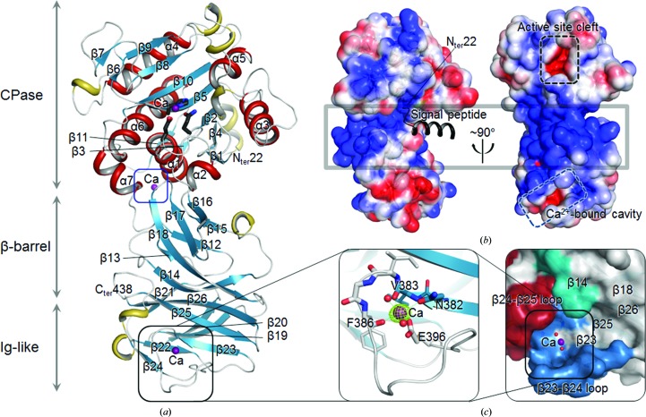
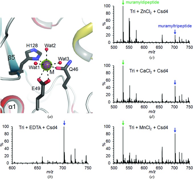
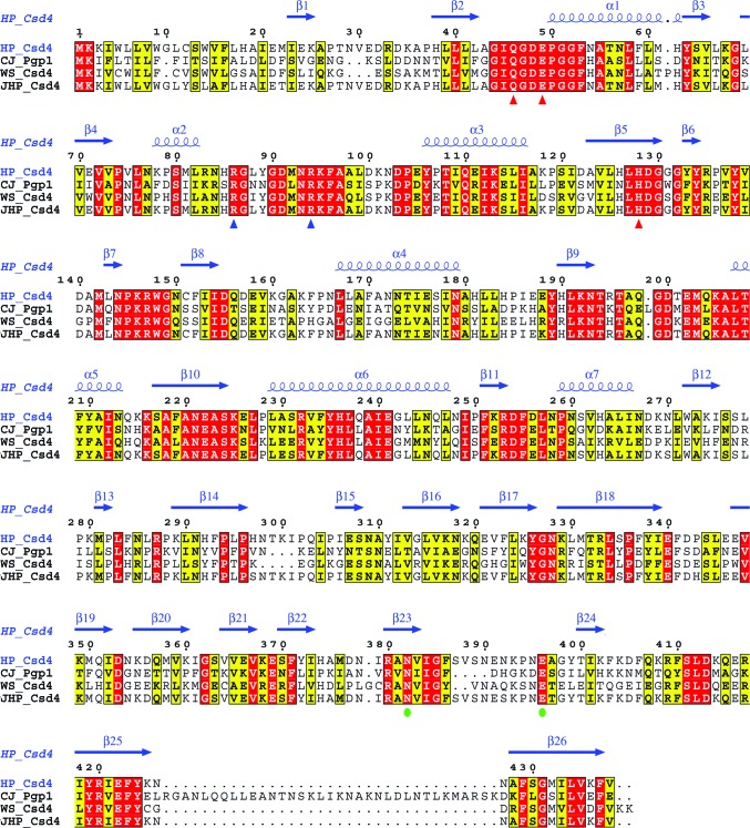
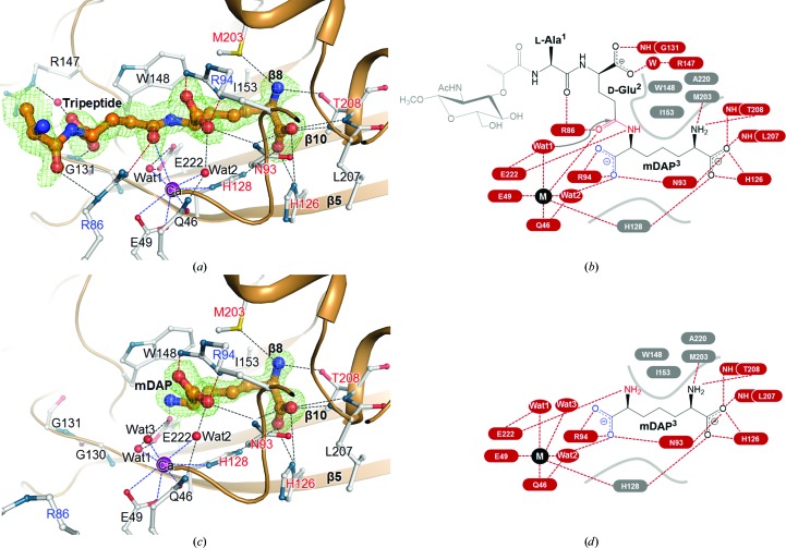
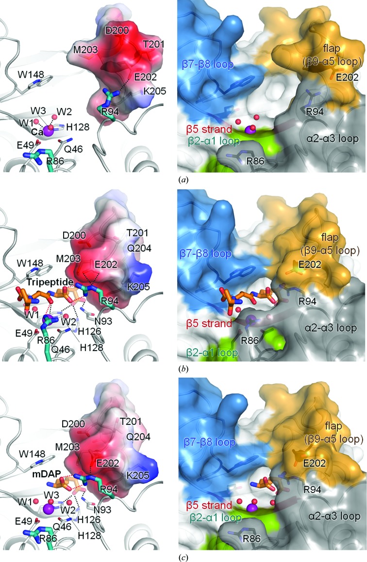
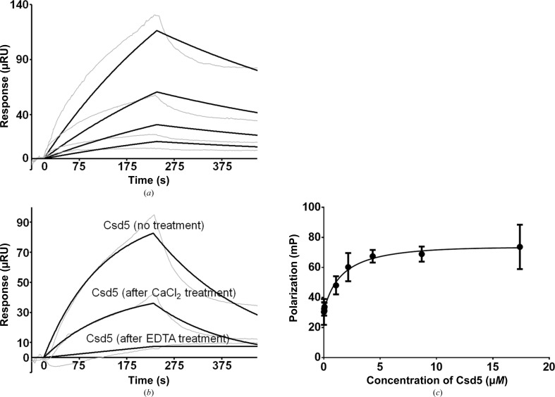
Helicobacter pylori infection causes a variety of gastrointestinal diseases, including peptic ulcers and gastric cancer. Its colonization of the gastric mucosa of the human stomach is a prerequisite for survival in the stomach. Colonization depends on its motility, which is facilitated by the helical shape of the bacterium. In H. pylori, cross-linking relaxation or trimming of peptidoglycan muropeptides affects the helical cell shape. Csd4 has been identified as one of the cell shape-determining peptidoglycan hydrolases in H. pylori. It is a Zn(2+)-dependent D,L-carboxypeptidase that cleaves the bond between the γ-D-Glu and the mDAP of the non-cross-linked muramyltripeptide (muramyl-L-Ala-γ-D-Glu-mDAP) of the peptidoglycan to produce the muramyldipeptide (muramyl-L-Ala-γ-D-Glu) and mDAP. Here, the crystal structure of H. pylori Csd4 (HP1075 in strain 26695) is reported in three different states: the ligand-unbound form, the substrate-bound form and the product-bound form. H. pylori Csd4 consists of three domains: an N-terminal D,L-carboxypeptidase domain with a typical carboxypeptidase fold, a central β-barrel domain with a novel fold and a C-terminal immunoglobulin-like domain. The D,L-carboxypeptidase domain recognizes the substrate by interacting primarily with the terminal mDAP moiety of the muramyltripeptide. It undergoes a significant structural change upon binding either mDAP or the mDAP-containing muramyltripeptide. It it also shown that Csd5, another cell-shape determinant in H. pylori, is capable of interacting not only with H. pylori Csd4 but also with the dipeptide product of the reaction catalyzed by Csd4.

Keywords: HP1075; Helicobacter pylori; cell shape; csd4; csd5; d,l-carboxypeptidase; meso-diaminopimelate; peptidoglycan; pgp1.


Figures

Figure 1
Figure 1
Overall structure of H. pylori Csd4. (a) Ribbon diagram of the Csd4-unbound structure. β-Strands, α-helices, 310-helices and loops are shown in cyan, red, yellow and grey, respectively. Three calcium ions are shown as purple spheres. The metal-binding residues in the N-terminal CPase domain are shown as stick models. The blue and black boxes indicate the previously unknown Ca2+-binding sites of the CPase domain and the Ca2+-binding motif of the C-­terminal Ig-like domain. The secondary-structure elements were defined by DSSP (Kabsch & Sander, 1983 ▶). (b) Electrostatic surface diagram of Csd4-unbound. The signal peptide is modelled as a black ribbon diagram. The black and cyan dotted boxes indicate the active-site cleft of the CPase domain and the Ca2+-binding channel of the Ig-like domain. The grey box indicates the highly positively charged surface of the central β-barrel domain. Electrostatic potential at the molecular surface was calculated using APBS (Baker et al., 2001 ▶). (c) Ribbon diagram of the Ca2+-binding motif (left) and the surface representation of the Ca2+-bound channel (right) in the Ig-like domain. The OMIT mF oDF c map (contoured at 10σ) for the calcium ion is coloured green. The Ca2+-binding residues are shown as stick models. The calcium ion and the bound waters are shown as purple and red spheres, respectively. All figures representing the protein structure were drawn using PyMOL (v.1.3r1; Schrödinger).
Figure 2
Figure 2
Metal-dependency of H. pylori Csd4 as a carboxypeptidase. (a) Ribbon diagram of the active site in the Csd4-unbound structure coloured as in Fig. 1 ▶(a). The OMIT mF oDF c map (contoured at 5σ) for the calcium ion is coloured green. (be) Mass spectra of muramylpeptides after the reaction catalyzed by Csd4 in the presence of EDTA (b) or in the presence of Zn2+ (c), Ca2+ (d) or Mn2+ (e). Green and blue arrows indicate the peaks corresponding to the muramyldipeptide and muramyltripeptide (Tri), respectively.
Figure 3
Figure 3
Sequence alignment of four Csd4 homologues. Sequence alignment of Csd4 from H. pylori strain 26695 (HP_Csd4; SWISS-PROT accession code O25708), Pgp1 from Campylobacter jejuni (CJ_Pgp1; A1W0W1), Csd4 from Wolinella succinogenes (WS_Csd4; Q7MSQ3) and Csd4 from H. pylori strain J99 (JHP_Csd4; Q9ZM72) was performed and presented using ClustalX (Larkin et al., 2007 ▶) and ESPript (Gouet et al., 2003; http://espript.ibcp.fr). Red and blue triangles indicate the metal-binding site motif (Gln46, Glu49 and His128) and the two key Arg residues (Arg86 and Arg94) showing conformational changes in the active site of the CPase domain. Green circles indicate the Ca2+-binding motif in the C-terminal Ig-like domain.
Figure 4
Figure 4
Ligand interactions in the Csd4–muramyltripeptide and Csd4–mDAP structures. (a, c) Ribbon diagrams of the active site in the Csd4–muramyltripeptide (a) and Csd4–mDAP (c) structures. The tripeptide and mDAP are shown as stick models. Since the sugar moiety of the NAM-tripeptide was invisible, only the tripeptide portion has been modelled into the electron-density map. Residues interacting with the tripeptide or mDAP are shown as stick models. The OMIT mF oDF c maps for the tripeptide (contoured at 1.5σ) and mDAP (contoured at 2.5σ) are coloured green. The red and blue dotted lines indicate the interactions of the tripeptide or mDAP with the metal ion and two key Arg residues (Arg86 and Arg94), respectively. (b, d) Schematic diagrams of interactions of the bound muramyltripeptide (b) and mDAP (d) with Csd4. The red and grey labels represent electrostatic and hydrophobic residues interacting with these ligands, respectively.
Figure 5
Figure 5
Open and closed conformations of the flap. Electrostatic surface diagrams of the mobile flap (left panels) and surface diagrams showing the active-site cleft (right panels) in the Csd4-unbound (a), Csd4–muramyltripeptide (b) and Csd4–mDAP (c) structures.
Figure 6
Figure 6
Biophysical studies of the Csd4–Csd5 or the Csd5–dipeptide interaction in solution. (a) SPR experiments with immobilized Csd4 and Csd5 as an analyte at different concentrations (0.25, 0.50, 1.00 and 2.00 µM) are shown as grey-coloured traces. Black traces show the corresponding binding-model curves. (b) At the same concentration of Csd5 (2.00 µM), the interaction between Csd4 and Csd5 (‘no treatment’) was lost upon treatment of the Csd4-immobilized chip with 20 mM EDTA to remove intrinsic metal ions (‘after EDTA treatment’). Retreatment of the EDTA-treated Csd4-immobilized chip with 5 mM calcium chloride recovered the binding between two proteins (‘after CaCl2 treatment’). All sensorgrams were obtained by subtracting the nonspecific binding of the analyte to the BSA-immobilized chip. (c) Fluorescence polarization binding assay using an FITC-labelled dipeptide against increasing concentrations of Csd5.

References

    1. Adams, P. D. et al. (2010). Acta Cryst. D66, 213–221. - PubMed
    1. Baker, N. A., Sept, D., Joseph, S., Holst, M. J. & McCammon, J. A. (2001). Proc. Natl Acad. Sci. USA, 98, 10037–10041. - PMC - PubMed
    1. Berg, H. C. & Turner, L. (1979). Nature (London), 278, 349–351. - PubMed
    1. Bonis, M., Ecobichon, C., Guadagnini, S., Prévost, M. C. & Boneca, I. G. (2010). Mol. Microbiol. 78, 809–819. - PubMed
    1. Bunnage, M. E. et al. (2007). J. Med. Chem. 50, 6095–6103. - PubMed

Publication types

MeSH terms