. 2014 Nov;70(Pt 11):3043-56.
doi: 10.1107/S1399004714021099. Epub 2014 Oct 29.

Enzyme-adenylate structure of a bacterial ATP-dependent DNA ligase with a minimized DNA-binding surface

Affiliations

Enzyme-adenylate structure of a bacterial ATP-dependent DNA ligase with a minimized DNA-binding surface

Adele Williamson et al. Acta Crystallogr D Biol Crystallogr. 2014 Nov.

Abstract

DNA ligases are a structurally diverse class of enzymes which share a common catalytic core and seal breaks in the phosphodiester backbone of double-stranded DNA via an adenylated intermediate. Here, the structure and activity of a recombinantly produced ATP-dependent DNA ligase from the bacterium Psychromonas sp. strain SP041 is described. This minimal-type ligase, like its close homologues, is able to ligate singly nicked double-stranded DNA with high efficiency and to join cohesive-ended and blunt-ended substrates to a more limited extent. The 1.65 Å resolution crystal structure of the enzyme-adenylate complex reveals no unstructured loops or segments, and suggests that this enzyme binds the DNA without requiring full encirclement of the DNA duplex. This is in contrast to previously characterized minimal DNA ligases from viruses, which use flexible loop regions for DNA interaction. The Psychromonas sp. enzyme is the first structure available for the minimal type of bacterial DNA ligases and is the smallest DNA ligase to be crystallized to date.

Keywords: ATP-dependent DNA ligase; Psychromonas sp. strain SP041.


Figures

Figure 1
Figure 1
Overall fold of Psy-Lig chain A (purple) and chain B (blue). The covalently bound AMP is shown in green. (a) Chains in the asymmetric unit. (b) Superposition of chains by alignment of their AD domains. The dashed arrow indicates the distance between the Cα atom of Gly191 of each monomer.
Figure 2
Figure 2
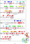
Sequence alignments of Psy-Lig with other ATP-dependent DNA ligases. Fully conserved residues are shaded red; partially or homologously conserved residues are shown in red text. Structural features discussed in the text and the conserved motifs of the nucleotidyltransferase enzymes are indicated in boxes, with numbering corresponding to the conventions given in Shuman & Lima (2004 ▶). α-Helices are indicated by curl symbols and β-strands by arrows. Filled boxes above the sequences give the numbering of the secondary-structural elements of Psy-Lig as referred to in the text, and correspond to the colours and numbering used in the ribbon diagram (inset). (a) Structure-based alignment between Psy-Lig and ChlV-­Lig constructed using PDBeFold. (b) Sequence-based alignment of Psy-Lig with biochemically characterized ligases from H. influenzae (Hin-Lig), N. meningitidis (Nme-Lig) and A. salmonicida (Vib-­Lig).
Figure 3
Figure 3
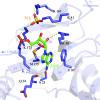
Active-site structure of Psy-Lig-A. The nucleotide cofactor is depicted and labelled in green and the sulfate ion is in yellow. Lys25 which is covalently attached to the AMP is labelled in bold; all residues making contacts with the cofactor and ion are shown as purple sticks. Secondary-structural elements of the AD domain are labelled in light purple and correspond to the notation used in Fig. 2 ▶.
Figure 4
Figure 4
Conformation of the linker between the AD and OB domains. (a) Chain A, (b) chain B.
Figure 5
Figure 5
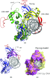
Comparison of the tertiary structure of Psy-Lig with that of ChlV-Lig. (a) Superposition of Psy-Lig chain A (purple) with ChlV-Lig–AMP (green, PDB entry 1fvi) and ChlV-Lig–DNA (yellow, PDB entry 2q2t). The DNA-binding latch of ChlV-Lig and the homologous loop of Psy-Lig are coloured red. The unstructured residues of ChlV-Lig–AMP are indicated as a dashed line. DNA is shown in grey. (b) Psy-Lig modelled in the DNA-bound conformation by superposition of the Psy-Lig OB domain with the ChlV-Lig–DNA OB domain–DNA complex. The original adenylated Psy-Lig structure (purple) and the re-oriented position (pink) are shown as ribbons. The DNA from the ChlV-Lig–DNA structure is shown in grey. (c) Surface of the Psy-Lig DNA-bound model (pink) superposed with the ChlV-Lig–DNA structure (yellow, transparent), showing incomplete encirclement of the DNA by Psy-Lig.
Figure 6
Figure 6
Psy-Lig-A coloured by electrostatic surface potential. The surface potential was generated using APBS (Dolinsky et al., 2007 ▶), with positively charged areas shown in blue and negatively charged areas in red. (a) Overview of the Psy-Lig structure with residues predicted to be involved in DNA interactions shown as sticks. (b) Surface and (c) important residues of the AD domain looking at the active site. The AMP cofactor and sulfate ion are shown as sticks. Green arrows indicate the approximate positions of the nicked DNA strands based on the model shown in Fig. 5 ▶. (d) Surface residues and (e) residues of the OB domain looking towards the DNA-binding site predicted by the model. The green arrow indicates the approximate position of the complementary DNA strand in the OB groove.
Figure 7
Figure 7
Ligation activity of Psy-Lig with different DNA substrates. (a) Schematic representation of DNA substrates and representative results of ligation on TBE–urea gels. The samples for the blunt substrate gel are for a 3.0 µM enzyme concentration over 8 h. The image contrast for the gapped substrate gel has been adjusted to allow visualization of the faint bands from ligation. Integration of bands was carried out prior to this adjustment. (b) Percentage of substrate ligated in 5 min for nicked, cohesive and mismatch substrates (main figure) and gapped substrate after 30 min (inset) as a function of enzyme concentration. (c) Percentage of blunt substrate ligated over 8 or 24 h for two Psy-Lig dilutions. Ligation activity was quantified by integration of band intensity and is expressed as the percentage of upper (ligated) band relative to the sum of the two bands. Measurements are the mean of three replicate experiments; error bars represent the standard deviation from the mean.
Figure 8
Figure 8
Kinetics of nick sealing by Psy-Lig and the effect of salt concentration measured using the MB assay. (a) The DNA (MB substrate) concentration was varied while the ATP concentration was kept at 0.1 µM. (b) The ATP concentration was varied while the DNA concentration was kept at 300 nM. (c) Activity as a function of MgCl2 concentration. The inset shows the low-concentration data range for clarity. (d) Activity as a function of NaCl concentration measured by the MB assay (solid line, circles) and the endpoint assay (dashed line, triangles). The measurements are the means of three replicate experiments; error bars represent the standard deviation from the mean. The values for the MB assay are the rates over the first 10 min of reaction, while the values for the endpoint assay are the ratio of ligated to unligated substrate. In (c) and (d) the data were normalized to the maximum rate under that condition and are expressed as a percentage.

Similar articles

Cited by

References

    1. Afonine, P. V., Grosse-Kunstleve, R. W., Echols, N., Headd, J. J., Moriarty, N. W., Mustyakimov, M., Terwilliger, T. C., Urzhumtsev, A., Zwart, P. H. & Adams, P. D. (2012). Acta Cryst. D68, 352–367. - PMC - PubMed
    1. Akey, D., Martins, A., Aniukwu, J., Glickman, M. S., Shuman, S. & Berger, J. M. (2006). J. Biol. Chem. 281, 13412–13423. - PubMed
    1. Cheng, C. & Shuman, S. (1997). Nucleic Acids Res. 25, 1369–1374. - PMC - PubMed
    1. Cherepanov, A. V. & de Vries, S. (2003). Eur. J. Biochem. 270, 4315–4325. - PubMed
    1. Cotner-Gohara, E., Kim, I.-K., Hammel, M., Tainer, J. A., Tomkinson, A. E. & Ellenberger, T. (2010). Biochemistry, 49, 6165–6176. - PMC - PubMed

Publication types