. 2014 Nov 5:4:6912.
doi: 10.1038/srep06912.

Citric acid-based hydroxyapatite composite scaffolds enhance calvarial regeneration

Affiliations

Citric acid-based hydroxyapatite composite scaffolds enhance calvarial regeneration

Dawei Sun et al. Sci Rep. .

Abstract

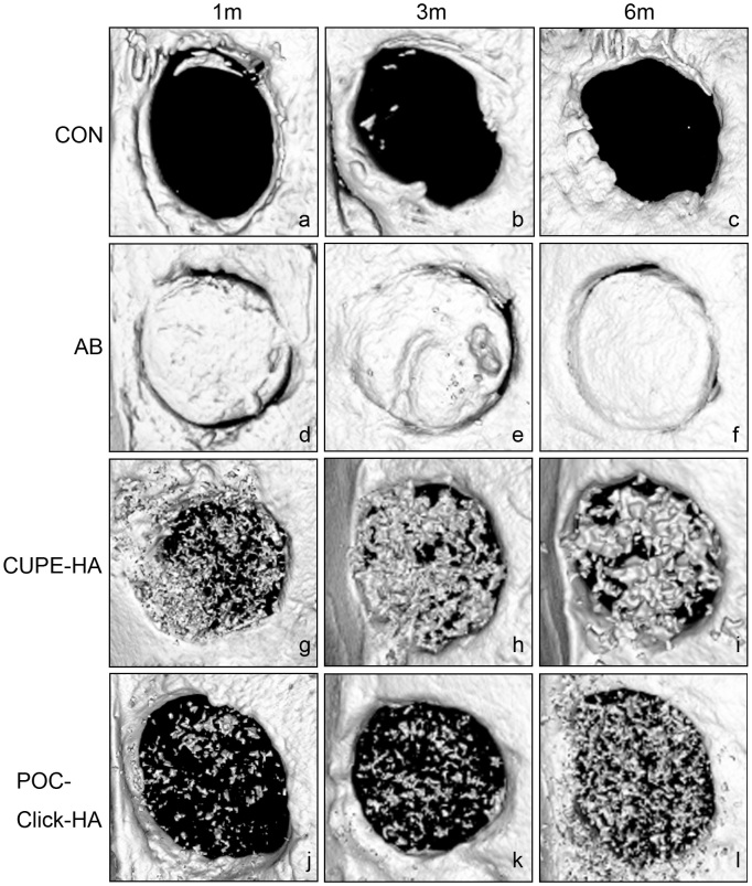
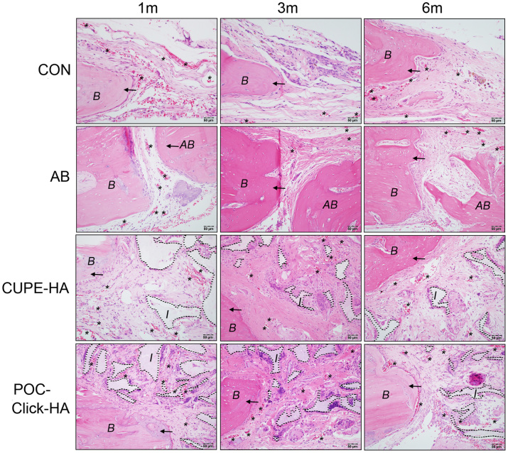
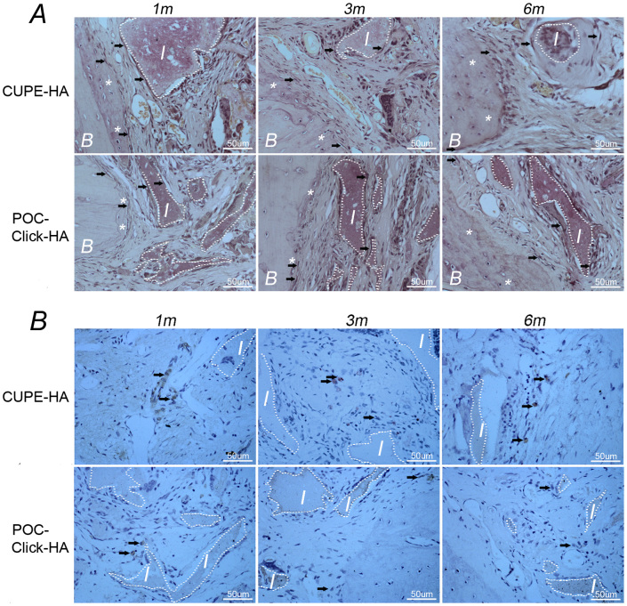
Citric acid-based polymer/hydroxyapatite composites (CABP-HAs) are a novel class of biomimetic composites that have recently attracted significant attention in tissue engineering. The objective of this study was to compare the efficacy of using two different CABP-HAs, poly (1,8-octanediol citrate)-click-HA (POC-Click-HA) and crosslinked urethane-doped polyester-HA (CUPE-HA) as an alternative to autologous tissue grafts in the repair of skeletal defects. CABP-HA disc-shaped scaffolds (65 wt.-% HA with 70% porosity) were used as bare implants without the addition of growth factors or cells to renovate 4 mm diameter rat calvarial defects (n = 72, n = 18 per group). Defects were either left empty (negative control group), or treated with CUPE-HA scaffolds, POC-Click-HA scaffolds, or autologous bone grafts (AB group). Radiological and histological data showed a significant enhancement of osteogenesis in defects treated with CUPE-HA scaffolds when compared to POC-Click-HA scaffolds. Both, POC-Click-HA and CUPE-HA scaffolds, resulted in enhanced bone mineral density, trabecular thickness, and angiogenesis when compared to the control groups at 1, 3, and 6 months post-trauma. These results show the potential of CABP-HA bare implants as biocompatible, osteogenic, and off-shelf-available options in the repair of orthopedic defects.


Figures

Figure 1
Figure 1. Representative image depicting the anatomical region used to define the volume of interest (VOI) used for bone mineral density (BMD) and trabecular thickness (Tb.Th) measurements in axial view.
A) A hollow VOI-1 (red circular area) was defined as the space between outer circle (4 mm in diameter; red arrow) and inner circle (2.8 mm in diameter; blue arrow) with a depth of 300 μm to measure mineralized tissue at the periphery of the implant, and B) a solid cylindrical VOI-2 (red circular area) was defined as the entire defect area (4 mm in diameter; red arrow) with a depth of 300 μm.
Figure 2
Figure 2
Representative scanning electron microscope (SEM) images of bare crosslinked urethane-doped polyester – hydroxyapatite (CUPE-HA) scaffolds (A) and (B) poly (octanediol citrate) – click – hydroxyapatite (POC-Click-HA) scaffolds (60 wt.-HA and 70% porosity); and C) CUPE explants and D) POC-click-HA explants 6 months after implantation in a 4 mm rat calvarial defect (magnification 50×) (I: implant; B: bone).
Figure 3
Figure 3
Microcomputer tomography (micro CT) reconstructed 3D images of 4 mm rat calvarial defects treated with a–c) negative control (untreated defects) (CON), d–f) autologous bone (AB), g–i) crosslinked urethane-doped polyester-hydroxyapatite scaffolds (CUPE-HA), and j–l) poly (octanediol citrate)-click-hydroxyapatite scaffolds (POC-Click-HA) 1, 3, and 6 months post-implantation.
Figure 4
Figure 4. Morphometric analysis of A and C) bone mineral density (BMD) and B and D) trabecular thickness (Tb.Th) in 4 mm rat calvarial defects after 1, 3, and 6 months of implantation Data are presented as mean ± SD (n = 3) (*p<0.05, **p<0.01, ***p<0.001).
CON: negative control group; AB: autologous bone group; CUPE-HA: crosslinked urethane-doped polyester-hydroxyapatite composite scaffold treated group; POC-Click-HA: poly (octanediol citrate)-click-hydroxyapatite composite scaffold treated group.
Figure 5
Figure 5. Microscopic images of hematoxylin and eosin (H&E) stained tissue sections from 4 mm rat calvarial defects after 1, 3, and 6 months of implantation.
New bone (arrows) and blood vessel formation (asterisks) were presented in the crosslinked urethane-doped polyester-hydroxyapatite composite scaffold (CUPE-HA) and poly (octanediol citrate)-click-hydroxyapatite composite scaffold (POC-Click-HA) treated animals after 3 and 6 months (magnification 200×). CON: control group; AB: autologous bone group (autogenous bone implant); 1 m, 3 m, and 6 m: 1, 3, and 6 months post-surgery, respectively; I: implant (black dotted lines); B: bone; Arrow: new bone; Asterisks: blood vessel.
Figure 6
Figure 6
A) Alkaline phosphatase (ALP) and B) osteocalcin (OCN) specific stained tissue sections from 4 mm rat calvarial defects treated with crosslinked urethane-doped polyester-hydroxyapatite composite scaffolds (CUPE-HA) and poly (octanediol citrate)-click-hydroxyapatite composite scaffolds (POC-Click-HA) 1, 3, and 6 months after surgery. Magnification: 400×; Arrow: ALP-positive (dark brown) or OCN-positive (brown) osteoblasts; I: implant (white dotted lines); B: bone.
Figure 7
Figure 7. Vascular endothelial growth factor (VEGF) immunohistochemical stained tissue sections from 4 mm rat calvarial defects treated with crosslinked urethane-doped polyester-hydroxyapatite composite scaffolds (CUPE-HA) and poly (octanediol citrate)-click-hydroxyapatite composite scaffolds (POC-Click-HA) 1, 3, and 6 months of implantation.
Both citric acid-based-hydroxyapatite scaffold treated groups (CUPE-HA and POC-Click-HA) displayed larger VEGF-positive areas (brown) in defect sites compared to empty defects (CON) and autologous implant (AB) groups in the same time period (magnification 200×). CON: control group; AB: autogenous bone group; 1 m, 3 m, and 6 m: 1, 3, and 6 months post-surgery, respectively; I: implant I: implant (black dotted lines); B: bone.
Figure 8
Figure 8. Number of blood vessels presented in critical sized cranial defects treated with crosslinked urethane-doped polyester-hydroxyapatite composite scaffolds (CUPE-HA), poly (octanediol citrate)-click-hydroxyapatite composite scaffolds (POC-Click-HA), autologous bone (AB), or left empty as a control (CON) 1, 3, and 6 months post-surgery.
Data are presented as mean ± SD (n = 5). (*p< 0.05). CON: negative control group; AB: autogenous bone group.

References

    1. Giannoudis P. V., Dinopoulos H. & Tsiridis E. Bone substitutes: an update. Injury 36 Suppl 3S20–27 (2005). - PubMed
    1. Watson J. T. Overview of biologics. J. Orthop. Trauma. 19, S14–16 (2005). - PubMed
    1. Friedlander A. H. The physiology, medical management and oral implications of menopause. J. Am. Dent. Assoc. 133, 73–81 (2002). - PubMed
    1. Bauer T. W. & Muschler G. F. Bone graft materials. An overview of the basic science. Clin. Orthop. Relat. R. 371, 10–27 (2000). - PubMed
    1. Acarturk T. O. & Hollinger J. O. Commercially available demineralized bone matrix compositions to regenerate calvarial critical-sized bone defects. Plast. Reconstr. Srug. 118, 862–873 (2006). - PubMed

Publication types

MeSH terms