. 2014 Nov 5;9(11):e109080.
doi: 10.1371/journal.pone.0109080. eCollection 2014.

Lymphocyte Activation Gene 3 (LAG-3) modulates the ability of CD4 T-cells to be suppressed in vivo

Affiliations

Lymphocyte Activation Gene 3 (LAG-3) modulates the ability of CD4 T-cells to be suppressed in vivo

Nicholas M Durham et al. PLoS One. .

Abstract

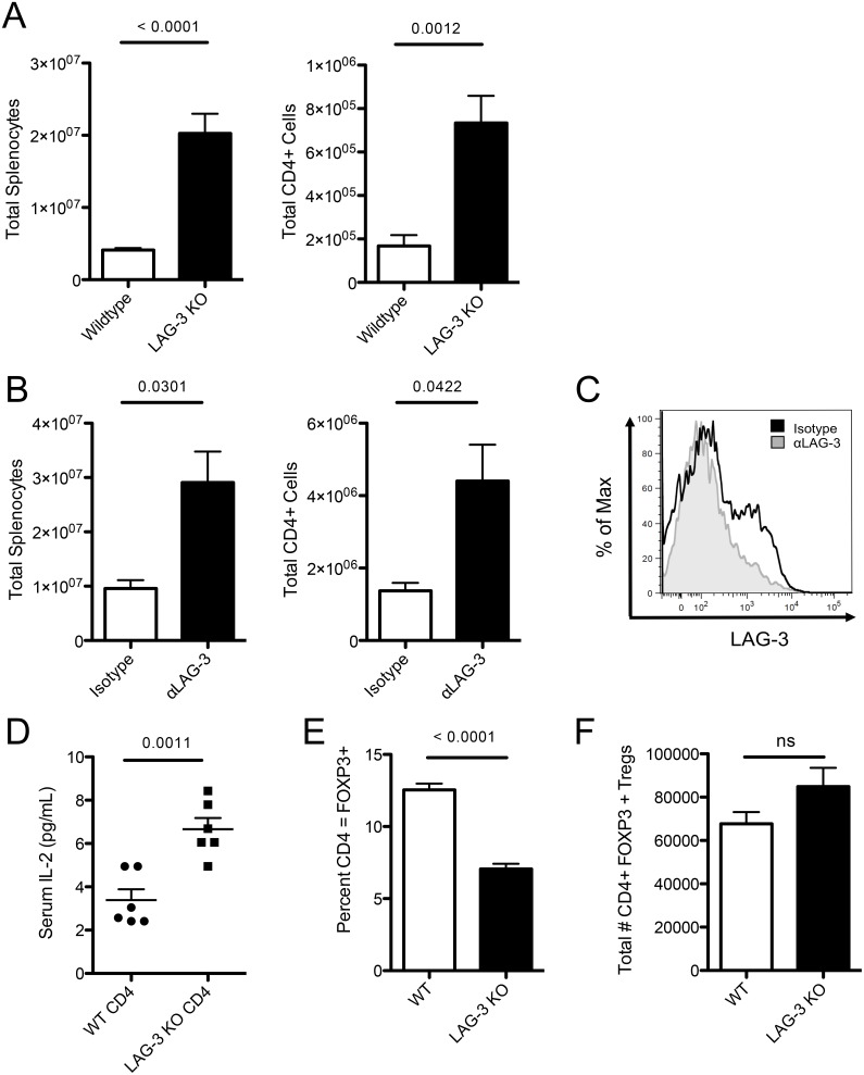
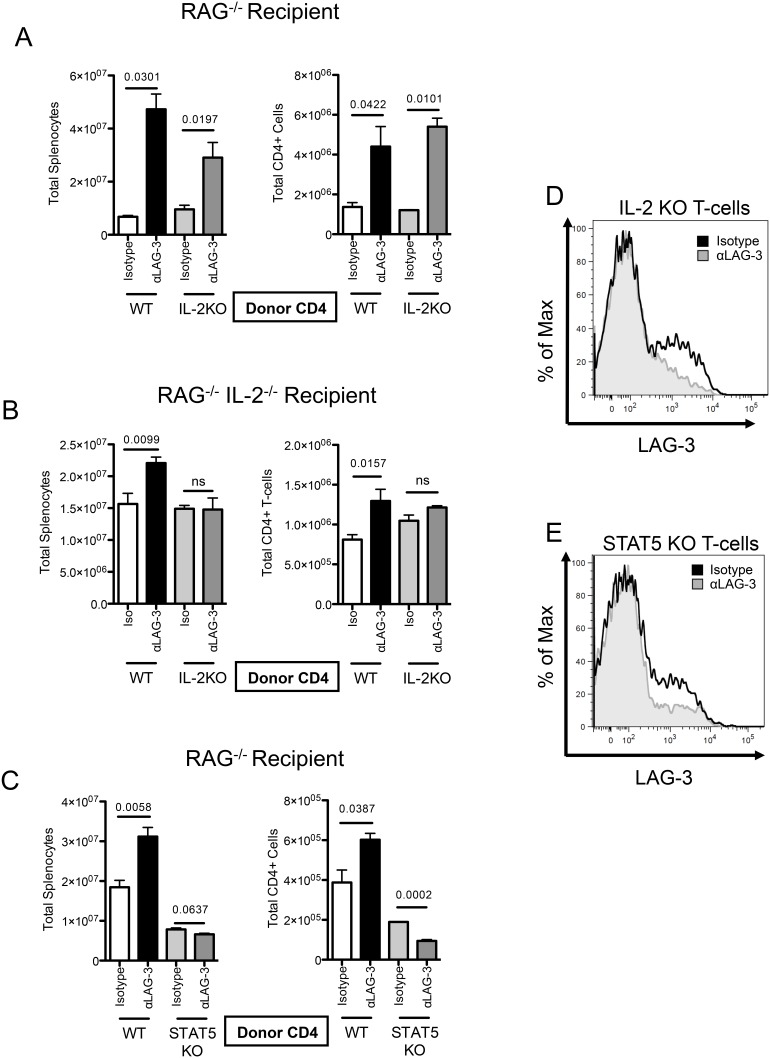
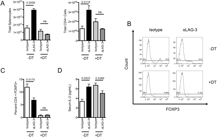
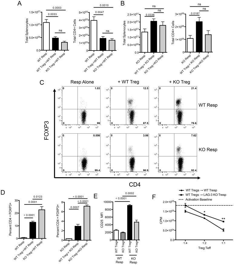
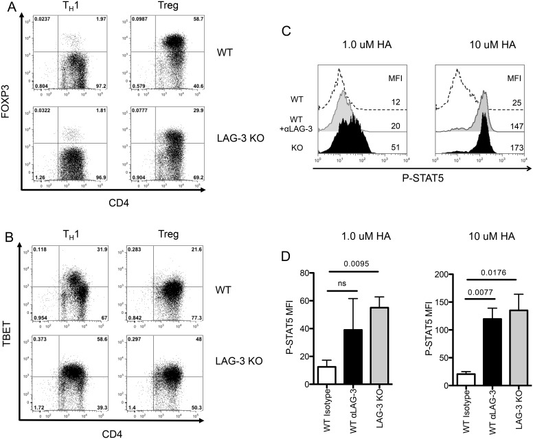
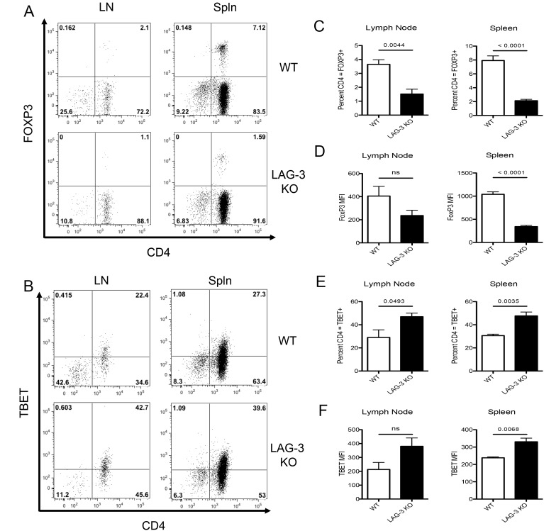
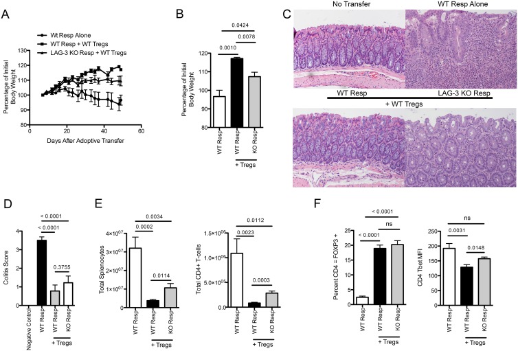
Lymphocyte Activation Gene - 3 (LAG-3) is an immune checkpoint molecule that regulates both T-cell activation and homeostasis. However, the molecular mechanisms underlying LAG-3's function are generally unknown. Using a model in which LAG-3 blockade or absence reliably augmented homeostatic proliferation in vivo, we found that IL-2 and STAT5 are critical for LAG-3 function. Similarly, LAG-3 blockade was ineffective in the absence of regulatory T-cells (Treg), suggesting an important role for LAG-3 in either the responsiveness of conventional T-cells (Tconv) to regulation, or a relative defect in the ability of LAG-3 KO regulatory T-cells (Treg) to suppress the proliferation of Tconv. In this model, LAG-3 KO Treg suppressed proliferation in a manner fairly similar to wild-type (WT) Treg, but LAG-3 KO Tconv were relatively resistant to suppression. Further studies also identified a role for LAG-3 in the induction/expansion of Treg. Finally, we found that LAG-3 blockade (or knockout) led to a relative skewing of naïve CD4 T-cells toward a TH1 phenotype both in vitro and in in vivo. Together, these data suggest that LAG-3 expression on Tconv cells makes them more susceptible to Treg based suppression, and also regulates the development of a TH1 T-cell response.


Conflict of interest statement

Competing Interests: CGD has served as a paid consultant for Bristol Myers Squibb, Compugen and Roche/Genentech. CGD is a co-inventor on a patent entitled: “Manipulation of Regulatory T Cells Using LAG-3 Antibodies, Genes, Agonists and Antagonists”, filing case number: C04255. This patent is currently un-licensed. This does not alter the authors’ adherence to PLOS ONE policies on sharing data and materials.

Figures

Figure 1
Figure 1. LAG-3 blockade augments homeostatic proliferation in vivo.
A) 1E6 WT or LAG-3 KO CD4+ T-cells were adoptively transferred into RAG KO mice and harvested on day 10. Splenocytes were then counted and analyzed. B) 1E6 WT CD4+ T-cells were transferred into RAG KO mice. Isotype control antibody or LAG-3 blocking antibody given every 2 days. Splenocytes were then counted and analyzed. C) LAG-3 antibody staining of LAG-3 in vivo. D) Serum IL-2 from RAG KO mice with 1E6 WT or LAG-3 KO CD4+ T-cells on Day 7. E) Percentage of CD4+ T-cells that express FOXP3. F) Total number of adoptively transferred CD4+ T-cells expressing FoxP3. Data shown are representative of at least two independent experiments with n = 3–6 mice per group.
Figure 2
Figure 2. IL-2 is required for LAG-3 blockade to augment homeostatic proliferation in vivo.
A) 1E6 WT or IL-2 KO CD4 T-cells transferred into RAG KO mice. Isotype control antibody or LAG-3 blocking antibody given every 2 days. Splenocytes counted and analyzed. B) 1E6 WT or IL-2 KO CD4+ T-cells were transferred into RAG KO/IL-2 KO mice. C) 1E6 WT or STAT5 KO CD4+ T-cells transferred into RAG KO mice. Isotype control antibody or LAG-3 blocking antibody was given every 2 days. Splenocytes were then counted and analyzed. D) LAG-3 antibody staining of LAG-3 on IL-2 KO cells in vivo. E) LAG-3 antibody staining of LAG-3 on STAT5 KO cells in vivo. Data shown are representative of at least two independent experiments with n = 3 mice per group.
Figure 3
Figure 3. FOXP3 Treg are required for LAG-3 Blockade to augment homeostatic proliferation in vivo.
A) 1E6 FOXP3 DTR CD4+ T-cells transferred into RAG KO mice. Isotype control antibody or LAG-3 blocking antibody was given every 2 days with either PBS or Diphtheria Toxin. B) FOXP3 expression by flow cytometry ICS. C) Percent of CD4+ T-cells expressing FOXP3 D) Serum IL-2 levels by ELISA. Data shown are representative of at least two independent experiments with n = 4 mice per group.
Figure 4
Figure 4. LAG-3 KO Treg suppress homeostatic proliferation, but LAG-3 KO responders are resistant to suppression.
A) WT or LAG-3 KO Treg were transferred into RAG KO mice at a ratio of 1∶4 with WT responders. Responders alone received 4E6 WT Tresp with no Treg. B) WT or LAG-3 KO Treg were transferred into RAG KO mice at a ratio of 1∶4 with KO responders. Responders alone received 4E6 LAG3 KO Tresp with no Treg. C) Representative plots of FOXP3 expression in adoptively transferred cells. D) Summary of FOXP3 expression in adoptively transferred cells (n = 4). E) CD25 MFI expression on Treg with either WT or LAG-3 KO Responders. F) WT or LAG-3 KO CD4 Tresp were mixed at a 4∶1 ratio with FOXP3 GFP Treg, stimulated with CD3/CD28, and pulsed with H3-Thymadine after 72 hours. Total CPM counts are shown. Baseline activation of WT or LAG-3 KO Tresp without Treg was not different and is reported by a dashed line. Data shown are representative of at least two independent experiments with n = 4–5 mice per group.
Figure 5
Figure 5. Decreased FOXP3 Treg induction in LAG-3 KO cells in vitro.
WT or LAG-3 KO 6.5 TCR Transgenic CD4+ T-cells skewed in Th1 or Treg Conditions and analyzed for A) FOXP3 or B) TBET Expression. C) 6.5 TCR transgenic CD4+ T-cells were isolated and mixed with matched splenocytes 1∶3 and stimulated with 1 or 10 µM HA peptide for 3 days. WT or LAG-3 KO 6.5 CD4+ T-cells were treated with αLAG-3 or isotype control antibody at 50 µg/mL and cells were stained for P-STAT5. D) Summary graph of two experiments. Data shown are representative of at least two independent experiments.
Figure 6
Figure 6. Decreased FOXP3 Treg induction in LAG-3 KO T-cells in an in vivo self-tolerance.
A) WT or LAG-3 KO 6.5 TCR Transgenic CD4+ T-cells adoptively transferred into C3-HA expressing mice. B) FOXP3 and C) Tbet expression was analyzed and representative graphs shown. C–D) Summary of FOXP3 expression in adoptively transferred cells. E–F) Summary of TBET expression in adoptively transferred cells. Data shown are representative of at least two independent experiments where n = 4 mice per group.
Figure 7
Figure 7. WT Treg Cannot Completely Protect against LAG-3 KO Tresp in a Colitis Model.
A) WT or LAG-3 KO Tresp were transferred into RAG KO mice at a ratio of 4∶1 with WT Treg. Mice were weighed 3 times weekly for 50 days. Percentage of initial body weight is reported. B) Percentage of initial body weight at Day 49. C) H & E staining of histological sections of colons from the 4 groups of mice. D) Blinded histological score of colitis in mouse groups. E) Total splenocytes as well as total CD4+ T-cells were counted and analyzed. F) Percentage of CD4+ T-cells that were FOXP3 or TBET positive. Data shown are representative of at least two independent experiments with n = 8–10 mice per group.

References

    1. Sakaguchi S, Toda M, Asano M, Itoh M, Morse SS, et al. (1996) T cell-mediated maintenance of natural self-tolerance: its breakdown as a possible cause of various autoimmune diseases. J Autoimmun 9: 211–220 10.1006/jaut.1996.0026 - DOI - PubMed
    1. Sprent J, Cho J-H, Boyman O, Surh CD (2008) T cell homeostasis. Immunol Cell Biol 86: 312–319 10.1038/icb.2008.12 - DOI - PubMed
    1. Boyman O, Sprent J (2012) The role of interleukin-2 during homeostasis and activation of the immune system. Nat Rev Immunol 12: 180–190 10.1038/nri3156 - DOI - PubMed
    1. Sprent J, Surh CD (2011) Normal T cell homeostasis: the conversion of naive cells into memory-phenotype cells. Nat Immunol 131: 478–484 10.1038/ni.2018 - DOI - PMC - PubMed
    1. Surh CD (2000) Homeostatic T Cell Proliferation: How Far Can T Cells Be Activated to Self-Ligands? J Exp Med 192: 9F–14 10.1084/jem.192.4.F9 - DOI - PMC - PubMed

Publication types