. 2015 Feb;23(2):330-8.
doi: 10.1038/mt.2014.219. Epub 2014 Nov 6.

Selective inhibition of tumor growth by clonal NK cells expressing an ErbB2/HER2-specific chimeric antigen receptor

Affiliations

Selective inhibition of tumor growth by clonal NK cells expressing an ErbB2/HER2-specific chimeric antigen receptor

Kurt Schönfeld et al. Mol Ther. 2015 Feb.

Abstract

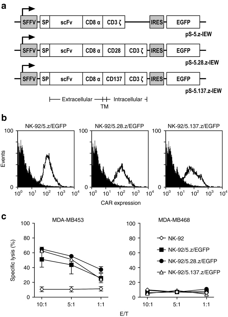
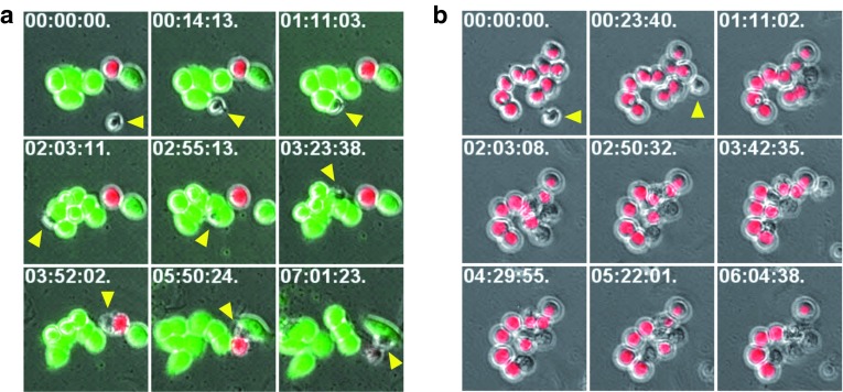
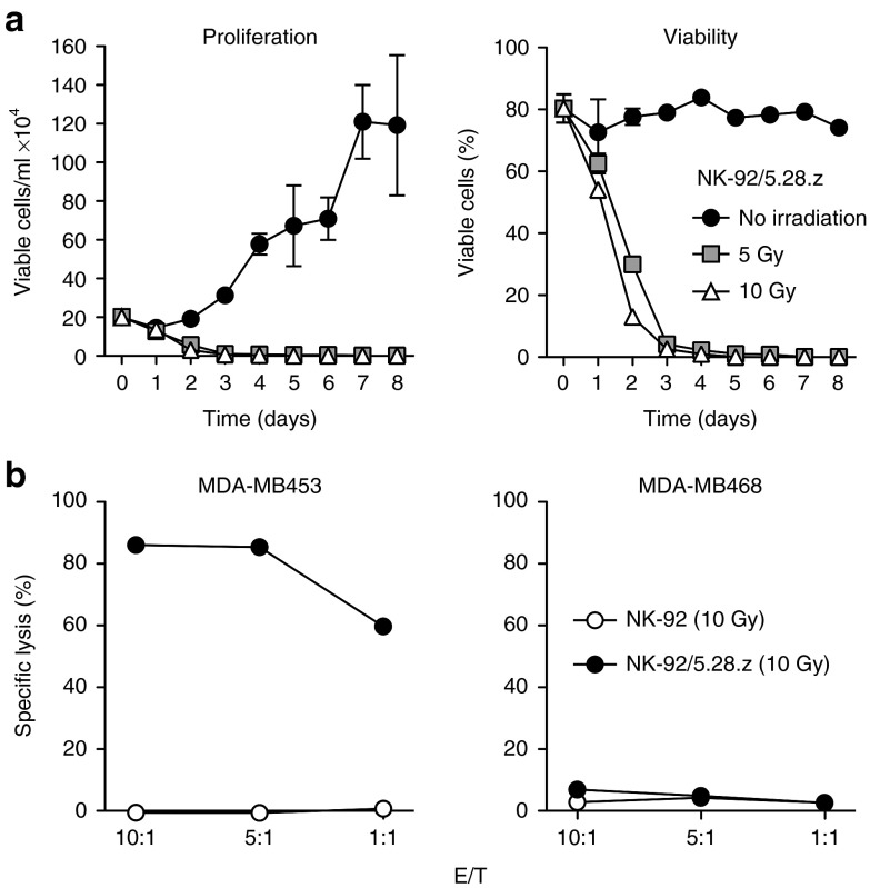
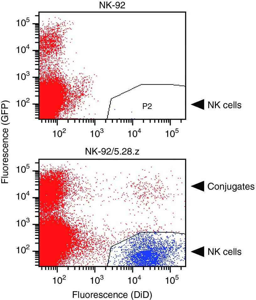
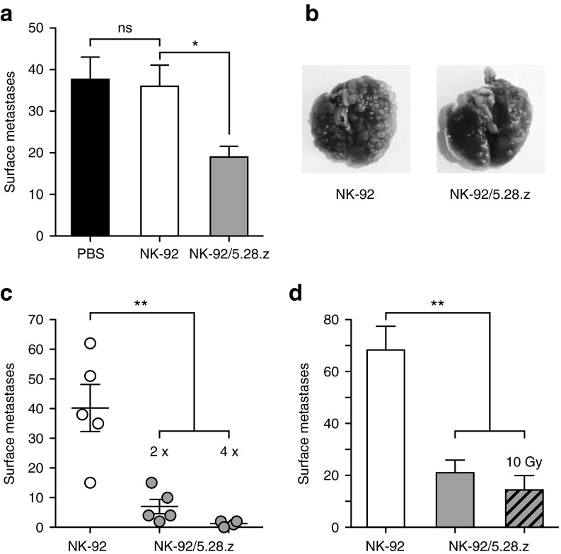
Natural killer (NK) cells are an important effector cell type for adoptive cancer immunotherapy. Similar to T cells, NK cells can be modified to express chimeric antigen receptors (CARs) to enhance antitumor activity, but experience with CAR-engineered NK cells and their clinical development is still limited. Here, we redirected continuously expanding and clinically usable established human NK-92 cells to the tumor-associated ErbB2 (HER2) antigen. Following GMP-compliant procedures, we generated a stable clonal cell line expressing a humanized CAR based on ErbB2-specific antibody FRP5 harboring CD28 and CD3ζ signaling domains (CAR 5.28.z). These NK-92/5.28.z cells efficiently lysed ErbB2-expressing tumor cells in vitro and exhibited serial target cell killing. Specific recognition of tumor cells and antitumor activity were retained in vivo, resulting in selective enrichment of NK-92/5.28.z cells in orthotopic breast carcinoma xenografts, and reduction of pulmonary metastasis in a renal cell carcinoma model, respectively. γ-irradiation as a potential safety measure for clinical application prevented NK cell replication, while antitumor activity was preserved. Our data demonstrate that it is feasible to engineer CAR-expressing NK cells as a clonal, molecularly and functionally well-defined and continuously expandable cell therapeutic agent, and suggest NK-92/5.28.z cells as a promising candidate for use in adoptive cancer immunotherapy.


Figures

Figure 1
Figure 1
Specific cytotoxicity of CAR-engineered NK-92 cells against ErbB2-expressing cancer cells. (a) Schematic representation of lentiviral transfer plasmids encoding under the transcriptional control of the spleen focus forming virus promoter (SFFV) different chimeric antigen receptors, followed by an internal ribosome entry site (IRES) and enhanced green fluorescent protein (EGFP) cDNA as a marker. In pS-5.z-IEW, the CAR consists of an immunoglobulin heavy chain signal peptide (SP), the ErbB2-specific scFv(FRP5) antibody fragment (scFv), a modified CD8α hinge region (CD8α), followed by transmembrane and intracellular domains of CD3ζ (CAR 5.z). In pS-5.28.z-IEW, the hinge region is followed by transmembrane and intracellular domains of CD28 and the intracellular domain of CD3ζ (CAR 5.28.z), while in pS-5.137.z-IEW, the hinge region is followed by transmembrane and intracellular domains of CD137 (4-1BB) and the intracellular domain of CD3ζ (CAR 5.137.z). (b) CAR-expression by NK-92/5.z/EGFP, NK-92/5.28.z/EGFP and NK-92/5.137.z/EGFP cells generated by transduction of NK-92 cells with the lentiviral vectors shown in a was determined by flow cytometry with ErbB2-Fc fusion protein (open area). Parental NK-92 cells served as control (filled area). (c) Cell killing by NK-92/5.z/EGFP (filled squares), NK-92/5.28.z/EGFP (filled circles), and NK-92/5.137.z/EGFP cells (open triangles) was investigated in FACS-based cytotoxicity assays at different effector to target ratios (E/T) using human ErbB2-positive MDA-MB453 (left panel) and ErbB2-negative MDA-MB468 breast carcinoma cells (right panel) as targets. Parental NK-92 cells were included for comparison (open diamonds). Mean values ± SEM are shown; n = 3.
Figure 2
Figure 2
Molecular and functional characterization of clonal NK-92/5.28.z cells. (a) CAR-expression by the clonal NK-92/5.28.z cell line generated under GMP conditions by transduction with lentiviral vector S-5.28.z-W was determined by flow cytometry with ErbB2-Fc fusion protein (open area). Parental NK-92 cells served as control (gray area). (b) Three-color fluorescence in situ hybridization (FISH) was performed on metaphase spreads of the NK-92/5.28.z cell clone using the CAR-encoding fragment of pS-5.28.z-W as probe together with whole painting probes to specifically stain chromosomes 2 (wcp2) and 9 (wcp9). Probes were labeled using biotin-, digoxigenin-, and FITC-conjugated nucleotides, respectively. For biotin- and digoxigenin-labeled probes, immunological detection was performed using AMCA (blue) and Cy3 (red) fluorescent dyes. Integrated copies of CAR-encoding lentiviral vector S-5.28.z-W (red signals; indicated by white arrowheads) were found at the terminal regions of the long arms of one copy of chromosomes 2 (blue) and 9 (green), respectively. (c) Cell killing by NK-92/5.28.z cells (filled circles) was investigated in FACS-based cytotoxicity assays at different effector to target ratios (E/T) using human MDA-MB453 (ErbB2-positive) and MDA-MB468 breast carcinoma cells (ErbB2-negative) as targets. Parental NK-92 cells were included as a control (open circles). For comparison, MDA-MB468 cells which are EpCAM-positive were also treated with NK-92/31.28.z cells that express an EpCAM-specific CAR. Mean values ± SEM are shown; n = 3. (d) To confirm specificity of cell killing, similar experiments were performed with murine renal cell carcinoma cells as targets that stably express human ErbB2 (Renca-lacZ/ErbB2) or human EGFR (Renca-lacZ/EGFR). Mean values ± SEM are shown; n = 3. (e) Reactivity with normal tissues was investigated using primary human cardiomyocytes (CM), lung epithelial cells (LEC), lung fibroblasts (LF), and peripheral blood mononuclear cells (PBMC) as targets. Mean values ± SEM are shown; n ≥ 3.
Figure 3
Figure 3
Kinetics of target cell killing by NK-92/5.28.z cells. (a) To investigate selectivity and kinetics of target cell killing, live cell imaging experiments were performed with cocultures of clonal NK-92/5.28.z cells and mixtures of tdTOMATO-expressing MDA-MB453 and EGFP-expressing MDA-MB468 breast carcinoma cells. Serial images of a microscopic field with a single NK-92/5.28.z cell (yellow arrowhead), a single MDA-MB453 cell (red fluorescence), and initially five MDA-MB468 cells (green fluorescence) are shown. The first contact between the NK cell and tumor cells occurred 14 minutes after beginning the observation (second image of the series). MDA-MB468 cells were not affected in their growth despite multiple contacts with the NK cell and continued to divide during the observation time. Merged phase-contrast and fluorescence microscopy images are shown. The time stamp indicates hours:minutes:seconds from the beginning of observation. (b) Serial images of a microscopic field with a single NK-92/5.28.z cell (yellow arrowhead) and 10 MDA-MB453 cells (red fluorescence). The first contact between the NK cell and tumor cells occurred 23 minutes after beginning the observation (second image of the series). Serial killing of five MDA-MB453 target cells by the single NK-92/5.28.z cell was completed ~5 hours and 40 minutes after initial contact (last image of the series).
Figure 4
Figure 4
Growth and cytotoxic activity of NK-92/5.28.z cells upon γ-irradiation. (a) To investigate the effect on viability, NK-92/5.28.z cells were irradiated with 5 or 10 Gy and cultured for up to 8 days. Proliferation (left panel) and percentage of viable cells (right panel) were analyzed by counting viable cells at the indicated time points using trypan blue exclusion. Mean values ± SEM are shown; n = 3. (b) Cytotoxic activity of NK-92/5.28.z cells 24 hours after irradiation with 10 Gy against ErbB2-positive MDA-MB453 and ErbB2-negative MDA-MB468 breast carcinoma cells was determined in FACS-based cytotoxicity assays at different effector to target ratios (E/T) as indicated (filled circles). Parental NK-92 cells 24 hours after irradiation were included for comparison (open circles). Mean values ± SEM are shown; n = 3.
Figure 5
Figure 5
Accumulation of NK-92/5.28.z cells in ErbB2-positive breast carcinomas in vivo. Parental NK-92 (upper panel) or ErbB2-specific NK-92/5.28.z cells (lower panel) were labeled with fluorescent DiD labeling reagent and intravenously injected into NSG mice carrying established orthotopic MDA-MB453/EGFP breast carcinoma xenografts. Twenty-four hours after injection, tumors were excised, single cell suspensions were prepared, and analyzed for the presence of EGFP-expressing and DiD-labeled cells. DiD-positive NK cells are indicated in blue (lower right quadrants). EGFP-positive breast carcinoma cells (upper left quadrants) and double-negative murine stromal cells (lower left quadrants) are indicated in red. Double-positive events (upper right quadrant) represent conjugates of CAR-expressing NK-92/5.28.z and MDA-MB453/EGFP target cells. Representative flow cytometric data from one animal of each group are shown (n = 3).
Figure 6
Figure 6
In vivo antitumor activity of NK-92/5.28.z cells. (a) To investigate antitumor activity, NSG mice were intravenously injected with Renca-lacZ/ErbB2 renal cell carcinoma cells. Then, animals were treated twice by i.v. injection of parental NK-92 or clonal NK-92/5.28.z cells at days 1 and 3 after tumor cell injection. Control mice received PBS. Four weeks after tumor challenge, lungs were excised and tumor nodules on the lung surface were counted. Mean values ± SEM are shown; n = 5. ns, P > 0.05; *P < 0.05. (b) Representative images of lungs at day 28 from animals treated with NK-92 or NK-92/5.28.z cells. (c) To evaluate the effect of multiple treatments, NSG mice injected with Renca-lacZ/ErbB2 cells were treated with NK-92 or NK-92/5.28.z cells twice as described in panel a (n = 5), or four times at days 1, 3, 6, and 8 (n = 4). Data points for each animal and mean values ± SEM are shown. **P < 0.01. (d) In a separate experiment, NSG mice injected with Renca-lacZ/ErbB2 cells were treated twice as described in panel a with nonirradiated NK-92 or NK-92/5.28.z cells, or NK-92/5.28.z cells irradiated with 10 Gy as indicated. Mean values ± SEM are shown; n = 5. **P < 0.01.

References

    1. Kalos M, Levine BL, Porter DL, Katz S, Grupp SA, Bagg A.et al. (2011T cells with chimeric antigen receptors have potent antitumor effects and can establish memory in patients with advanced leukemia Sci Transl Med 395ra73 - PMC - PubMed
    1. Grupp SA, Kalos M, Barrett D, Aplenc R, Porter DL, Rheingold SR.et al. (2013Chimeric antigen receptor-modified T cells for acute lymphoid leukemia N Engl J Med 3681509–1518. - PMC - PubMed
    1. Brentjens RJ, Davila ML, Riviere I, Park J, Wang X, Cowell LG.et al. (2013CD19-targeted T cells rapidly induce molecular remissions in adults with chemotherapy-refractory acute lymphoblastic leukemia Sci Transl Med 5177ra38 - PMC - PubMed
    1. Kochenderfer JN, Dudley ME, Carpenter RO, Kassim SH, Rose JJ, Telford WG.et al. (2013Donor-derived CD19-targeted T cells cause regression of malignancy persisting after allogeneic hematopoietic stem cell transplantation Blood 1224129–4139. - PMC - PubMed
    1. Kalos M, June CH. Adoptive T cell transfer for cancer immunotherapy in the era of synthetic biology. Immunity. 2013;39:49–60. - PMC - PubMed

Publication types

MeSH terms