. 2014 Oct 23;9(2):674-87.
doi: 10.1016/j.celrep.2014.09.019. Epub 2014 Oct 16.

ESCRT-II/Vps25 constrains digit number by endosome-mediated selective modulation of FGF-SHH signaling

Affiliations

ESCRT-II/Vps25 constrains digit number by endosome-mediated selective modulation of FGF-SHH signaling

Karen Handschuh et al. Cell Rep. .

Abstract

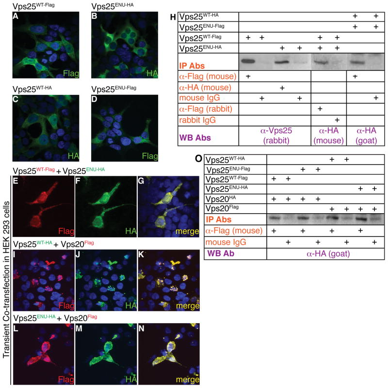
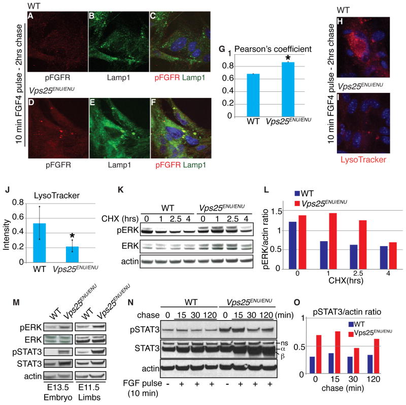
Sorting and degradation of receptors and associated signaling molecules maintain homeostasis of conserved signaling pathways during cell specification and tissue development. Yet, whether machineries that sort signaling proteins act preferentially on different receptors and ligands in different contexts remains mysterious. Here, we show that Vacuolar protein sorting 25, Vps25, a component of ESCRT-II (Endosomal Sorting Complex Required for Transport II), directs preferential endosome-mediated modulation of FGF signaling in limbs. By ENU-induced mutagenesis, we isolated a polydactylous mouse line carrying a hypomorphic mutation of Vps25 (Vps25(ENU)). Unlike Vps25-null embryos we generated, Vps25(ENU/ENU) mutants survive until late gestation. Their limbs display FGF signaling enhancement and consequent hyperactivation of the FGF-SHH feedback loop causing polydactyly, whereas WNT and BMP signaling remain unperturbed. Notably, Vps25(ENU/ENU) Mouse Embryonic Fibroblasts exhibit aberrant FGFR trafficking and degradation; however, SHH signaling is unperturbed. These studies establish that the ESCRT-II machinery selectively limits FGF signaling in vertebrate skeletal patterning.


Figures

Figure 1
Figure 1. Identification of polydactylous mouse line 04-014 by ENU mutagenesis and cloning of the ESCRT-II/Vps25 hypomorphic mutation
(A–D) Gross morphology shows different expressivity of the polydactyly in E14.5 Vps25ENU/ENU hindlimbs, including a widened autopod with six (B) or seven (C) digits, or synpolydactyly (D) (domain of additional digits; white asterisk). (E,F) Sequencing of Vps25 genomic (g) and complementary (c) DNA detects a mutation (asterisk) within intron 3, five nucleotides downstream to the exon-intron border, resulting in a 27 nucleotide insertion in the mutant Vps25 mRNA, visualized by PCR (F). (G) Alignment of Vps25WT (top) (Protein UniProt/NCBI number: Q9CQ80) and Vps25ENU mutant (bottom) proteins shows a 9 amino acid insertion starting at amino acid 86. Vps25WT protein contains two winged helix domains WH1 (amino acids 1-84) and WH2 (amino acids 84-176) (Langelier et al., 2006). The additional 9 amino acids, indicated by roman numbers, reside within the Vps25 WH2 domain. (H,I) qRT-PCR and Western blot (WB) from E10.5 whole embryos demonstrate presence of WT Vps25 mRNA and protein, respectively, in Vps25ENU/ENU mutants. qRT-PCR, mean of 2 samples run in triplicate ± SEM; Tbp used for normalization. (J–M) X-gal staining reveals ubiquitous Vps25 expression, including forelimb (FL) and hindlimb (HL) (black arrowheads), in E9.5 Vps25LacZ/+ embryos, versus lack of staining (white arrowheads) in WT. s, somites; nt, neural tube. (N–Q) Delayed and abnormal development of Vps25LacZ/ENU double heterozygote (arrowheads indicate hemorrhaging) as early as E9.5 versus WT morphology of Vps25LacZ/+ and Vps25ENU/+ single heterozygotes proves that the Vps25ENU mutation is not complemented by the Vps25 LOF allele. As predicted for a hypomorphic allele, Vps25ENU/ENU embryos survive until later in gestation and appear normal at E10.5 (Q). See also Figure S1, Tables S1,S2,S3.
Figure 2
Figure 2. Pre-axial polydactyly, proliferation and apoptosis defects in Vps25ENU/ENU mutant hindlimb
(A,B) Shorter and thicker hindlimb (HL) zeugopod (Z) and pre-axial polydactyly (asterisk) in E14.5 Vps25ENU/ENU autopod (A) by alcian blue staining. (C,D) OPT shows polydactyly (asterisk) in E14.5 Vps25ENU/ENU HL. (E–L) BrdU and cleaved Caspase- 3 IF reveals increased proliferation (white arrowhead) and decreased apoptosis (empty arrowhead) in E12.5 Vps25ENU/ENU anterior HL. (M–P) In situ hybridization shows loss of Msx2 (empty arrowhead) and increased Sox9 (black arrowhead) mRNA in E13.5 Vps25ENU/ENU anterior HL. See also Figure S2, Movies S1,S2.
Figure 3
Figure 3. Selective enhancement of the FGF-SHH cross-regulatory loop in Vps25ENU/ENU early HL buds
(A–F) Expression of Fgf4, Shh, Fgf8, and Patched1 by in situ hybridization of E10.5-E11.5 WT and Vps25ENU/ENU HL buds. (G,H) qRT-PCR of Shh and Patched1 mRNAs from E11.5 WT and Vps25ENU/ENU HL buds; Rpl19 as internal control. Bars; mean of 3 HL bud pairs run in triplicate ± SD (*, p<0.05). (I) WB demonstrates altered Gli3 processing in E10.75 Vps25ENU/ENU HL, wherein levels of Gli3R are reduced. FL,full-length; R, repressor. (J–Q) Expression of Hand2, HoxD13, Gli1, Gli3, Shh, Fgf4, Fgf8, and Sprouty2 by in situ hybridization of E11.5-E12.5 WT and Vps25ENU/ENU HL buds. Anterior, top; proximal, left; d, dorsal; v, ventral; a′, anterior view. Black arrowheads; spatially expanded, temporally protracted, and ectopic gene expression in mutant buds. Empty arrowheads; absence of signal. See also Figure S3.
Figure 4
Figure 4. ESCRT-II/Vps25WT and ESCRT-II/Vps25ENU immunoprecipitate with ESCRT-III/Vps20 protein; the WT protein complex is mostly detected within puncta, while the mutant complex is diffuse throughout the cytoplasm
(A–G) Transient transfection of Vps25WT-Flag, Vps25WT-HA, Vps25ENU-HA or Vps25ENU-Flag alone or in combination, in HEK293 cells. IF with Flag or HA antibodies (Ab) reveals that Vps25WT and Vps25ENU mutant proteins co-localize within the cytoplasm, also within puncta. (H) Co-immunoprecipitation (IP) of Vps25 WT and mutant proteins from cells transfected with Vps25WT-Flag and Vps25ENU-HA or with Vps25WT-HA and Vps25ENU-Flag. IP with anti-Flag or anti-HA Ab; WB with anti-Vps25 or an anti-HA Ab. (I–O) Both Vps25WT-HA and Vps25ENU-HA co-localize and co-immunoprecipitate with Vps20Flag in transfected HEK293 cells. However, subcellular localization of the mutant Vps25ENU-Vps20 complex is diffuse throughout the cytoplasm, unlike the WT complex, which localizes to puncta. Abs used for IP and WB labeled orange and purple, respectively. See also Figure S4.
Figure 5
Figure 5. Vps25 ENU-induced mutation yields low-levels of both WT and structurally altered mutant protein and is associated with engorged MVBs in cultured cells and limb AER and mesenchyme in vivo, resulting in perturbed endosomal trafficking
(A) Structural representation of WT and Vps25ENU proteins reveals a conserved Glu88-Arg151 hydrogen bond between WH1 (green) and WH2 (magenta) domains. In the Vps25ENU protein a new strong hydrogen bond (<2Å) Ser(IV)-Glu(105+9) is formed, due to the 9 amino acid insertion (orange). See also Figure S5. (B–D) Transmission Electron Microscopy (TEM) reveals a significant increase in the diameters of multivesicular bodies (MVB) in E13.5 mutant versus WT MEFs treated with HRP. The box-and-whisker plot (D) depicts MVB diameters (in nanometers, nm) on the Y axis and WT and mutant (Vps25ENU/ENU) MVB sample populations on the X axis. Blue boxes represent the middle 50% of the values of the sample range; red lines represent the value of the median sample; legs and bars represent upper and lower limits of the diameter sample. Red cross indicates an outlier. Diameters measured in 16 WT and 12 mutant MVBs (***p<0.0001). (E) 100 MVBs and 100 lysosomes counted in HRP-treated WT and mutant MEFs. In WT MEFs, HRP-positive MVBs (green) and lysosomes (blue) are present in approximately equal numbers, while in Vps25ENU/ENU MEFs HRP-positive lysosomes are negligible. (F) Numbers of total (purple) and HRP-positive (red) lysosomes (100) counted and reported per surface areas (mm2) in both WT and Vps25ENU/ENU MEFs, showing that in mutant MEFs HRP is not escorted to the lysosomes. (G) WB analysis of MEFs treated with a 30 min HRP pulse, and chased for different times (hours, hrs), demonstrates abnormal HRP degradation in mutant MEFs. (H–K) TEM reveals a significant increase in MVB diameters within the limb AER and mesenchyme compartments of E11.5 Vps25ENU/ENU (N=5) versus WT (N=2) embryos. The box-and-whisker plots (J,K), as detailed above (in D), depict MVB diameters (in nanometers, nm) on the Y axis and WT and Vps25ENU/ENU MVB sample populations on the X axis. Diameters measured in 13 WT and 12 mutant MVBs, in limb AER (*p<0.05). In limb mesenchyme diameters measured in 15 WT and 21 mutant MVBs (*p<0.05).
Figure 6
Figure 6. Vps25 ENU-induced mutation results in FGF receptor degradation defects and enhancement of FGF signaling in both cultured cells and limb buds in vivo
(A–G) IF for phospho(p)-FGFR following FGF4 pulse-chase in MEFs shows increased co-localization of pFGFR and Lamp1 in late endosomes (or MVBs) of mutant MEFs versus WT, quantified by Pearson’s coefficient (*p < 0.05), indicating MVB sequestration of pFGFR. (H–J) After a 10 min FGF4 pulse, WT and Vps25ENU/ENU MEFs were chased for 2 hours (hrs) and vesicular pH measured by LysoTracker staining. Quantification of signal intensity reveals significantly lower levels of staining in Vps25ENU/ENU MEFs, indicating lysosomal functionality defects (*p < 0.05). (K,L) Assessment of pERK protein stability in WT and Vps25ENU/ENU MEFs treated with cycloheximide (CHX) for different times, indicated in hours, demonstrates abnormal protein degradation in mutant cells. ERK and actin as controls. Quantification of pERK levels over actin by ImageJ64 in WT (purple) and Vps25ENU/ENU (red) MEFs (L). (M–O) WB of whole embryos (E13.5) and limbs (E11.5), as well as FGF4 pulse-chase experiment in MEFs, show increase of FGF second messengers and decreased pSTAT3 degradation, quantified by ImageJ64, in Vps25ENU/ENU (red) versus WT (purple) MEFs. Actin as control. min, minutes; ns, non-specific band.
Figure 7
Figure 7. Vps25 ENU-induced mutation does not cause intrinsic enhancement of SHH signaling in cultured cells, whereas in Vps25ENU/ENU embryos polydactyly is partially rescued by limiting the FGF-SHH feedback loop
(A,B) qRT-PCR of Gli1 and Patched1 in WT (green) and Vps25ENU/ENU (red) MEFs, with (dashed bar) and without (solid bar) SAG treatment, demonstrates lack of statistically significant changes of both transcripts in WT versus mutant MEFs. (A) Independent MEF populations derived from 3 WT and 3 Vps25ENU/ENU embryos analyzed for biological replicates. (B) Independent MEF populations derived from 1 WT and 1 Vps25ENU/ENU embryo analyzed in technical triplicates. (C,D) IF for Smoothened (Smo) (green) and Lamp1 (red) shows no accumulation of Smo in the engorged late endosomes of mutant MEFs, untreated or stimulated with SAG. Pearson’s coefficient analysis of Smo trafficking confirms lack of co-localization of Smo and engorged Lamp1-positive endosomes in Vps25ENU/ENU MEFs untreated or stimulated with SAG (D). Engorged Lamp1-positive endosomes identified based on a mask selecting for pixels that are among the top 55% in Lamp1 signal intensity. (E, E′) Partial rescue of digit number and reduction of anterior AER Fgf4 expansion in Vps25ENU/ENU;Shh+/− mutant HL buds. Normal Fgf4 expression domain; white arrowheads. Fgf4 spatial expansion; purple arrowheads. (F,G) ESCRT-II/Vps25 constrains digit number during development by endosome-mediated selective modulation of the FGF-SHH signaling loop. Substantial loss of Vps25 WT and concomitant presence of Vps25 mutant protein, resulting from the ENU-induced mutation, lead to abnormal accumulation of pFGFR within engorged MBVs and perturbed trafficking to the lysosomes, producing increased levels of FGF second messengers. This triggers hyper-activation of FGF signaling that enhances the FGF-SHH feedback loop, yielding increased proliferation and decreased apoptosis in the limb bud. Overall, this causes polydactyly. See also Figure S7.

References

    1. Anderson E, Peluso S, Lettice LA, Hill RE. Human limb abnormalities caused by disruption of hedgehog signaling. Trends Genet. 2012;28:364–373. - PubMed
    1. Anderson KV, Ingham PW. The transformation of the model organism: a decade of developmental genetics. Nat Genet. 2003;33:285–293. - PubMed
    1. Arnold CN, Xia Y, Lin P, Ross C, Schwander M, Smart NG, Muller U, Beutler B. Rapid identification of a disease allele in mouse through whole genome sequencing and bulk segregation analysis. Genetics. 2011;187:633–641. - PMC - PubMed
    1. Babst M, Katzmann DJ, Estepa-Sabal EJ, Meerloo T, Emr SD. Escrt-III: an endosome-associated heterooligomeric protein complex required for mvb sorting. Dev Cell. 2002a;3:271–282. - PubMed
    1. Babst M, Katzmann DJ, Snyder WB, Wendland B, Emr SD. Endosome-associated complex, ESCRT-II, recruits transport machinery for protein sorting at the multivesicular body. Dev Cell. 2002b;3:283–289. - PubMed

Publication types

MeSH terms

Substances