. 2014:2014:986264.
doi: 10.1155/2014/986264. Epub 2014 Oct 13.

LPS from P. gingivalis and hypoxia increases oxidative stress in periodontal ligament fibroblasts and contributes to periodontitis

Affiliations

LPS from P. gingivalis and hypoxia increases oxidative stress in periodontal ligament fibroblasts and contributes to periodontitis

L Gölz et al. Mediators Inflamm. 2014.

Abstract

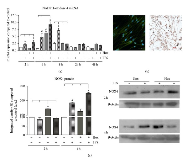
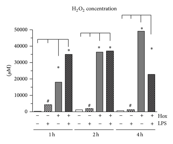
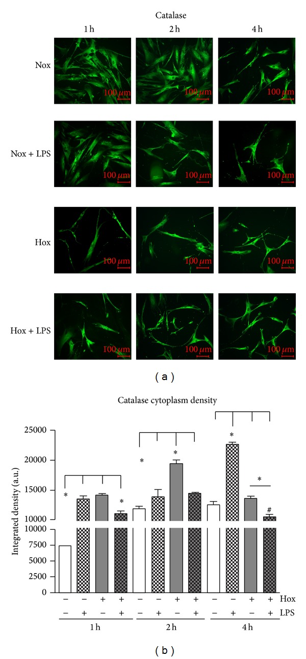
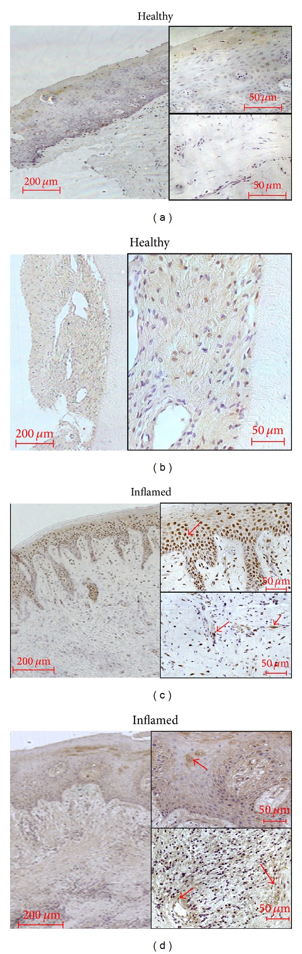
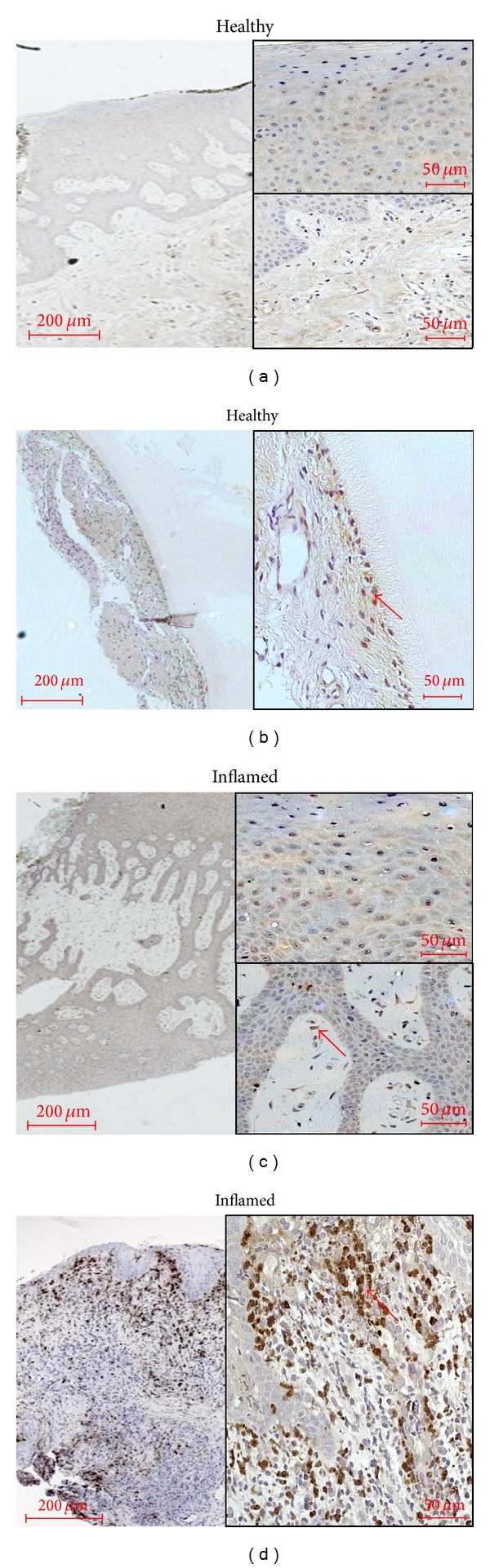
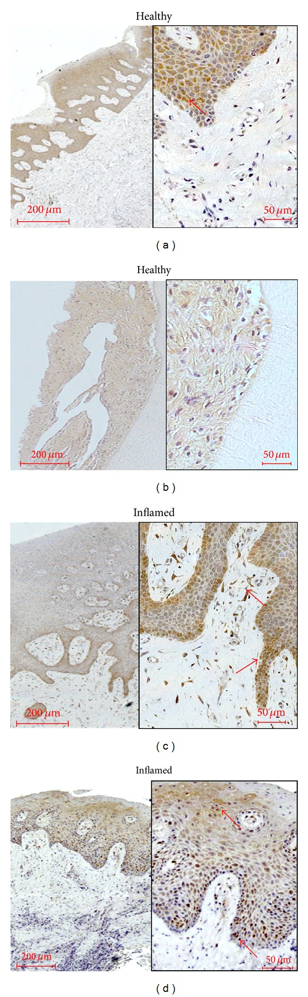
Oxidative stress is characterized by an accumulation of reactive oxygen species (ROS) and plays a key role in the progression of inflammatory diseases. We hypothesize that hypoxic and inflammatory events induce oxidative stress in the periodontal ligament (PDL) by activating NOX4. Human primary PDL fibroblasts were stimulated with lipopolysaccharide from Porphyromonas gingivalis (LPS-PG), a periodontal pathogen bacterium under normoxic and hypoxic conditions. By quantitative PCR, immunoblot, immunostaining, and a specific ROS assay we determined the amount of NOX4, ROS, and several redox systems. Healthy and inflamed periodontal tissues were collected to evaluate NOX4 and redox systems by immunohistochemistry. We found significantly increased NOX4 levels after hypoxic or inflammatory stimulation in PDL cells (P < 0.001) which was even more pronounced after combination of the stimuli. This was accompanied by a significant upregulation of ROS and catalase (P < 0.001). However, prolonged incubation with both stimuli induced a reduction of catalase indicating a collapse of the protective machinery favoring ROS increase and the progression of inflammatory oral diseases. Analysis of inflamed tissues confirmed our hypothesis. In conclusion, we demonstrated that the interplay of NOX4 and redox systems is crucial for ROS formation which plays a pivotal role during oral diseases.


Figures

Figure 1
Figure 1
NOX4 in human primary periodontal ligament (PDL) cells. (a) PDL cells were cultured under normoxic or hypoxic condition and stimulated with or without LPS of Porphyromonas gingivalis (1 μg/mL). NOX4 mRNA expression was analyzed after 2, 4, 8, 24, and 48 h. Statistical differences were analyzed by one-way ANOVA and post hoc Dunnett and Tukey's multiple comparison test; # P < 0.05 difference to control; ∗P < 0.05 difference between groups (means ± SD; n = 9). (b) NOX4 protein was visualized by immunofluorescence as well as immunohistochemical staining in unstimulated PDL fibroblasts (1 : 50; antibody from Abcam). (c) Immunoblot data of NOX4 in PDL cells cultured under normoxic or hypoxic condition (Hox) and stimulated with or without LPS of Porphyromonas gingivalis (1 μg/mL). NOX4 protein levels were analyzed after 2 and 4 h. Statistical analysis of western blot data was performed using the freely available image-processing software ImageJ 1.43 (http://rsb.info.nih.gov/ij/). Statistical differences were analyzed by one-way ANOVA and post hoc Dunnett and Tukey's multiple comparison test; # P < 0.05 difference to control; ∗P < 0.05 difference between groups (means ± SD; n = 3). The scale bars indicate 200 μm in the bright field survey and 50 μm in the immunofluorescence image, respectively.
Figure 2
Figure 2
Determination of ROS formation (H2O2) using Amplex Red assay. PDL cells were cultured under normoxic (Nox) or hypoxic condition (Hox) and stimulated with or without LPS of Porphyromonas gingivalis (1 μg/mL). H2O2 was analyzed after 1, 2 and 4 hours. Statistical differences were analyzed by one-way ANOVA and post hoc Dunnett and Tukey's multiple comparison test; # P < 0.05 difference to control; ∗P < 0.05 difference between groups (means ± SD; n = 6).
Figure 3
Figure 3
Catalase (CAT) in PDL cells. (a) CAT was visualized by immunofluorescence staining (1 : 50; antibody from Abcam). PDL cells were cultured under normoxic (Nox) or hypoxic condition (Hox) and stimulated with or without LPS of Porphyromonas gingivalis (1 μg/mL). Catalase cytoplasm density (b) was determined in relation to cell area using the freely available image-processing software ImageJ 1.43 (http://rsb.info.nih.gov/ij/). Statistical analysis of immunofluorescence data was analyzed by one-way ANOVA and post hoc Dunnett and Tukey's multiple comparison test; # P < 0.05 difference to control; ∗P < 0.05 difference between groups (means ± SD; n = 6). The scale bars indicate 100 μm in the immunofluorescence images.
Figure 4
Figure 4
NADPH-oxidase 4 (NOX4) occurrence in healthy and inflamed human tissue. Healthy gingival epithelium (a), periodontal ligament (b), and samples of gingivitis (c) or periodontitis (d) were obtained after the approval of the Ethics Committee of the University of Bonn and parental as well as patient's allowance (n = 3). Monoclonal primary antibody against NOX4 (Abcam, Cambridge, UK) was used in a concentration of 1 : 100. In gingival and PDL fibroblasts as well as in immune cells like leukocytes and in vessels, the immunoreactivity was primarily restricted to the cytosol and cell walls. Red arrows indicate examples for the typical distribution (Figures 4(c) and 4(d)). The scale bars indicate 200 μm in the surveys and 50 μm in the higher magnifications, respectively.
Figure 5
Figure 5
Catalase (CAT) occurrence in healthy and inflamed human tissue. Healthy gingival epithelium (a), periodontal ligament (b), and samples of gingivitis (c) or periodontitis (d) were obtained after the approval of the Ethics Committee of the University of Bonn and parental as well as patient's allowance (n = 3). Polyclonal primary antibody against catalase (Abcam, Cambridge, UK) was used in a concentration of 1 : 100. No immunostaining could be found in the healthy gingival epithelium. In the healthy PDL, predominantly PDL cells located near to the tooth root surfaces were stained (Figure 5(b), red arrow). In gingivitis tissue, a weak staining of gingival keratinocytes similar to healthy tissue could be seen (Figure 5(c)). Instead, in the tissue samples of patients with periodontitis a strong immunoreactivity was observed in the subepithelial layer probably referring to nuclei staining of immune cells like leukocytes. Red arrows indicate examples for the typical localisation (Figure 5(d)). The scale bars indicate 200 μm in the surveys and 50 μm in the higher magnifications, respectively.
Figure 6
Figure 6
Superoxide dismutase (SOD) occurrence in healthy and inflamed human tissue. Healthy gingival epithelium (a), periodontal ligament (b), and samples of gingivitis (c) or periodontitis (d) were obtained after the approval of the Ethics Committee of the University of Bonn and parental as well as patient's allowance (n = 3). Polyclonal primary antibody against SOD (Abcam, Cambridge, UK) was used in a concentration of 1 : 100. Gingival epithelium showed a moderate to strong staining of SOD in keratinocytes which was primarily limited to the cytosol (Figure 6(a) and red arrow depicts the typical localization). In the healthy periodontium, gingival and PDL fibroblasts were weakly stained (Figure 6(b)). In inflamed tissues, basal epithelial cells seemed to be stained more intensively in the gingivitis specimen than in periodontitis (Figures 6(c) and 6(d)). In addition, the subepithelium revealed immunoreactivity in local cells like gingival fibroblasts and scattered immune cells (Figure 6(c), see red arrow). In periodontitis, SOD immunostaining seemed to be limited to the gingival epithelium (Figure 6(d); see red arrow). The scale bars indicate 200 μm in the surveys and 50 μm in the higher magnifications, respectively.

Similar articles

Cited by

References

    1. Reuter S, Gupta SC, Chaturvedi MM, Aggarwal BB. Oxidative stress, inflammation, and cancer: how are they linked? Free Radical Biology and Medicine. 2010;49(11):1603–1616. - PMC - PubMed
    1. Sone H, Akanuma H, Fukuda T. Oxygenomics in environmental stress. Redox Report. 2010;15(3):98–114. - PMC - PubMed
    1. Wellen KE, Thompson CB. Cellular metabolic stress: considering how cells respond to nutrient excess. Molecular Cell. 2010;40(2):323–332. - PMC - PubMed
    1. Poyton RO, Ball KA, Castello PR. Mitochondrial generation of free radicals and hypoxic signaling. Trends in Endocrinology and Metabolism. 2009;20(7):332–340. - PubMed
    1. Weinberg F, Chandel NS. Reactive oxygen species-dependent signaling regulates cancer. Cellular and Molecular Life Sciences. 2009;66(23):3663–3673. - PMC - PubMed

Publication types

MeSH terms