. 2014 Oct 21:8:306.
doi: 10.3389/fncel.2014.00306. eCollection 2014.

The connexin43 mimetic peptide Gap19 inhibits hemichannels without altering gap junctional communication in astrocytes

Affiliations

The connexin43 mimetic peptide Gap19 inhibits hemichannels without altering gap junctional communication in astrocytes

Verónica Abudara et al. Front Cell Neurosci. .

Abstract

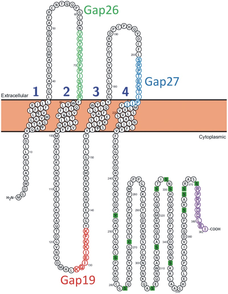
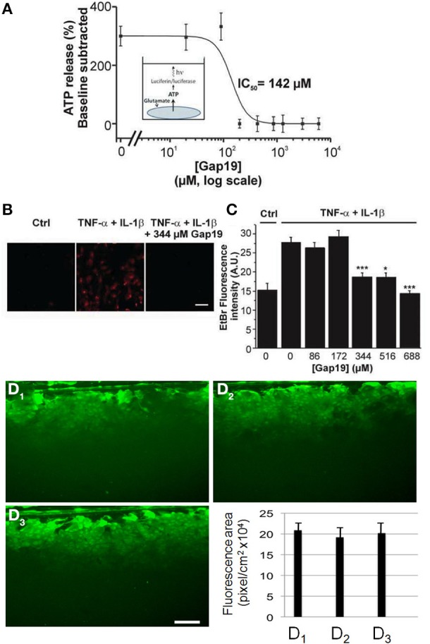
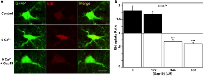
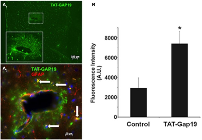
In the brain, astrocytes represent the cellular population that expresses the highest amount of connexins (Cxs). This family of membrane proteins is the molecular constituent of gap junction channels and hemichannels that provide pathways for direct cytoplasm-to-cytoplasm and inside-out exchange, respectively. Both types of Cx channels are permeable to ions and small signaling molecules allowing astrocytes to establish dynamic interactions with neurons. So far, most pharmacological approaches currently available do not distinguish between these two channel functions, stressing the need to develop new specific molecular tools. In astrocytes two major Cxs are expressed, Cx43 and Cx30, and there is now evidence indicating that at least Cx43 operates as a gap junction channel as well as a hemichannel in these cells. Based on studies in primary cultures as well as in acute hippocampal slices, we report here that Gap19, a nonapeptide derived from the cytoplasmic loop of Cx43, inhibits astroglial Cx43 hemichannels in a dose-dependent manner, without affecting gap junction channels. This peptide, which not only selectively inhibits hemichannels but is also specific for Cx43, can be delivered in vivo in mice as TAT-Gap19, and displays penetration into the brain parenchyma. As a result, Gap19 combined with other tools opens up new avenues to decipher the role of Cx43 hemichannels in interactions between astrocytes and neurons in physiological as well as pathological situations.

Keywords: astroglia; connexins; gap junctions; glial cells; mimetic peptide.


Figures

Figure 1
Figure 1
Position of the Gap19 sequence in the intracellular cytoplasmic loop domain of human Cx43. One identified interaction site is located in the last 9 AAs of the CT-tail marked in purple (Wang et al., 2013a). The sequences of Gap19 (red) on the intracellular loop, Gap26 (green) and Gap27 (blue) on the extracellular loops are indicated on the drawing. The CT residues marked as green squares are sites of posttranslational modifications and have been added for illustration purposes [Illustration generated with the Protter tool (Omasits et al., 2014)].
Figure 2
Figure 2
Dose-dependent inhibition of ATP release and Etd+ uptake through Cx43 hemichannels by Gap19 with lack of effect on gap junctional communication in cultured astrocytes. (A) Concentration-dependent inhibition by Gap19 (30 min pre-incubation) of ATP release in cultured cortical astrocytes triggered by glutamate (100 μM, 15 min application) (n = 6 independent experiments). (B) Representative images showing Etd+ uptake (red) in cultured astrocytes under control conditions (Ctrl) and after TNF-α/IL-1β or TNF-α/IL-1β + Gap19 treatment. Scale bar: 20 μm. (*p < 0.05; ***p < 0.001). (C) Summary data of Etd+ uptake studies in astrocytes, demonstrating inhibition by Gap19 (n = 5–8 independent experiments). Statistical comparisons refer to the stimulus condition without Gap19 (zero Gap19 concentration). (D1–D3) Representative images of scrape-loading dye transfer experiment in confluent cultures of astrocytes. Compared to control condition (D1), with Gap19 344 μM (D2, 30 min pre-incubation) or 688 μM (D3, 30 min pre-incubation). Lower graph: Quantification of scrape-loading data indicating that Gap19 did not influence gap junctional coupling as measured in confluent cultures of astrocytes (from left to right, bars are from control and the two tested concentrations of Gap19, respectively). (n = 3–5 independent experiments).
Figure 3
Figure 3
Gap19 inhibits hemichannel activity in astrocytes studied in acute hippocampal slices. (A) Representative images of Etd+ uptake (red) in astrocytes (green) in hippocampal slices from GFAP-eGFP transgenic mice, under control conditions (upper row) and after 10 min exposure to a Ca2+-free solution without (middle row) or with (lower row) 344 μM Gap19 treatment. Scale bar: 10 μm. (B) Summary graph demonstrating significant Etd+ uptake in astrocytes that was inhibited by Gap19 used at concentration of 344 and 688 μM. Statistical comparisons in (B) were done with the stimulus condition without Gap19 (n = 3 independent experiments; ***p < 0.001).
Figure 4
Figure 4
Detection of TAT-Gap19 in the cortex of the mouse. (A1–A2) One hour after carotid injection of TAT-Gap19 the brain displayed clear TAT immunoreactivity compared to mice that received vehicle (PBS) only. (A1), Area in small box is enlarged in the lower right box of the panel; (A2), Double labeling with anti-TAT and anti-GFAP antibodies indicates that some GFAP-positive astrocytes (white arrows) have taken up the TAT peptide. Note that the green fluorescence is concentrated in the white box area probably because this represents a large vessel that would have more passage of TAT-Gap19 into the brain parenchyma surrounding the vessel. (B) A single i.v. injection of TAT-Gap19 gave significant immune signal in the brain 24 h later. Taken together, these experiments indicate that TAT-Gap19 traverses the blood-brain barrier and is retained in the cells. Fluorescence intensities of Alexa 488 signal of the secondary antibody determined in slices immunostained with a primary antibody directed against the TAT sequence. Control represents experiments in mice injected with PBS vehicle (n = 5 for control and TAT-Gap19; *p < 0.05).

References

    1. Chever O., Lee C. Y., Rouach N. (2014). Astroglial connexin43 hemichannels tune Basal excitatory synaptic transmission. J. Neurosci. 34, 11228–11232. 10.1523/JNEUROSCI.0015-14.2014 - DOI - PMC - PubMed
    1. Cina C., Maass K., Theis M., Willecke K., Bechberger J. F., Naus C. C. (2009). Involvement of the cytoplasmic C-terminal domain of connexin43 in neuronal migration. J. Neurosci. 29, 2009–2021. 10.1523/JNEUROSCI.5025-08.2009 - DOI - PMC - PubMed
    1. D'hondt C., Iyyathurai J., Wang N., Gourdie R. G., Himpens B., Leybaert L., et al. . (2013). Negatively charged residues (Asp378 and Asp379) in the last ten amino acids of the C-terminal tail of Cx43 hemichannels are essential for loop/tail interactions. Biochem. Biophys. Res. Commun. 432, 707–712. 10.1016/j.bbrc.2013.01.066 - DOI - PubMed
    1. De Bock M., Wang N., Decrock E., Bol M., Gadicherla A. K., Culot M., et al. . (2013). Endothelial calcium dynamics, connexin channels and blood-brain barrier function. Prog. Neurobiol. 108, 1–20. 10.1016/j.pneurobio.2013.06.001 - DOI - PubMed
    1. De Vuyst E., Wang N., Decrock E., De Bock M., Vinken M., Van Moorhem M., et al. . (2009). Ca(2+) regulation of connexin 43 hemichannels in C6 glioma and glial cells. Cell Calcium 46, 176–187. 10.1016/j.ceca.2009.07.002 - DOI - PubMed