

Oxidative phosphorylated neurofilament protein M protects spinal cord against ischemia/reperfusion injury
- PMID: 25374588
- PMCID: PMC4211187
- DOI: 10.4103/1673-5374.141803
Oxidative phosphorylated neurofilament protein M protects spinal cord against ischemia/reperfusion injury
Abstract
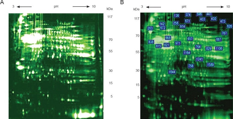
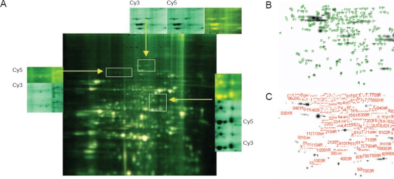
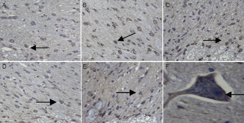
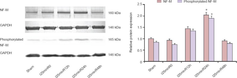
Previous studies have shown that neurofilament protein M expression is upregulated in the early stage of spinal cord ischemia/reperfusion injury, indicating that this protein may play a role in the injury process. In the present study, we compared protein expression in spinal cord tissue of rabbits after 25 minutes of ischemia followed by 0, 12, 24, or 48 hours of reperfusion with that of sham operated rabbits, using proteomic two-dimensional gel electrophoresis and mass spectrometry. In addition, the nerve repair-related neurofilament protein M with the unregulated expression was detected with immunohistochemistry and western blot analysis. Two-dimensional gel electrophoresis and mass spectrometry showed that, compared with the sham group, upregulation of protein expression was most significant in the spinal cords of rabbits that had undergone ischemia and 24 hours of reperfusion. Immunohistochemical analysis revealed that neurofilament protein M was located in the membrane and cytoplasm of neuronal soma and axons at each time point after injury. Western blot analysis showed that neurofilament protein M expression increased with reperfusion time until it peaked at 24 hours and returned to baseline level after 48 hours. Furthermore, neurofilament protein M is phosphorylated under oxidative stress, and expression changes were parallel for the phosphorylated and non-phosphorylated forms. Neurofilament protein M plays an important role in spinal cord ischemia/reperfusion injury, and its functions are achieved through oxidative phosphorylation.
Keywords: NSFC grant; ischemia/reperfusion; nerve regeneration; neural regeneration; neurofilament protein M; neuroprotection; phosphorylation; proteomics; spinal cord injury.
Conflict of interest statement
Figures
References
-
- Blizzard CA, King AE, Vickers J, Dickson T. Cortical murine neurons lacking the neurofilament light chain protein have an attenuated response to injury in vitro. J Neurotrauma. 2013;30:1908–1918. - PubMed
-
- Bradford MM. A rapid and sensitive method for the quantitation of microgram quantities of protein utilizing the principle of protein-dye binding. Anal Biochem. 1976;72:248–254. - PubMed
-
- Carter J, Gragerov A, Konvicka K, Elder G, Weinstein H, Lazzarini RA. Neurofilament (NF) assembly; divergent characteristics of human and rodent NF-L subunits. J Biol Chem. 1998;273:5101–5108. - PubMed
-
- de Groot DM, Coenen AJ, Verhofstad A, van Herp F, Martens GJ. In vivo induction of glial cell proliferation and axonal outgrowth and myelination by brain-derived neurotrophic factor. Mol Endocrinol. 2006;20:2987–2998. - PubMed
LinkOut - more resources
Full Text Sources
Other Literature Sources