Review
. 2014 Nov 6;3(4):122-42.
doi: 10.5527/wjn.v3.i4.122.

Nephropathy in dietary hyperoxaluria: A potentially preventable acute or chronic kidney disease

Affiliations
Review

Nephropathy in dietary hyperoxaluria: A potentially preventable acute or chronic kidney disease

Robert H Glew et al. World J Nephrol. .

Abstract

Hyperoxaluria can cause not only nephrolithiasis and nephrocalcinosis, but also renal parenchymal disease histologically characterized by deposition of calcium oxalate crystals throughout the renal parenchyma, profound tubular damage and interstitial inflammation and fibrosis. Hyperoxaluric nephropathy presents clinically as acute or chronic renal failure that may progress to end-stage renal disease (ESRD). This sequence of events, well recognized in the past in primary and enteric hyperoxalurias, has also been documented in a few cases of dietary hyperoxaluria. Estimates of oxalate intake in patients with chronic dietary hyperoxaluria who developed chronic kidney disease or ESRD were comparable to the reported average oxalate content of the diets of certain populations worldwide, thus raising the question whether dietary hyperoxaluria is a primary cause of ESRD in these regions. Studies addressing this question have the potential of improving population health and should be undertaken, alongside ongoing studies which are yielding fresh insights into the mechanisms of intestinal absorption and renal excretion of oxalate, and into the mechanisms of development of oxalate-induced renal parenchymal disease. Novel preventive and therapeutic strategies for treating all types of hyperoxaluria are expected to develop from these studies.

Keywords: Acute oxalate nephropathy; Acute tubular necrosis; Calcium oxalate nephrolithiasis; Chronic oxalate nephropathy; Dietary hyperoxaluria; Inflammasomes; Interstitial nephritis; Nephrocalcinosis; Oxalate transporters.


Figures

Figure 1
Figure 1
Biosynthesis of calcium oxalate. Glyoxylate is the main precursor of oxalate which combines spontaneously with calcium ions to form calcium oxalate. Names of enzymes: 1, serine hydroxymethyltransferase; 2, D-amino acid oxidase; 3, alanine:glyoxylate aminotransferase (AGT); 4, glyoxylate reductase-hydroxypyruvate reductase (GRHPR); 5, glycolate oxidase; 6, alcohol dehydrogenase; 7, aldehyde dehydrogenase; 8, lactate dehydrogenase; and 9, five enzyme-catalyzed reactions. PH1 results from mutations in AGT which is a hepatic peroxisomal enzyme. PH2 results from mutations in GRHPR which is a cytosolic enzyme found in several tissues, but primarily the liver. PH3 results from defects in the hepatic mitochondrial enzyme 4-hydroxy-2-oxoglutarate (HOG) aldolase which converts HOG and glyoxylate to pyruvate (reaction not shown), the last step in hydroxyproline catabolism. The reason why a deficiency of HOG aldolase activity increases oxalate production is obscure.
Figure 2
Figure 2
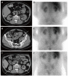
Sequential imaging studies of a not yet reported patient with chronic kidney disease from dietary hyperoxaluria. Axial computed tomography (CT) images obtained two years before the hyperoxaluria diagnosis show (A) mild left hydronephrosis (arrowheads) caused by (B) a left distal ureteral calculus (arrow). Axial CT image obtained around the time of the hyperoxaluria diagnosis shows (C) bilateral nephrolithiasis (arrows). Nuclear medicine gallium-67 citrate scan images were also obtained around the time of diagnosis, including (D) 4-, (E) 24-, and (F) 48 h after administration. These show abnormal, persistent bilateral renal activity at all time points, indicative of interstitial nephritis. Gallium scanning has classically been used to distinguish acute interstitial nephritis from acute tubular necrosis and other causes of acute renal failure[216-218]. In this patient chronic interstitial nephritis associated with hyperoxaluria led to this positive scan. The patient’s diet for several years was based on nuts with estimated oxalate consumption ≥ 800 mg daily. During high oxalate intake, urine oxalate excretion was > 200 mg/24-h in several measurements obtained at serum creatinine levels > 3.5 mg/dL. After resumption of a diet low in oxalate and improvement of renal function to serum creatinine levels < 3.0 mg/dL, urine oxalate excretion decreased to normal levels.
Figure 3
Figure 3
Renal histology in the patient depicted in Figure 2. A: Low power view of kidney showing two complete glomeruli and expansion of the interstitium by lymphocytes and edema. Periodic acid-Schiff (PAS) stain highlights the basement membranes of the tubules and Bowman’s capsule. PAS stain; B: Low power view of renal parenchyma showing tubulointerstitial nephritis (solid arrow) and oxalate crystal within tubule (open arrow). H and E stain; C: High power view showing interstitium expanded by lymphocytic infiltrates and tubular atrophy. PAS stain; D: High power view of calcium oxalate crystal under polarized light. H and E stain.

Similar articles

Cited by

References

    1. Ogilvie D, McCollum JP, Packer S, Manning J, Oyesiku J, Muller DP, Harries JT. Urinary outputs of oxalate, calcium, and magnesium in children with intestinal disorders. Potential cause of renal calculi. Arch Dis Child. 1976;51:790–795. - PMC - PubMed
    1. Pak CY, Britton F, Peterson R, Ward D, Northcutt C, Breslau NA, McGuire J, Sakhaee K, Bush S, Nicar M, et al. Ambulatory evaluation of nephrolithiasis. Classification, clinical presentation and diagnostic criteria. Am J Med. 1980;69:19–30. - PubMed
    1. Larking P, Lovell-Smith CJ, Hocken AG. Urine oxalate levels in a New Zealand reference population and renal stone formers. N Z Med J. 1983;96:606–607. - PubMed
    1. Lindsjö M, Fellström B, Danielson BG, Kasidas GP, Rose GA, Ljunghall S. Hyperoxaluria or hypercalciuria in nephrolithiasis: the importance of renal tubular functions. Eur J Clin Invest. 1990;20:546–554. - PubMed
    1. Laminski NA, Meyers AM, Kruger M, Sonnekus MI, Margolius LP. Hyperoxaluria in patients with recurrent calcium oxalate calculi: dietary and other risk factors. Br J Urol. 1991;68:454–458. - PubMed