A system for evaluating magnetic resonance imaging of prostate cancer using patient-specific 3D printed molds
- PMID: 25374914
- PMCID: PMC4219304
A system for evaluating magnetic resonance imaging of prostate cancer using patient-specific 3D printed molds
Abstract
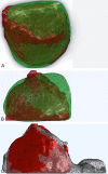
We have developed a system for evaluating magnetic resonance imaging of prostate cancer, using patient-specific 3D printed molds to facilitate MR-histology correlation. Prior to radical prostatectomy a patient receives a multiparametric MRI, which an expert genitourinary radiologist uses to identify and contour regions suspicious for disease. The same MR series is used to generate a prostate contour, which is the basis for design of a patient-specific mold. The 3D printed mold contains a series of evenly spaced parallel slits, each of which corresponds to a known MRI slice. After surgery, the patient's specimen is enclosed within the mold, and all whole-mount levels are obtained simultaneously through use of a multi-bladed slicing device. The levels are then formalin fixed, processed, and delivered to an expert pathologist, who identifies and grades all lesions within the slides. Finally, the lesion contours are loaded into custom software, which elastically warps them to fit the MR prostate contour. The suspicious regions on MR can then be directly compared to lesions on histology. Furthermore, the false-negative and false-positive regions on MR can be retrospectively examined, with the ultimate goal of developing methods for improving the predictive accuracy of MRI. This work presents the details of our analysis method, following a patient from diagnosis through the MR-histology correlation process. For this patient MRI successfully predicted the presence of cancer, but true lesion volume and extent were underestimated. Most cancer-positive regions missed on MR were observed to have patterns of low T2 signal, suggesting that there is potential to improve sensitivity.
Keywords: 3D printing; MRI; pathology; prostate cancer; registration; whole mount.
Figures


References
-
- Hodge K, McNeal J, Terris M, Stamey T. Random systematic versus directed ultrasound guided transrectal core biopsies of the prostate. J Urol. 1989;1:71–4. - PubMed
-
- Hoeks CMA, Barentsz JO, Hambrock T, Yakar D, Somford DM, Heijmink SW, Scheenen TWJ, Vos PC, Huisman H, Van Oort IM, Witjes JA, Heerschap A, Fütterer JJ. Prostate Cancer: Multiparametric MR Imaging for Detection, Localization, and Staging. Radiology. 2011;1:46–66. - PubMed
-
- Moore CM, Robertson NL, Arsanious N, Middleton T, Villers A, Klotz L, Taneja SS, Emberton M. Image-guided prostate biopsy using magnetic resonance imaging--derived targets: a systematic review. Eur Urol. 2013;1:125–140. - PubMed
-
- Kozlowski P, Chang SD, Jones EC, Berean KW, Chen H, Goldenberg SL. Combined diffusion-weighted and dynamic contrast-enhanced MRI for prostate cancer diagnosis---Correlation with biopsy and histopathology. J Magn Reson Imaging. 2006;1:108–113. - PubMed
Grants and funding
LinkOut - more resources
Full Text Sources
Other Literature Sources
