. 2014 Nov 6;10(11):e1004454.
doi: 10.1371/journal.ppat.1004454. eCollection 2014 Nov.

Co-dependence of HTLV-1 p12 and p8 functions in virus persistence

Affiliations

Co-dependence of HTLV-1 p12 and p8 functions in virus persistence

Cynthia A Pise-Masison et al. PLoS Pathog. .

Abstract

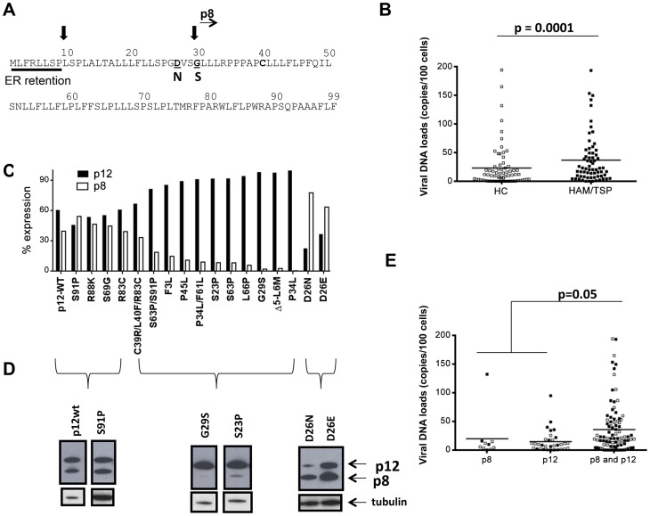
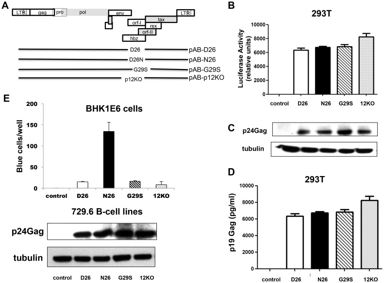
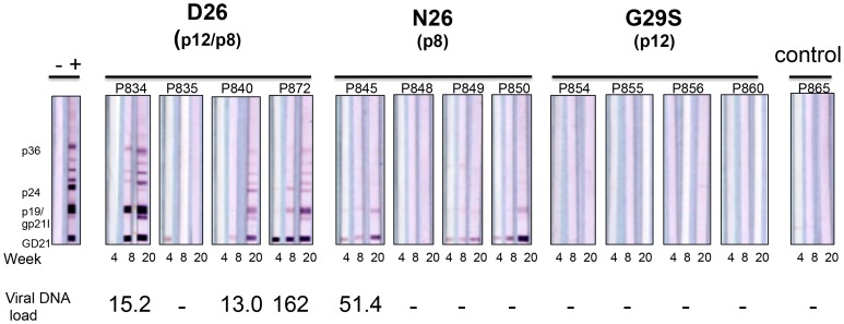
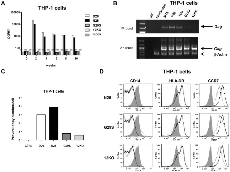
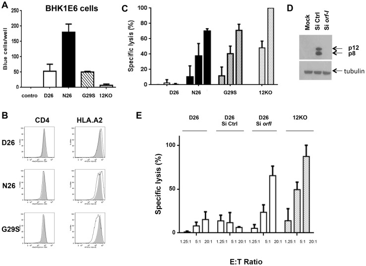
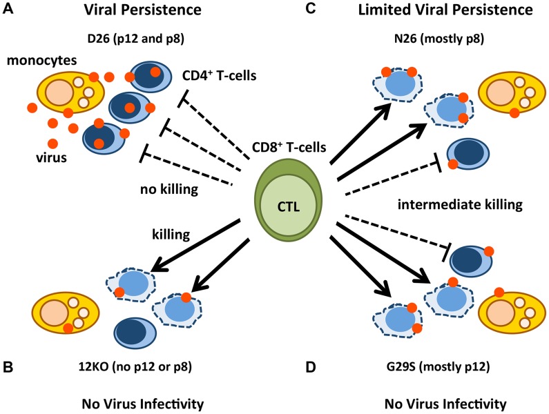
HTLV-1 orf-I is linked to immune evasion, viral replication and persistence. Examining the orf-I sequence of 160 HTLV-1-infected individuals; we found polymorphism of orf-I that alters the relative amounts of p12 and its cleavage product p8. Three groups were identified on the basis of p12 and p8 expression: predominantly p12, predominantly p8 and balanced expression of p12 and p8. We found a significant association between balanced expression of p12 and p8 with high viral DNA loads, a correlate of disease development. To determine the individual roles of p12 and p8 in viral persistence, we constructed infectious molecular clones expressing p12 and p8 (D26), predominantly p12 (G29S) or predominantly p8 (N26). As we previously showed, cells expressing N26 had a higher level of virus transmission in vitro. However, when inoculated into Rhesus macaques, cells producing N26 virus caused only a partial seroconversion in 3 of 4 animals and only 1 of those animals was HTLV-1 DNA positive by PCR. None of the animals exposed to G29S virus seroconverted or had detectable viral DNA. In contrast, 3 of 4 animals exposed to D26 virus seroconverted and were HTLV-1 positive by PCR. In vitro studies in THP-1 cells suggested that expression of p8 was sufficient for productive infection of monocytes. Since orf-I plays a role in T-cell activation and recognition; we compared the CTL response elicited by CD4+ T-cells infected with the different HTLV-1 clones. Although supernatant p19 levels and viral DNA loads for all four infected lines were similar, a significant difference in Tax-specific HLA.A2-restricted killing was observed. Cells infected with Orf-I-knockout virus (12KO), G29S or N26 were killed by CTLs, whereas cells infected with D26 virus were resistant to CTL killing. These results indicate that efficient viral persistence and spread require the combined functions of p12 and p8.


Conflict of interest statement

The authors have declared that no competing interests exist.

Figures

Figure 1
Figure 1. Analysis of orf-I from the PBMCs of HTLV-1 infected individuals.
(A) Schematic diagram of the Orf-I protein. The non-canonical endoplasmic reticulum (ER) retention sequence is underlined by a solid bar. Black arrows indicate the putative cleavage sites, as well as the start of the p8 isoform. Mutations which identify cleavage variants at position 26 and 29 are indicated in bold below the sequence. (B) Comparison of viral DNA levels in PBMCs from HTLV-1 infected individuals by disease association, HC: healthy carrier (open symbols) and HAM/TSP: HTLV-1 associated myelopathy/tropical spastic paraparesis (filled symbols). The data from 70 healthy carriers (HC) (n = 70) and 66 HAM/TSP individuals (n = 66) were analyzed using the Mann-Whitney Test stratified by disease status. The statistically significant difference is marked with the p value. The horizontal lines represent the mean viral DNA load. (C) Cloned orf-I cDNA constructs were transfected into 293T-cells and protein expression analyzed 48 hours after transfection. The density of p12 and p8 bands was measured using AlphaView Software on an AlphaImager (ProteinSimple, San Leandro, CA). Expression of p12 and p8 were added to give 100% expression. The percent of total Orf-I expression for each clone was graphed. The black bars represent the percentage of p12 expressed and the lighter bar represents the percentage of p8 expressed. The clone is indicated at the bottom of the graph. Expression patterns for each clone were examined in independent transfection experiments where n = 20 for D26, n = 8 for G29S; P45L, n = 7 for P34L/F61L, n = 6 for S69G; S23P; S63P; D26E; P34L, n = 5 for C39R/L40F/R83C; F3L; L66P; Δ5-L6M, n = 4 for R83C, D26N, n = 3 for S91P, n = 2 for R88K; S63P/S91P. The expression patterns could be divided into three groups: p12 and p8, p12 mainly (p12) or p8 mainly (p8). (D) Representative western blot analysis of cell lysates for Orf-I expression, using anti-HA (upper panel) or a loading control (anti-tubulin, lower panel) was performed. Amino acid changes are indicated above each lane. The p12 or p8 isoform is indicated by arrows at the right. (E) Viral DNA levels in PBMCs from individuals with the indicated orf-1 gene expression patterns are indicated in the x-axis. The data obtained was a total of 136 individuals using the same assay (n = 10 individuals with mainly p8 expression, n = 32 individuals with mainly p12 expression and n = 94 individuals with similar p12 and p8 expression) and analyzed by an exact Wilcoxon rank sum test stratified by disease status. The horizontal lines represent the mean viral DNA levels. The open symbols identify healthy carriers and the filled symbols HAM/TSP patients. The statistical significance is indicated by the p value.
Figure 2
Figure 2. Mutant viruses produce equivalent levels of Gag protein but the virus N26 is transmitted better.
(A) The schematic diagram of the HTLV-1 molecular clones indicates the amino acid change in each clone. The initiation codon for Orf-I is mutated in p12KO such that no Orf-I protein is made. The changes did not affect the sequence and/or function of the overlapping pX region genes. Infectious molecular clones or control DNA were co-transfected with an HTLV-1-LTR-luciferase construct and the renilla-luciferase transfection efficiency control into 293T-cells and culture supernatants or protein lysates prepared 48 hours after transfection. (B) The HTLV-1 promoter activity induced by the HTLV-1 mutant was measured by assaying luciferase activity from transfected cell lysates. Luciferase activity for each clone (indicated on the x-axis) from three independent transfection experiments was graphed (n = 3). LTR-luciferase activity was normalized using the transfection efficiency control renilla-luciferase activity. Error bars indicate the standard deviation. (C) Western blot analysis of protein lysates from transfected cells was assayed for intracellular p24Gag expression (top panel) or the loading control, tubulin (bottom panel). (D) Culture supernatants from transfected 293T-cells were collected, spun to remove debris and assayed for p19Gag levels using an HTLV-1 ELISA kit. The values graphed are from three independent experiments (n = 3). (E) Stable producer 729.6 B-cell lines were cloned and used to quantify the transmission of the viral mutants. The 729-HTLV-1-producing cells or parental control cells were co-cultured with BHK1E6 cells and 48 hours later, adherent cells were stained for -galactosidase activity. Graphed is the number of blue cells per well for the indicated clone from three independent wells (n = 3). Error bars indicate standard deviation. By ANOVA and t- test, transmission of WT, D26N and G29S was significantly different than control (p<0.0001). Transmission of D26N was significantly different than WT, G29S and p12KO (p = 0.0007). There was no significant difference among transmission of WT, G29S and p12KO. Western blot analysis for HTLV-1 p24Gag was performed on whole cell extracts from 729-HTLV-1 producing cell lines. The housekeeping gene tubulin is shown for a loading control (lower panels).
Figure 3
Figure 3. D26, N26, and G29S infectivity in macaques.
Sera from inoculated male Rhesus macaques were assayed for reactivity to HTLV-1 antigens. The animal number and inoculation group are indicated above each sample. Indicated below each western blot strip is the time of sera collection. The presence of HTLV-1 viral DNA was measured from PBMC DNA isolated at the designated time points by PCR analysis for HTLV-1 integrase; (-) indicates PCR negative. Viral DNA loads were normalized to the macaque albumin gene and expressed as the number of HTLV-1 viral DNA copies per 106 PBMCs. The value of the viral DNA load provided at the bottom of the figure is the highest measured for the indicated animal.
Figure 4
Figure 4. HTLV-1 infection of the monocytic cell line THP-1.
(A) THP-1 cells were infected with supernatants from 729-HTLV-1 producing or parental 729.6 cell lines (concentrated by ultracentrifugation). Culture supernatants were monitored by ELISA for p19Gag levels. Graphed is the log scale of p19Gag in picograms per milliliter over a 16 week period for one set of cultures. THP-1 infected cultures: D26 (white bar); N26 (black bar); G29S (slanted bar); 12KO (dotted bar); Mock (gray bar). The dashed line indicates assay background level. (B) PCR analysis was performed on genomic DNA isolated at week 16. The first (upper panel) and second (lower panel) rounds of nested PCR were separated by electrophoresis and stained with ethidium bromide to visualize products for the indicated cell cultures. Arrows designate the Gag and the control β-actin fragments. (C) The viral DNA copy number for each cell culture at week 18 was determined by quantitative real-time PCR. The human albumin gene was used for normalization. (D) Histogram plots show the phenotype of HTLV-1 infected THP-1 cells for the cell surface monocytic markers: CD14, HLA-DR and CCR7. Each viral mutant (gray line) was compared to the wild-type (D26, un-shaded, black line) and the mock (shaded) infected THP-1 cells.
Figure 5
Figure 5. Susceptibility of HTLV-1 producing CD4+ cell lines to CTL killing.
(A) CD4+ T-cells infected with the D26, N26, G29S and 12KO viruses were incubated with BHKE16 indicator cells for 48 hours. Un-infected Jurkat T-cells (control) were used as a negative control. The number of blue cells per well for three independent experiments is graphed (n = 3). Error bars indicate standard deviation. (B) A comparison of the surface expression of CD4 (left panels) and HLA.A2 (right panels) are shown for the indicated virus-infected CD4+ T-cells (black line) in comparison to the 12KO CD4+ T-cell line (shaded). (C) Cytoxic T-lymphocyte killing assays were done to evaluate specific lysis of CD4+ T-cells infected with the D26, N26, G29S and 12KO viruses. A long term HLA.A2 restricted CD8+ T-cell line from an HAM/TSP patient was used as the effector cell (see Materials and Methods). Graphed is the percent of specific lysis at varying effector-to-target cell ratios (1.25∶1; 5∶1; 20∶1). The graph represents data from at least two independent experiments done in triplicate (n≥2). Lysis of 12KO at the 20∶1 ratio (highest specific lysis) was set to 100%. All samples were normalized to maximal killing obtained with the 12KO virus. Error bars indicate standard deviation. (D) Western blot analysis of protein lysates from transfected cells was assayed for p12 and p8 expression (top panel) or the loading control, tubulin (bottom panel) to determine the effectiveness of the siRNA constructs. Cells were co-transfected with p12WT cDNA or control expression constructs in the presence or absence of siRNA (Si Ctrl or Si orf-I). (E) CD4+ D26-infected cells were transfected with siRNA control (Si CTRL) or siRNA to orf-I (Si orf-I) and used as target cells in CTL killing assays. CTL lysis of the cells at increasing effector-to-target cell ratios is graphed. The graph represents data from at least two independent experiments done in triplicate (n≥2). Error bars indicate standard deviation.
Figure 6
Figure 6. Model of p12 and p8 functions on monocyte, T-cell infection, and their susceptibility to CTL killing.
The red dots represent HTLV-1 virions/proteins and the solid arrows represent effective CTL killing of CD4+-infected T-cells. The dashed lines indicate no CTL killing. Lysed cells are represented by misshapen, dashed lines. Cell types are indicated in the figure. D26-infected CD4+ T-cells expressing balanced levels of p12 and p8 (A); 12KO-infected CD4+ T-cells expressing neither p12 nor p8 (B); N26-infected CD4+ T-cells expressing mainly p8 (C); and G29S-infected CD4+ T-cells expressing mainly p12 (D).

References

    1. Poiesz BJ, Ruscetti FW, Gazdar AF, Bunn PA, Minna JD, et al. (1980) Detection and isolation of type C retrovirus particles from fresh and cultured lymphocytes of a patient with cutaneous T-cell lymphoma. Proceedings of the National Academy of Sciences of the United States of America 77: 7415–7419. - PMC - PubMed
    1. Poiesz BJ, Ruscetti FW, Mier JW, Woods AM, Gallo RC (1980) T-cell lines established from human T-lymphocytic neoplasias by direct response to T-cell growth factor. Proc Natl Acad Sci USA 77: 6815–6819. - PMC - PubMed
    1. Gessain A, Barin F, Vernant J-C, Gout O, Maurs L, et al. (1985) Antibodies to human T-lymphotropic virus type I in patients with tropical spastic paraparesis. Lancet ii: 407–410. - PubMed
    1. Osame M, Usuku K, Izumo S, Ijichi N, Amitani H, et al. (1986) HTLV-I associated myelopathy, a new clinical entity. Lancet 1: 1031–1032. - PubMed
    1. Franchini G, Lairmore MD (2007) Human T-cell leukemia/lymphoma virus types 1 and 2. In: Fields Virology. Philadelphia LWW, editor. pp. 2071–2106.

Publication types

MeSH terms

Substances