. 2014 Nov 6;10(11):e1004757.
doi: 10.1371/journal.pgen.1004757. eCollection 2014 Nov.

The CSN/COP9 signalosome regulates synaptonemal complex assembly during meiotic prophase I of Caenorhabditis elegans

Affiliations

The CSN/COP9 signalosome regulates synaptonemal complex assembly during meiotic prophase I of Caenorhabditis elegans

Heather Brockway et al. PLoS Genet. .

Abstract

The synaptonemal complex (SC) is a conserved protein structure that holds homologous chromosome pairs together throughout much of meiotic prophase I. It is essential for the formation of crossovers, which are required for the proper segregation of chromosomes into gametes. The assembly of the SC is likely to be regulated by post-translational modifications. The CSN/COP9 signalosome has been shown to act in many pathways, mainly via the ubiquitin degradation/proteasome pathway. Here we examine the role of the CSN/COP9 signalosome in SC assembly in the model organism C. elegans. Our work shows that mutants in three subunits of the CSN/COP9 signalosome fail to properly assemble the SC. In these mutants, SC proteins aggregate, leading to a decrease in proper pairing between homologous chromosomes. The reduction in homolog pairing also results in an accumulation of recombination intermediates and defects in repair of meiotic DSBs to form the designated crossovers. The effect of the CSN/COP9 signalosome mutants on synapsis and crossover formation is due to increased neddylation, as reducing neddylation in these mutants can partially suppress their phenotypes. We also find a marked increase in apoptosis in csn mutants that specifically eliminates nuclei with aggregated SC proteins. csn mutants exhibit defects in germline proliferation, and an almost complete pachytene arrest due to an inability to activate the MAPK pathway. The work described here supports a previously unknown role for the CSN/COP9 signalosome in chromosome behavior during meiotic prophase I.


Conflict of interest statement

The authors have declared that no competing interests exist.

Figures

Figure 1
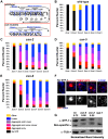
Figure 1. SC central element assembly defects in csn mutants.
A) CSN alleles used in this study: black rectangles represent exons, black lines introns, gray areas represent UTR regions, red lines region deleted and purple, green and blue rectangles the protein domains. PAM and PINT are subdomains of the PCI domain, B) Micrographs of SYP-1 (red or grey scaled) and DAPI (blue) stained wild-type and csn-5 mutants nuclei representing the various stages of the C. elegans gonad. Images are projections through half of a three-dimensional data stacks. Scale bar is 2 µm. PMT = pre-meiotic tip, TZ = transition zone, EP = early pachytene, MP = mid pachytene, LP = late pachytene. SYP-1 aggregates appear in the TZ-like stage of the gonad and persist through the LP-like stage. C) Whole gonad from wild-type and csn mutants SYP-1 and DAPI stained. Images show smaller gonads in csn mutants and lack of oocytes progressing through diakinesis. Scale Bar 16 µm. SYP-1 (grayscale) staining only of gonads showing aggregation throughout the gonad, starting at transition zone.
Figure 2
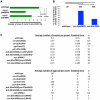
Figure 2. Quantification of the SYP-1 aggregates.
A) Schematic representation of the zones of the C. elegans gonad. PMT = pre-meiotic tip, TZ = transition zone, EP = early pachytene, MP = mid pachytene, LP = late pachytene. B–E) Quantification of SYP-1 aggregates in zones of the gonad. Percent of nuclei with: no SYP-1 (black), linear SYP-1 (blue), aggregated SYP-1 (purple pink and red) and other (yellow), zones as in A, n nuclei scored wild type: 1123, csn-2: 868, csn-5: 1020, csn-6: 85, p<0.0005 for pairwise comparisons; Fisher's Exact Test F) Representative images of nuclei scored in C–D all taken from the same gonad in late pachytene of csn-2 mutants, G) Western Blot confirming the reduction of expression of SYP-1 in csn mutants. Normalization values (α-SYP-1/α-TUB) shown are the average of 2 different experiments. Normalized intensities: wild-type 0.72±0.26, csn-2 0.24±0.20 and csn-5 0.58±0.11.
Figure 3
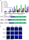
Figure 3. Quantification of the lack of oocytes and fecundity test.
A) Relative sizes of the pre-meiotic tips for wild-type and the csn mutants. The size of the mitotic zone is reduced in csn mutants. n = 10 for each strain p<0.0005 for wild-type vs. csn-2; p = 0.005 for wild-type vs. csn-5 and p<0.05 for csn-2 vs. csn-5; Mann Whitney Test B) Quantification of the number of gonads that contained oocytes in diakinesis for the csn mutants, *p MW<0.0005 and **p MW<0.005, Mann Whitney Test C) Top: the average number of oocytes in diakinesis for the csn mutants and the csn mutant, apoptosis checkpoint double mutants. Bottom: the average number of eggs laid for csn mutants and apoptosis checkpoint double mutants. csn mutants have a severe reduction in the number of oocytes and lay no eggs.
Figure 4
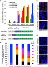
Figure 4. Pairing stabilization is affected in csn mutants.
A) Analysis of pairing stabilization between wild-type, syp-1(me17), csn mutants. A schematic representation of the timing of meiotic stages relative to the zones in the C elegans gonad. zone 1 = pre-meiotic tip, zone 2 and 3 = transition from mitosis to meiosis, zone 4–6 = pachytene. The black arrow represents the movement of nuclei through the stages (zones) of meiosis. csn mutants show defects in pairing stabilization number of nuclei counted and p-values can be found in Sup.Tables 1 and 2. B) High magnification micrographs of individual nuclei. Images are projections through three-dimensional data stacks. 5S FISH probe foci are in green and DAPI stained chromosomes are in blue. zone 4 = early pachytene, zone 5 = mid pachytene, zone 6 = late pachytene. Scale bar is 2 µm.
Figure 5
Figure 5. Accumulation of recombination intermediates and reduced crossover formation in csn mutants.
A) Analysis of RAD-51 foci in wild-type compared to csn mutants. Position along the x-axis refers to the zone in the gonad (Figure 4). RAD-51 foci accumulate upon entrance to meiosis in csn mutants. Numbers of nuclei counted and p-values can be found in Sup. Table 3. Schematic representation of the timing of meiotic stages relative to zones scored in the csn mutants compared to wild-type. B) Quantitative analysis of COSA-1 foci in late pachytene of the wild-type and zone 6-like section of the csn mutants color code for number on COSA-1 is at right. Number of designated crossovers marked by COSA-1 is reduced. Number of nuclei scored: wild-type n = 123, csn-2 n = 111, csn-5 n = 94, csn-6 n = 78 p<0.0005 for comparison between wild-type and mutants and csn-2 to csn-5 or csn-6, p = 0.13 for csn-5 to csn-6, Mann Whitney Test, C) Micrograph images of COSA-1 foci (green), chromosomes DAPI (blue), and SYP-1 (red) in wild-type and csn mutants. D) Micrograph images of RAD-51 foci (red), COSA-1 foci (green), and chromosomes DAPI (blue) in wild-type and csn mutants. Images are projections through three-dimensional data stacks. Scale bar is 2 µm.
Figure 6
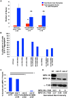
Figure 6. Apoptosis and MPK-1 expression are altered in csn mutants.
A) Quantification of the number of nuclei with CED-1::GFP present in late pachytene of the C. elegans gonad. Red bars represent the total number of apoptotic nuclei in the late pachytene region. The blue bars represent the total number of nuclei in the late pachytene region. Apoptosis is increased in csn-2 mutants, but not in csn-5 mutants, *p MW<0.0005. There is also a reduction of overall nuclei in the late pachytene region of the gonad in both csn mutants, **p MW<0.0005. wild-type n = 25 gonads; csn-2 n = 19; and csn-5, n = 18, B) Analysis of SYP-1 aggregate phenotype in csn mutants and apoptosis checkpoint double mutants. Bypassing the apoptotic checkpoints reduces the number of nuclei with aggregates. Total number of nuclei counted and p-values can be found in Sup. Table 5. C) Quantification of dpMPK-1 expression via IF analyses. csn mutants lack MPK-1 staining in late pachytene and in diakinesis, *p FET<0.0005. wild-type n = 75, csn-2 n = 32, csn-5, n = 27, syp-1(me17) n = 32, D) Western Blot confirming the lack of expression of MPK-1B in csn mutants. MPK-1A is mostly somatic and MPK-1B is germline specific. Normalization values (α-MPK-1/α-TUB) shown are the average of 3 different experiments. Normalized intensities: wild-type 2.27±1.03, csn-2 0.96±0.42 and csn-5 0.99±0.09.
Figure 7
Figure 7. csn mutant genetically interact with the ubiquitination-neddylation pathway in the regulation of SC assembly and recombination.
A–D) Quantification of SYP-1 aggregates. A and C are data from all gonad, while B and D is from late pachytene nuclei. Percent of nuclei with: no SYP-1 (black), linear SYP-1 (blue), aggregated SYP-1 (purple pink and red) and other (yellow), zones as in Figure 2A, n nuclei scored for whole gonad csn-2: with pL4440 = 2023 with ned-8(RNAi) = 430, with uba-1(RNAi) = 1121, csn-5: with pL4440 = 2096, with ned-8(RNAi) = 441, with uba-1(RNAi) = 1014. E) Quantitative analysis of COSA-1 foci in late pachytene wild-type: Percent of nuclei with: zero (black) one (orange), two (red), three (pink) four (magenta), five (purple), six (blue) and seven (gray), n nuclei scored for pL4440 = 57, with ned-8(RNAi) = 47, with uba-1(RNAi) = 25, csn-2: with pL4440 = 168, with ned-8(RNAi) = 66, with uba-1(RNAi) = 126, csn-5: with pL4440 = 152, with ned-8(RNAi) = 47 with, uba-1(RNAi) = 83, pL4440 = empty vector control vs. ned-8(RNAi) on csn-2 or csn-5 p = 0.002, p<0.001, Fisher's Exact Test, pL4440 vs. uba-1(RNAi) on csn-2 or csn-5 p<0.001 Mann Whitney Test). F–H) Quantification of SYP-1 aggregates in zones of the gonad for the indicated genotypes, as performed in figure, number of total nuclei scored: wild-type 25 n = 641, rfl-1 at 25 n = 1674, cul-4 n = 542 2, I) Schematic representation of the pathway examined in the experiment.

References

    1. Hassold T, HUNT P (2001) To err (meiotically) is human: the genesis of human aneuploidy. Nature Reviews Genetics 2: 280–291 10.1038/35066065 - DOI - PubMed
    1. Hirose Y, Suzuki R, Ohba T, Hinohara Y, Matsuhara H, et al. (2011) Chiasmata Promote Monopolar Attachment of Sister Chromatids and Their Co-Segregation toward the Proper Pole during Meiosis I. PLoS Genet 7: e1001329 10.1371/journal.pgen.1001329.s010 - DOI - PMC - PubMed
    1. Page SL, Hawley RS (2004) the genetics and molecular biology of the synaptonemal complex. Annu Rev Cell Dev Biol 20: 525–558 10.1146/annurev.cellbio.19.111301.155141 - DOI - PubMed
    1. Colaiácovo MP, MacQueen AJ, Martinez-Perez E, McDonald K, Adamo A, et al. (2003) Synaptonemal Complex Assembly in C. elegans Is Dispensable for Loading Strand-Exchange Proteins but Critical for Proper Completion of Recombination. Developmental Cell 5: 463–474 10.1016/S1534-5807(03)00232-6 - DOI - PubMed
    1. Zetka M, Kawasaki I, Strome S (1999) Synapsis and chiasma formation in Caenorhabditis elegans require HIM-3, a meiotic chromosome core component that functions in chromosome segregation. Genes & … - PMC - PubMed

Publication types

Substances

LinkOut - more resources