. 2014 Nov 6;5(11):e1512.
doi: 10.1038/cddis.2014.470.

IFN-γ induces aberrant CD49b⁺ NK cell recruitment through regulating CX3CL1: a novel mechanism by which IFN-γ provokes pregnancy failure

Affiliations

IFN-γ induces aberrant CD49b⁺ NK cell recruitment through regulating CX3CL1: a novel mechanism by which IFN-γ provokes pregnancy failure

Z-Y Li et al. Cell Death Dis. .

Abstract

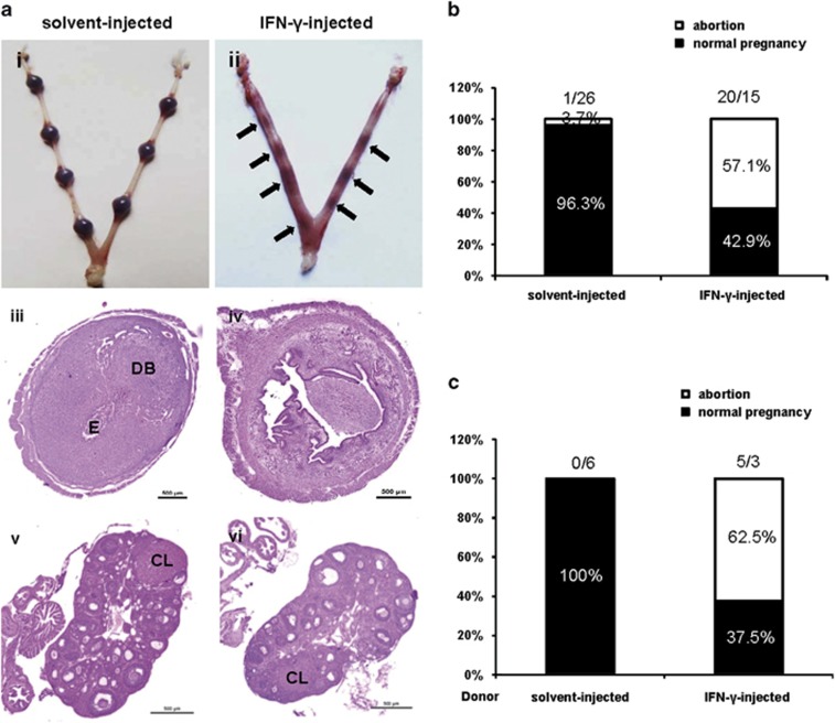
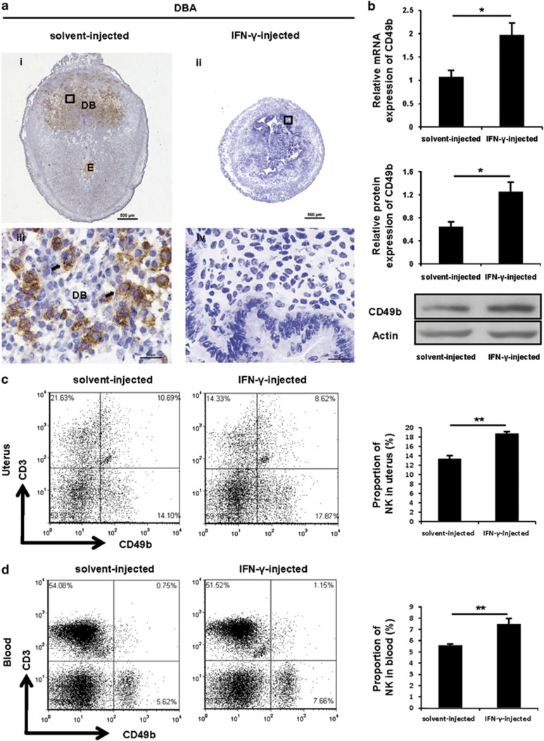
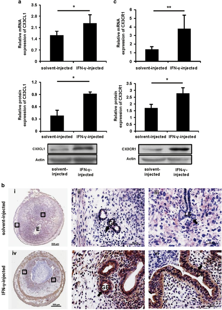
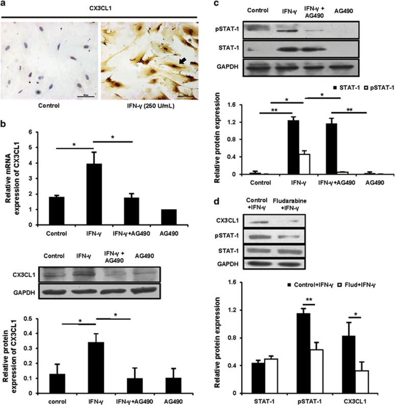
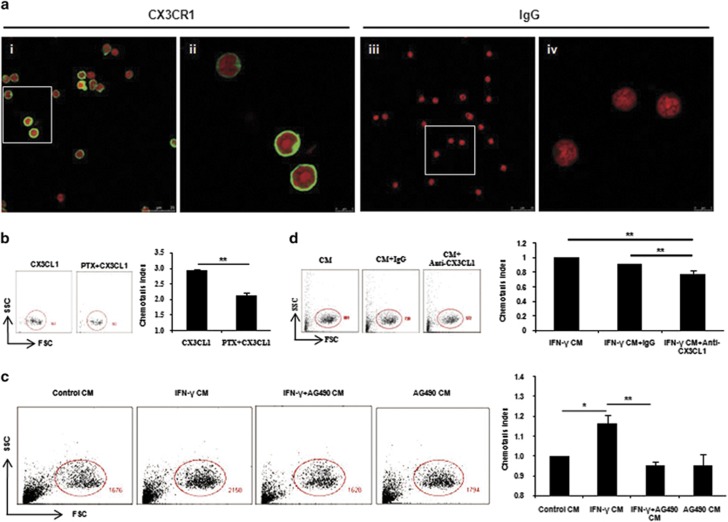
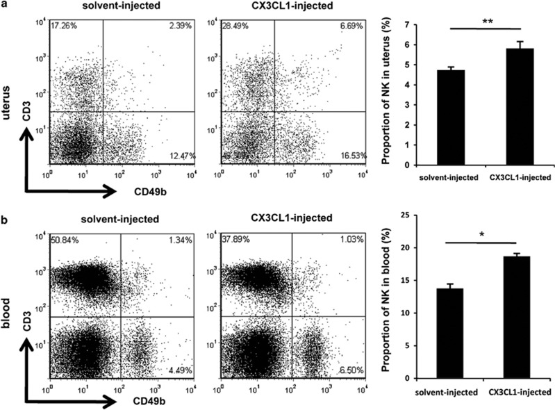
Interferon-γ (IFN-γ), a pleiotropic lymphokine, has important regulatory effects on many cell types. Although IFN-γ is essential for the initiation of uterine vascular modifications and maintenance of decidual integrity, IFN-γ administration can also cause pregnancy failure in many species. However, little is known about the effector mechanisms involved. In this study, using an IFN-γ-induced abortion mouse model, we reported that no Dolichos biflorus agglutinin lectin-positive uterine natural killer (uNK) cells were observed in the uteri from IFN-γ-induced abortion mice. By contrast, the percentage of CD3(-)CD49b(+) NK cells in the uterus and blood from a foetal resorption group was significantly higher than that of the control group. Similarly, significantly upregulated expression of CD49b (a pan-NK cell marker), CX3CL1 and CX3CR1 (CX3CL1 receptor) was detected in the uteri of IFN-γ-induced abortion mice. Using isolated uterine stromal cells, we showed that upregulated expression of CX3CL1 by IFN-γ was dependent on a Janus family kinase 2-signal transducers and activators of transcription 1 (JAK2-STAT1) pathway. We further demonstrated the chemotactic activity of CX3CL1 in uterine stromal cell conditioned medium on primary splenic NK cells. Finally, we observed increased recruitment of CD49b(+) NK cells into the endometrium after exogenous CX3CL1 administration. Collectively, our findings indicate that IFN-γ can significantly increase uterine CX3CL1 expression via activation of the JAK2-STAT1 pathway, thus inducing CD49b(+) NK cell uterine homing, and eventually provoke foetal loss. Thus, we provide a new line of evidence correlating the deleterious effects of IFN-γ on pregnancy with the aberrant regulation of CX3CL1 and CD49b(+) NK cells.


Figures

Figure 1
Figure 1
IFN-γ administration resulted in foetal resorption. Syngeneically mated BALB/c female mice were injected with solvent or IFN-γ intraperitoneally on GD6 and killed on GD8. (a) Representative macroscopic views of a healthy uterine horn from a solvent-injected mouse (i) and an aborted uterine horn from an IFN-γ-injected mouse (ii) are shown. H&E staining of uterine (iii, iv) and ovarian (v, vi) paraffin sections from solvent-injected mice and IFN-γ-injected mice are shown. Arrows indicate the corpus luteum. Photomicrographs are representative of at least three mice in each group. Scale bar: 500 μm. (b) Ratio of foetal abortions induced by IFN-γ is shown. The numbers above the bars indicate the number of mice with abortion/normal pregnancy. The ratio of foetal abortions was calculated from the following formula: (no. of abortion/no. of abortion plus no. of normal pregnancy) × 100%. ***P<0.001 by χ2. (c) Splenic cells of placebo-treated or IFN-γ-induced abortion mice after erythrocyte lysis were transferred into syngeneically mated BALB/c mice on GD6, and mice were killed 2 days posttransfer. The ratio of foetal abortions is shown, and the numbers above the bars indicate the number of mice with abortion/normal pregnancy. The ratio of foetal abortions was calculated as above. CL, corpus luteum; DB, decidua basalis; E, embryo; no., number
Figure 2
Figure 2
IFN-γ treatment enhanced the accumulation of the CD49b+ NK cell subset. Syngeneically mated BALB/c female mice were injected with solvent or IFN-γ intraperitoneally on GD6 and killed on GD8. (a) Analysis of DBA lectin-stained uNK cells in the uteri by immunohistochemistry. Arrows indicate DBA lectin-positive cells. Photomicrographs are representative of five mice in each group. Panels iii and iv are higher magnifications of areas marked by the black rectangles in panels i and ii, respectively. Scale bar: 500 μm (i and ii) and 25 μm (iii and iv). (b) CD49b expression in uteri was analysed by quantitative PCR (top panel) and western blotting (bottom panel). Data show the mean±S.E.M. of four independent experiments and are obtained from four mice of each group, respectively. *P<0.05 by independent samples T-test. Flow cytometric analysis of cell suspensions from uteri (c) and peripheral blood (d). See Supplementary Figures 1B and C for the gating strategy and the percentages of CD3CD49b+ NK cells (lower-right quadrant). Data show the mean±S.E.M. of four (uteri) or five (blood) independent experiments and are obtained from four (uteri) or five (blood) mice of each group, respectively. **P<0.01 by independent samples T-test. DB, decidua basalis; E, embryo
Figure 3
Figure 3
IFN-γ significantly increased uterine CX3CL1 and CX3CR1 expression. Syngeneically mated BALB/c female mice were injected with solvent or IFN-γ intraperitoneally on GD6 and killed on GD8. (a) CX3CL1 expression was analysed by quantitative PCR (top panel) and western blotting (bottom panel) in uteri. Data show the mean±S.E.M. of four independent experiments and are obtained from four mice of each group, respectively. *P<0.05 by independent samples T-test. (b) CX3CL1 expression was analysed by immunohistochemistry in uteri. Arrows indicate that stronger staining is observed in the LE and GE of the uteri from IFN-γ-injected mice. Photomicrographs are representative of three mice in each group. Panels ii, iii and v, vi are higher magnifications of different areas marked by the black rectangles in panels i and iv, respectively. Scale bar: 500 μm (i and iv) and 50 μm (ii, iii, v and vi). (c) CX3CR1 expression was analysed by quantitative PCR (top panel) and western blotting (bottom panel) in uteri. Data show the mean±S.E.M. of five (quantitative PCR) or three (western blotting) independent experiments and are obtained from five (quantitative PCR) or three (western blotting) mice of each group, respectively. *P<0.05 and **P<0.01 by independent samples T-test. E, embryo; GE, glandular epithelium; LE, luminal epithelium
Figure 4
Figure 4
IFN-γ upregulated CX3CL1 via a JAK2-STAT1 pathway in uterine stromal cells. (a) The isolated uterine stromal cells were treated with or without IFN-γ at a dose of 250 U/ml for 12 h, and CX3CL1 protein expression was analysed by immunocytochemical staining. The arrow indicates that CX3CL1 protein expression is markedly induced in response to IFN-γ. Scale bar: 100 μm. (b) Uterine stromal cells were pretreated with AG490 at 10 μM for 2 h before IFN-γ treatment, and then CX3CL1 expression was analysed by quantitative PCR (top panel) and western blotting (bottom panel). Data show the mean±S.E.M. of three independent experiments, respectively. *P<0.05 by one-way analysis of variance (ANOVA). (c) The treatment was the same as described in (b). The STAT1 and pSTAT1 were analysed by western blotting and normalized to GAPDH and STAT1, respectively. Data show the mean±S.E.M. of three independent experiments. *P<0.05 and **P<0.01 by one-way ANOVA. (d) Uterine stromal cells were pretreated with fludarabine at 100 μM for 2 h before IFN-γ treatment, and then CX3CL1, pSTAT1 and STAT1 were analysed by western blotting. CX3CL1, STAT1 and pSTAT1 were normalized to GAPDH, GAPDH and STAT1, respectively. Data show the mean±S.E.M. of three independent experiments. *P<0.05 and **P<0.01 by independent samples T-test
Figure 5
Figure 5
CX3CL1 facilitated peripheral NK cell migration. (a) Isolated splenic CD3CD49b+ NK cells were spun onto slides, stained for CX3CR1 (green) or isotype IgG and counterstained with PI (red). Panels ii and vi show higher magnifications of the areas marked by the white rectangles in panels i and iii, respectively. Original magnification, × 63/1.40 (oil), zoom 1.00 (i and iii) and zoom 4.00 (ii and vi). Data shown are representative of two independent experiments from two mice. (b) To distinguish between chemotaxis and chemokinesis, NK cells were preincubated overnight with or without 500 ng/ml PTX before measuring cell migration. NK cells were gated on the basis of forward scatter and side scatter (FSC-SSC) and the numbers of NK cells were shown. Data show the mean±S.E.M. of three independent experiments. **P<0.01 by independent samples T-test. (c) After 12 h in culture, stromal cell CM (termed control CM), IFN-γ-treated stromal cell CM (termed IFN-γ CM), AG490-preincubated stromal cells before IFN-γ treatment CM (termed IFN-γ+AG490 CM) and AG490-treated stromal cell CM (termed AG490 CM) were collected and NK cell migration in response to them was measured as described above. For comparison, the chemotaxis index of NK cells toward control CM was set at 1. Data show the mean±S.E.M. of three independent experiments. *P<0.05 and **P<0.01 by one-way analysis of variance (ANOVA). (d) Five μg/ml anti-CX3CL1-neutralizing mAb or control rat IgG was added to the NK cell suspension and the migration of NK cells toward IFN-γ CM was measured as described above. The chemotaxis index of NK cells without any treatment toward IFN-γ CM was set at 1. Data show the mean±S.E.M. of three independent experiments. **P<0.01 by one-way ANOVA
Figure 6
Figure 6
CX3CL1 facilitated CD49b+ NK cell migration in vivo. Homozygously mated BALB/c female mice were injected intraperitoneally with 1 μg of CX3CL1 or placebo on GD6 and killed on GD8. Flow cytometric analysis of cell suspensions from the uteri (a) and peripheral blood (b). See Supplementary Figures 1B and C for the gating strategy and the percentages of CD3CD49b+ NK cells (lower-right quadrant). Data show the mean±S.E.M. of three independent experiments and are obtained from three mice of each group, respectively. *P<0.05 and **P<0.01 by independent samples T-test

References

    1. Murphy SP, Tayade C, Ashkar AA, Hatta K, Zhang J, Croy BA. Interferon gamma in successful pregnancies. Biol Reprod. 2009;80:848–859. - PMC - PubMed
    1. Liu Z, Chen Y, Yang Y, Peng JP. The effect on MHC class II expression and apoptosis in placenta by IFNgamma administration. Contraception. 2002;65:177–184. - PubMed
    1. Chaouat G, Menu E, Clark DA, Dy M, Minkowski M, Wegmann TG. Control of fetal survival in CBAxDBA/2 mice by lymphokine therapy. J Reprod Fertil. 1990;89:447–458. - PubMed
    1. Sun QH, Peng JP, Xia HF, Yang Y, Liu ML. Effect on expression of RT1-A and RT1-DM molecules of treatment with interferon-gamma at the maternal–fetal interface of pregnant rats. Hum Reprod. 2005;20:2639–2647. - PubMed
    1. Liu Z, Sun QH, Yang Y, Liu JM, Peng JP. Effect of IFNgamma on caspase-3, Bcl-2 and Bax expression, and apoptosis in rabbit placenta. Cytokine. 2003;24:201–209. - PubMed

Publication types

MeSH terms