. 2014 Nov 6;9(11):e111762.
doi: 10.1371/journal.pone.0111762. eCollection 2014.

Efficient uptake of blood-borne BK and JC polyomavirus-like particles in endothelial cells of liver sinusoids and renal vasa recta

Affiliations

Efficient uptake of blood-borne BK and JC polyomavirus-like particles in endothelial cells of liver sinusoids and renal vasa recta

Jaione Simon-Santamaria et al. PLoS One. .

Abstract

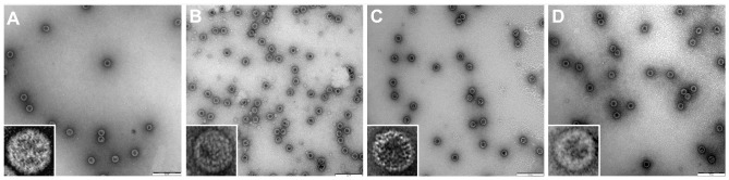
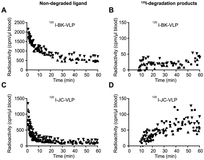
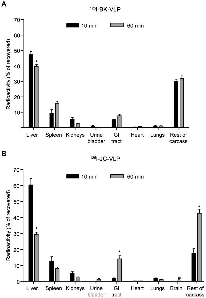
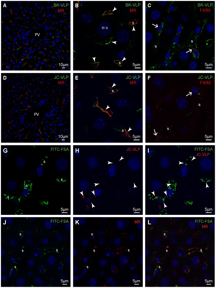
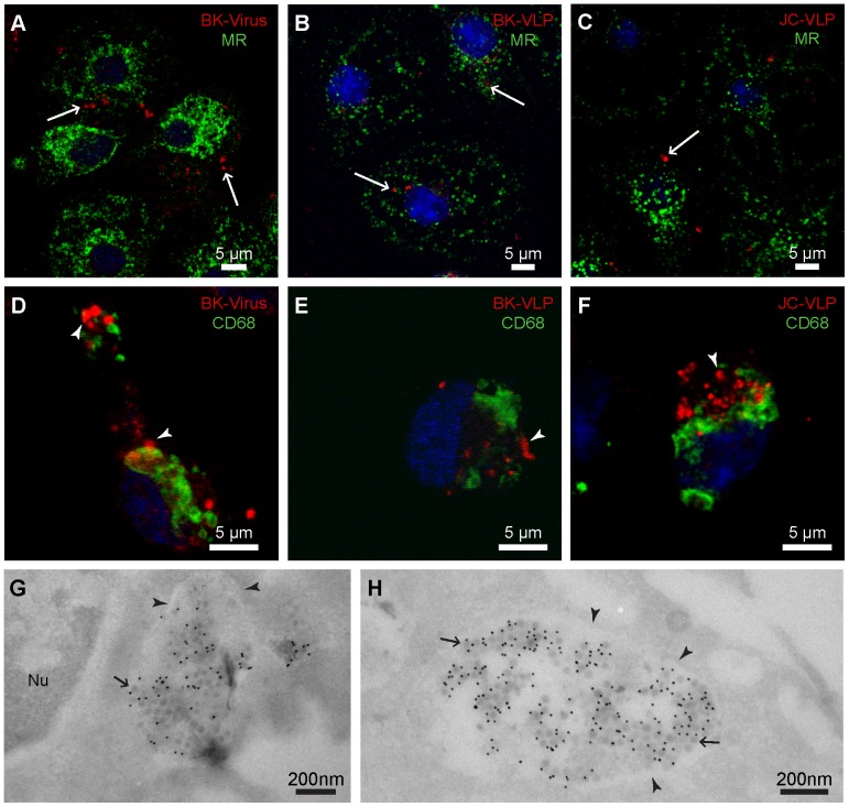
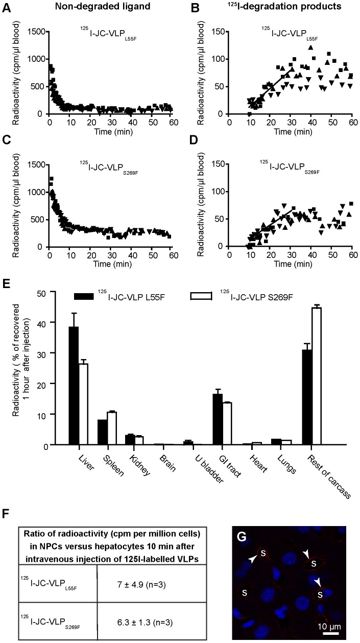
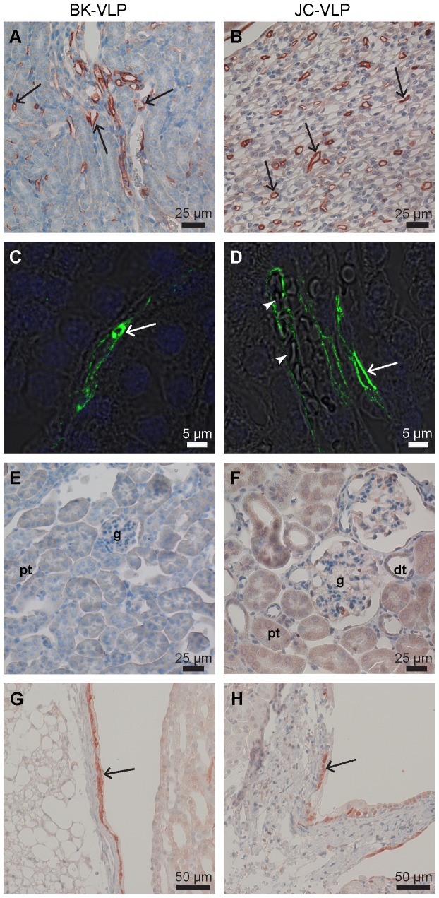
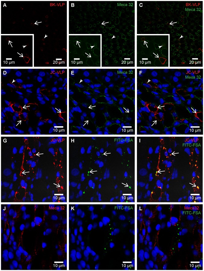
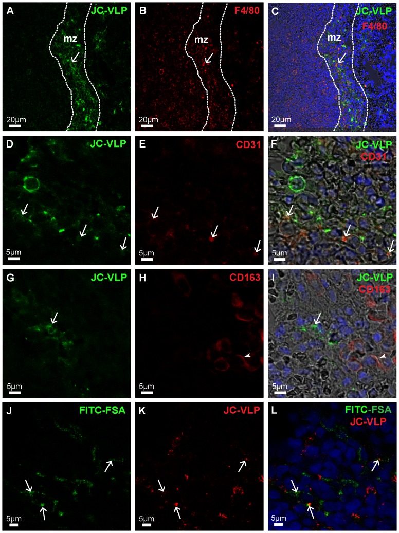
Liver sinusoidal endothelial cells (LSECs) are specialized scavenger cells that mediate high-capacity clearance of soluble waste macromolecules and colloid material, including blood-borne adenovirus. To explore if LSECs function as a sink for other viruses in blood, we studied the fate of virus-like particles (VLPs) of two ubiquitous human DNA viruses, BK and JC polyomavirus, in mice. Like complete virions, VLPs specifically bind to receptors and enter cells, but unlike complete virions, they cannot replicate. 125I-labeled VLPs were used to assess blood decay, organ-, and hepatocellular distribution of ligand, and non-labeled VLPs to examine cellular uptake by immunohisto- and -cytochemistry. BK- and JC-VLPs rapidly distributed to liver, with lesser uptake in kidney and spleen. Liver uptake was predominantly in LSECs. Blood half-life (∼1 min), and tissue distribution of JC-VLPs and two JC-VLP-mutants (L55F and S269F) that lack sialic acid binding affinity, were similar, indicating involvement of non-sialic acid receptors in cellular uptake. Liver uptake was not mediated by scavenger receptors. In spleen, the VLPs localized to the red pulp marginal zone reticuloendothelium, and in kidney to the endothelial lining of vasa recta segments, and the transitional epithelium of renal pelvis. Most VLP-positive vessels in renal medulla did not express PV-1/Meca 32, suggesting location to the non-fenestrated part of vasa recta. The endothelial cells of these vessels also efficiently endocytosed a scavenger receptor ligand, formaldehyde-denatured albumin, suggesting high endocytic activity compared to other renal endothelia. We conclude that LSECs very effectively cleared a large fraction of blood-borne BK- and JC-VLPs, indicating a central role of these cells in early removal of polyomavirus from the circulation. In addition, we report the novel finding that a subpopulation of endothelial cells in kidney, the main organ of polyomavirus persistence, showed selective and rapid uptake of VLPs, suggesting a role in viremic organ tropism.


Conflict of interest statement

Competing Interests: The authors have declared that no competing interests exist.

Figures

Figure 1
Figure 1. Electron microscopy of BK- and JC-virus like particles (VLPs).
The figure show transmission electron micrographs of negatively stained BK- and JC-VLPs constructed from BK-VP1 or JC-VP1 proteins: A) BK-VP1 VLPs (Dunlop strain); B) JC-VP1-Mad-1 VLPs; C) JC-VP1-L55F VLPs; D) JC-VP1-S269F VLPs. Scale bars: 200 nm.
Figure 2
Figure 2. Blood clearance and subsequent in vivo degradation of BK- and JC-virus like particles (VLPs).
Mice were injected intravenously with approximately 0.5 µg of 125I-BK-VLPs (n = 3; A, B), or 125I-JC-VLPs (n = 5; C, D), and blood samples taken at the indicated time points and analyzed for 125I-labeled degradation products and intact ligand . Panels A and C show decrease of intact ligand in blood as a function of time. Panels B and D show increase in degradation products released into the blood with time. Results are given as cpm per µl blood. Different symbols refer to separate animals. cpm, counts per minute. The slope of line drawn in panel D represents the average rate of release of degradation products in the 10–30 min period after ligand injection where this release followed approximate first order kinetics.
Figure 3
Figure 3. Anatomical distribution of 125I-labelled BK- and JC-VLPs.
Mice were injected intravenously with 125I-labelled BK-VLPs (A) or JC-VLPs (B), euthanized after 10 min (black bars; n = 3) or 60 min (grey bars; n = 3 in A; n = 5 in B), and organs and tissues analyzed for radioactivity. The mice used in the circulatory half-life study (Figure 2) were included in this anatomical distribution study (60 min values). Total recovered radioactivity in all tissues at the given time points was taken as 100%. Error bars represent SEM. *Statistically significant difference (p<0.01) between tissue radioactivity at 10 min versus 60 min. # Brain radioactivity was measured at 60 min, only. GI tract, gastrointestinal tract (stomach and intestines including mesenterium).
Figure 4
Figure 4. Liver cell distribution of BK- and JC-VLPs after uptake from blood.
Panels A–F: Mouse livers were perfusion fixed 15 min after intravenous injection of BK-VLPs (A–C), or JC-VLPs (D–F). Paraffin embedded tissue sections were double immune labeled using a rabbit antiserum against BK-VP1 , and antibodies against the mannose receptor (MR), which is an LSEC marker in mouse liver , or F4/80 (Kupffer cell marker). Antibodies are listed in Table 1. The VP1 staining (green), indicative of BK-/JC-VLP uptake, showed a typical LSEC pattern (A, D, overview; B, E, close ups), as evidenced by similar localization of green VP1 and red MR staining along the sinusoids (arrowheads in B, E; overlap of red and green fluorescence is shown in yellow). Some BK- and JC-VLP uptake was also observed in F4/80 positive (red) Kupffer cells (arrows in C, F). PV, portal vein; S, sinusoid; Hn, nucleus of hepatocyte. Panels G–L: In G–L, FITC-labeled formaldehyde-denatured serum albumin (FITC-FSA) was injected into the tail vein 5 min after intravenous injection of JC-VLPs to functionally label the endothelium of the liver sinusoids , . Ten min thereafter the animals were euthanized, tissues perfusion fixed, paraffin embedded and prepared for immune histochemistry. Panels G-I show the distribution of FITC-FSA (green) and JC-VLP (red, arrowheads) in liver sinusoids (s). Panels J–L show similar distribution of FITC-FSA (green) and MR expression (red) in the sinusoids, s.
Figure 5
Figure 5. In vitro endocytosis of BK- and JC-VLPs, and BK virions in liver sinusoidal cells.
Freshly isolated non-parenchymal liver cells (mixed LSEC and Kupffer cell cultures) were incubated at 37°C for 30 min with BK virions (A, D), or for 1 h with 10 µg/ml BK-VLPs (B, E), or JC-VLPs (C, F), fixed, and double immune labeled with a rabbit anti-BK-VP1 antiserum (red fluorescence) and antibodies against the mannose receptor (MR; LSEC marker; shown in green), or CD68 (Kupffer cell marker; green). Antibodies are listed in Table 1. Uptake of BK virions and BK-/JC-VLPs was observed both in isolated LSECs (arrows, A–C) and Kupffer cells (arrow heads, D–F). Panels G–H: Cryo-immune electron microscopy of BK virion uptake in LSEC membrane-bound vesicles. Purified LSEC cultures were incubated with BK virions for 2 h at 37°C. Positive BK-VP1 staining was visualized by protein-A-10 nm gold particles (black dots). Arrowheads points to vesicular membranes, and arrows to virus particles. Nu, nucleus.
Figure 6
Figure 6. Blood clearance and organ distribution of mutant JC-VLPs.
Mice were injected intravenously with 125I-JC-VLPL55F (n = 3, A–B), or 125I-JC-VLPS269F (n = 3, C–D), and blood samples taken at the indicated time points and analyzed for 125I-labeled degradation products and intact ligand . Panels A and C show the decrease of intact ligand in blood as a function of time. Panels B and D show the increase in degradation products released into the blood with time. Different symbols refer to separate animals, and cpm to counts per minute. The slopes of the lines drawn in B and D represent the average release rate of degradation products in the 10–30 min period after ligand injection where this release followed approximate first order kinetics. E) Tissue distribution 60 min post injection. The animals in A–D were sacrificed after 60 min, and organs and tissues analyzed for radioactivity. Recovered radioactivity in all tissues at this time point was taken as 100%. Error bars represent SEM. GI tract, gastrointestinal tract (stomach and intestines including mesenterium); U bladder, urine bladder. F) Hepatocellular distribution (±SD) of 125I-labeled JC-VLP mutants 10 min after intravenous injection; liver tissue was dispersed by collagenase perfusion, and radioactivity was measured in hepatocyte and non-parenchymal cell (NPC) fractions, respectively. G) Cellular distribution of JC-VLPS269F (anti-VP1 staining) 15 min after VLP injection. Uptake of JC-VLPS269F (red fluorescence; arrowheads) is seen along the sinusoids (s).
Figure 7
Figure 7. Kidney distribution of intravenously administered BK- and JC-VLPs.
Mouse tissues were perfusion fixed 7 or 15 min after injection of BK-VLPs (A, C, E, G), or JC-VLPs (B, D, F, H). Paraffin sections were labeled using a rabbit anti-BK-VP1 antiserum . VP1-labeling was visualized using HRP polymer/DAB reaction (brown color), or Alexa488-goat-anti-rabbit (green fluorescence). Specific uptake of VLPs was seen in the endothelial lining of microvessels in kidney medulla (arrows in A–D; with arrowheads in D pointing to blood filled medullary capillaries), whereas glomeruli (E, F), and tubular structures were negative (A–F). Epithelial cells in the transitional epithelium of renal pelvis also showed positive VP1 staining (arrows in G, H). g, glomerulus; pt, proximal tubulus; dt, distal tubulus.
Figure 8
Figure 8. Polyoma VLP and FITC-FSA uptake in renal medullary endothelial cells.
Panels A–F: Mouse tissues were perfusion fixed 7 min after intravenous injection of BK-VLP (A–C), or JC-VLP (D–F), and paraffin sections double immune labeled with anti-BK-VP1 (red) and anti-Meca 32 (green). Panels A–C: Arrows indicate endothelial uptake of BK-VLPs in Meca 32 negative vessels, and arrow heads to BK-VLP uptake in Meca 32 positive vessels. Panels D–F: Arrows indicate endothelial uptake of JC-VLPs in Meca 32 negative vessels, and arrow heads to JC-VLP uptake in Meca 32 positive vessels. Most BK-VLP or JC-VLP-positive vessels were Meca 32 negative or showed only weak Meca32 staining. Panels G–L: Tail vein injection of JC-VLPs was followed by injection of the scavenger receptor ligand FITC-FSA in the opposite tail vein and tissues perfusion fixed 15 min after the VLP administration. Paraffin sections were immune labeled with either anti-BK-VP1 (red; G–I) or anti-Meca 32 (red; J–L). G–I) The JC-VLP uptake in medullary vessel endothelia totally overlapped with FITC-FSA uptake (arrows). J–L) FITC-FSA uptake occurred mainly in Meca 32 negative vessels.
Figure 9
Figure 9. JC-VLP uptake in spleen.
Panels A–I: Mouse tissues were perfusion fixed 7 min after intravenous injection of JC-VLPs. Paraffin sections of spleen were double immune labeled using a rabbit antiserum against BK-VP1 (reacts also with JC-VP1), and antibodies against F4/80 (A–C), CD31 (D–F) or CD163 (G–I). Antibodies are listed in Table 1. The VP1 staining pattern (green) showed that the uptake of VLPs was concentrated in the red pulp marginal zone, mz (A), and here partly co-localized with F4/80 (C, arrow), and CD31 (F, arrows), but not with CD163 (I; arrow indicates VP1, and arrow head to CD163 staining). The F4/80 (B, C), CD31 (E, F), and CD163 (H, I) staining patterns are all shown in red. Panels J–L: In J–L, tail vein injection of JC-VLPs was followed by injection of FITC-FSA in the opposite tail vein and tissues perfusion fixed 15 min after the VLP administration. Both JC-VLP (K; red fluorescence) and FITC-FSA (J; green fluorescence) distributed to the reticuloendothelial network of the spleen red pulp marginal zone (L; arrows point to overlap of JC-VLP staining and FITC-FSA uptake).

References

    1. Virgin HW (2014) The Virome in Mammalian Physiology and Disease. Cell 157: 142–150. - PMC - PubMed
    1. Zhang L, Dailey PJ, He T, Gettie A, Bonhoeffer S, et al. (1999) Rapid clearance of simian immunodeficiency virus particles from plasma of rhesus macaques. J Virol 73: 855–860. - PMC - PubMed
    1. Mims CA (1959) The response of mice to large intravenous injections of ectromelia virus. II. The growth of virus in the liver. Br J Exp Pathol 40: 543–550. - PMC - PubMed
    1. Alemany R, Suzuki K, Curiel DT (2000) Blood clearance rates of adenovirus type 5 in mice. J Gen Virol 81: 2605–2609. - PubMed
    1. Inchley CJ (1969) The activity of mouse Kupffer cells following intravenous injection of T4 bacteriophage. Clin Exp Immunol 5: 173–187. - PMC - PubMed

Publication types

Substances

LinkOut - more resources