Is 3-Tesla Gd-EOB-DTPA-enhanced MRI with diffusion-weighted imaging superior to 64-slice contrast-enhanced CT for the diagnosis of hepatocellular carcinoma?
- PMID: 25375778
- PMCID: PMC4223069
- DOI: 10.1371/journal.pone.0111935
Is 3-Tesla Gd-EOB-DTPA-enhanced MRI with diffusion-weighted imaging superior to 64-slice contrast-enhanced CT for the diagnosis of hepatocellular carcinoma?
Abstract
Objectives: To compare 64-slice contrast-enhanced computed tomography (CT) with 3-Tesla magnetic resonance imaging (MRI) using Gd-EOB-DTPA for the diagnosis of hepatocellular carcinoma (HCC) and evaluate the utility of diffusion-weighted imaging (DWI) in this setting.
Methods: 3-phase-liver-CT was performed in fifty patients (42 male, 8 female) with suspected or proven HCC. The patients were subjected to a 3-Tesla-MRI-examination with Gd-EOB-DTPA and diffusion weighted imaging (DWI) at b-values of 0, 50 and 400 s/mm2. The apparent diffusion coefficient (ADC)-value was determined for each lesion detected in DWI. The histopathological report after resection or biopsy of a lesion served as the gold standard, and a surrogate of follow-up or complementary imaging techniques in combination with clinical and paraclinical parameters was used in unresected lesions. Diagnostic accuracy, sensitivity, specificity, and positive and negative predictive values were evaluated for each technique.
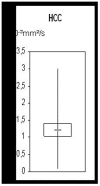
Results: MRI detected slightly more lesions that were considered suspicious for HCC per patient compared to CT (2.7 versus 2.3, respectively). ADC-measurements in HCC showed notably heterogeneous values with a median of 1.2±0.5×10⁻³ mm2/s (range from 0.07±0.1 to 3.0±0.1×10⁻³ mm2/s). MRI showed similar diagnostic accuracy, sensitivity, and positive and negative predictive values compared to CT (AUC 0.837, sensitivity 92%, PPV 80% and NPV 90% for MRI vs. AUC 0.798, sensitivity 85%, PPV 79% and NPV 82% for CT; not significant). Specificity was 75% for both techniques.
Conclusions: Our study did not show a statistically significant difference in detection in detection of HCC between MRI and CT. Gd-EOB-DTPA-enhanced MRI tended to detect more lesions per patient compared to contrast-enhanced CT; therefore, we would recommend this modality as the first-choice imaging method for the detection of HCC and therapeutic decisions. However, contrast-enhanced CT was not inferior in our study, so that it can be a useful image modality for follow-up examinations.
Conflict of interest statement
Figures


References
-
- Bruix J, Sherman M (2005) Management of hepatocellular carcinoma. Hepatology 42: 1208–36. - PubMed
-
- Kudo M (2010) The 2008 Okuda lecture: Management of hepatocellular carcinoma: from surveillance to molecular targeted therapy. J Gastroenterol Hepatol 25: 439–52. - PubMed
-
- El-Serag HB (2007) Epidemiology of hepatocellular carcinoma in USA. Hepatol Res 37: S88–94. - PubMed
-
- Tannapfel A, Wittekind C (2003) Pathology of hepatocellular carcinoma. Chir Gastroenterol 19: 225–230.
-
- Tanimoto A, Lee JM, Murakami T, Huppertz A, Kudo M, et al. (2009) Consensus report of the 2nd International Forum for Liver MRI. Eur Radiol 19: S975–89. - PubMed
Publication types
MeSH terms
Substances
LinkOut - more resources
Full Text Sources
Other Literature Sources
Medical

