

Comparison of optic disc morphology of optic nerve atrophy between compressive optic neuropathy and glaucomatous optic neuropathy
- PMID: 25375855
- PMCID: PMC4223062
- DOI: 10.1371/journal.pone.0112403
Comparison of optic disc morphology of optic nerve atrophy between compressive optic neuropathy and glaucomatous optic neuropathy
Abstract
Objectives: To compare the optic nerve head (ONH) structure between compressive optic neuropathy (CON) and glaucomatous optic neuropathy (GON), and to determine whether selected ONH quantitative parameters effectively discriminate between GON and CON, especially CON cases presenting with a glaucoma-like disc.
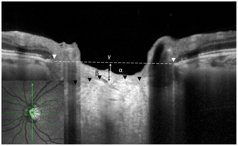
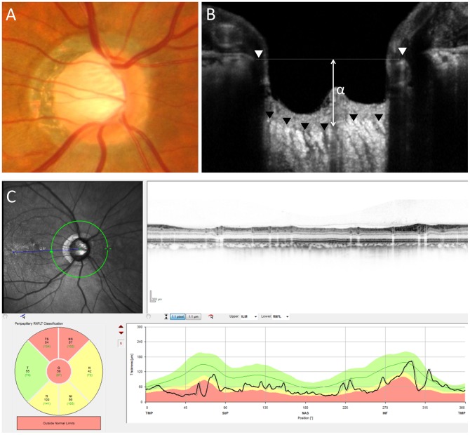
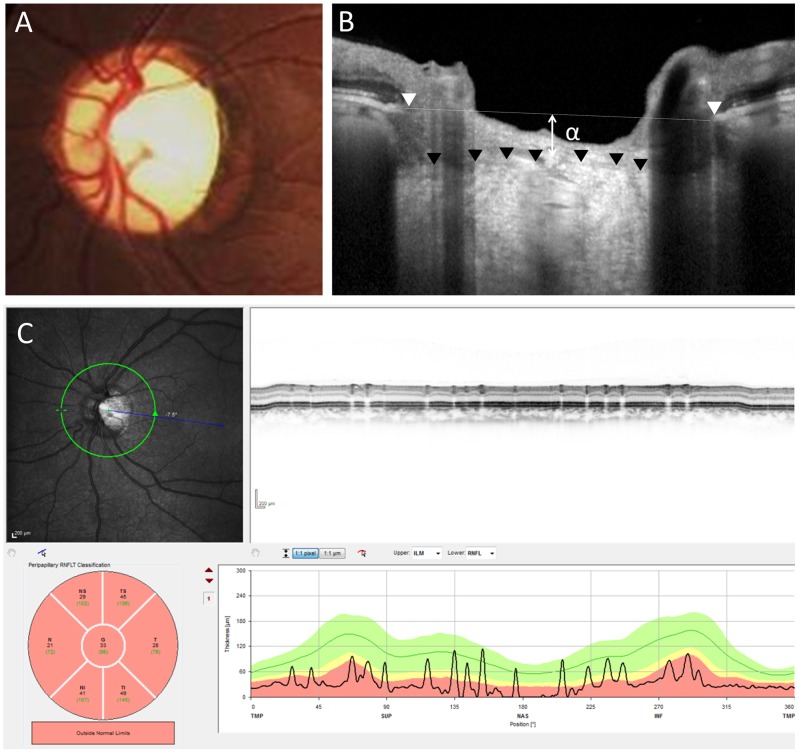
Methods: We prospectively assessed 34 patients with CON, 34 age-matched patients with moderate or severe GON, and 34 age-matched healthy control subjects. The quantitative parameters of ONH structure were compared using the Heidelberg Retina Tomograph 2 (HRT2) and Spectralis optical coherence tomography with an enhanced depth imaging method.
Results: The mean and maximum cup depths of CON were significantly smaller than those with GON (P < 0.001 and P < 0.001, respectively). The distance between Bruch's membrane opening and anterior surface of the lamina cribrosa (BMO-anterior LC) of CON was also significantly smaller than that of glaucoma but was similar to that of the healthy group (P < 0.001 and P = 0.47, respectively). Based on Moorfields regression analysis of the glaucoma classification of HRT2, 15 eyes with CON were classified with a glaucoma-like disc. The cup/disc area ratio did not differ between cases of CON with a glaucoma-like disc and cases of GON (P = 0.16), but the BMO-anterior LC and mean and maximum cup depths of CON cases with a glaucoma-like disc were smaller than those in GON (P = 0.005, P = 0.003, and P = 0.001, respectively).
Conclusions: Measurements of the cup depths and the LC depth had good ability to differentiate between CON with a glaucoma-like disc and glaucoma. There was no laminar remodeling detected by laminar surface position in the patients with CON compared to those with GON.
Conflict of interest statement
Figures
References
-
- Quigley H, Anderson DR (1977) Cupping of the optic disc in ischemic optic neuropathy. Trans Sect Ophthalmol Am Acad Ophthalmol Otolaryngol 83: 755–762. - PubMed
-
- Trobe JD, Glaser JS, Cassady J, Herschler J, Anderson DR (1980) Nonglaucomatous excavation of the optic disc. Arch Ophthalmol 98: 1046–1050. - PubMed
-
- Kupersmith MJ, Krohn D (1984) Cupping of the optic disc with compressive lesions of the anterior visual pathway. Ann Ophthalmol 16: 948–953. - PubMed
-
- Rebolleda G, Noval S, Contreras I, Arnalich-Montiel F, Garcia-Perez JL, et al. (2009) Optic disc cupping after optic neuritis evaluated with optic coherence tomography. Eye (Lond) 23: 890–894. - PubMed
-
- Fournier AV, Damji KF, Epstein DL, Pollock SC (2001) Disc excavation in dominant optic atrophy: differentiation from normal tension glaucoma. Ophthalmology 108: 1595–1602. - PubMed
Publication types
MeSH terms
LinkOut - more resources
Full Text Sources
Other Literature Sources
Medical
