. 2014 Nov 7:4:6962.
doi: 10.1038/srep06962.

PINK1-mediated phosphorylation of Miro inhibits synaptic growth and protects dopaminergic neurons in Drosophila

Affiliations

PINK1-mediated phosphorylation of Miro inhibits synaptic growth and protects dopaminergic neurons in Drosophila

Pei-I Tsai et al. Sci Rep. .

Abstract

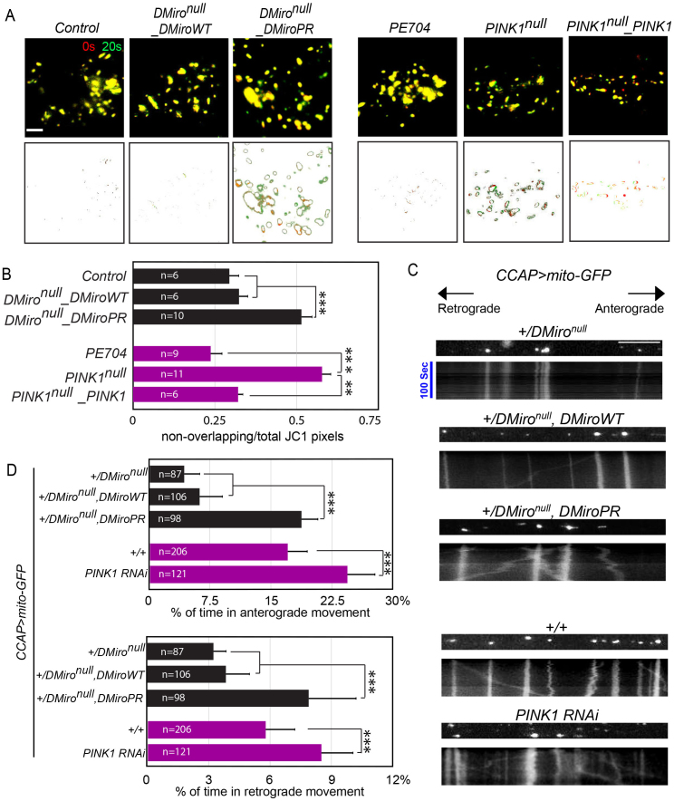
Mutations in the mitochondrial Ser/Thr kinase PINK1 cause Parkinson's disease. One of the substrates of PINK1 is the outer mitochondrial membrane protein Miro, which regulates mitochondrial transport. In this study, we uncovered novel physiological functions of PINK1-mediated phosphorylation of Miro, using Drosophila as a model. We replaced endogenous Drosophila Miro (DMiro) with transgenically expressed wildtype, or mutant DMiro predicted to resist PINK1-mediated phosphorylation. We found that the expression of phospho-resistant DMiro in a DMiro null mutant background phenocopied a subset of phenotypes of PINK1 null. Specifically, phospho-resistant DMiro increased mitochondrial movement and synaptic growth at larval neuromuscular junctions, and decreased the number of dopaminergic neurons in adult brains. Therefore, PINK1 may inhibit synaptic growth and protect dopaminergic neurons by phosphorylating DMiro. Furthermore, muscle degeneration, swollen mitochondria and locomotor defects found in PINK1 null flies were not observed in phospho-resistant DMiro flies. Thus, our study established an in vivo platform to define functional consequences of PINK1-mediated phosphorylation of its substrates.


Figures

Figure 1
Figure 1. Ser182, Ser324, and Thr325 of DMiro Mediate PINK1/Parkin-dependent Degradation.
(A) After transfection with wildtype or mutated forms of T7-DMiro and YFP-Parkin, HEK293T cell lysates were prepared and immunoprobed with anti-T7, anti-ATP5β and anti-GFP. The intensity of each T7-DMiro band was detected with a fluorescence scanner for quantification (B) after normalization to the mitochondrial matrix loading control ATP5β, and the control band without Parkin coexpression was set as 1. n = 5–25 transfections. The amounts of overexpressed YFP-Parkin did not significantly vary among different genotypes: P = 0.998 for normalized YFP-Parkin intensity to ATP5β (One-way ANOVA, n = 4 transfections). (C, D) HEK293T cells transfected with T7-DMiro were incubated with 10 μM CCCP for 1.5 hr, or 40 μM CCCP for 3 hr, prior to lysing the cells. Immunoblots of lysates were probed with anti-T7, and detected with a fluorescent scanner for quantification (D) after normalization to the mitochondrial loading control ATP5β and expressed as a fraction of the control value with no CCCP treatment in the same genotype. n = 4 transfections. 1–2 μg DNA of T7-DMiro and 2 μg of YFP-Parkin were expressed in (A, B), and 0.5–1 μg DNA of T7-DMiro was expressed in (C, D), in one well of a 6-well plate. (E) Lysates from 5 adult flies 5 days after eclosion were analyzed by immunoblotting as indicated. (F) The band intensity of T7-DMiro with PINK1 co-expression was normalized to that of ATP5β, and expressed as a fraction of the control value with mito-GFP co-expression. n = 3 independent experiments. Act > mito-GFP: UAS-mito-GFP;Actin-GAL4. Act > T7-DMiroWT&mito-GFP: UAS-mito-GFP,UAS-T7-DMirowildtype;Actin-GAL4. Act > T7-DMiroPR&mito-GFP: UAS-mito-GFP,UAS-T7-DMiroS182A,S324A,T325A;Actin-GAL4. Act > T7-DMiroWT&PINK1: UAS-PINK1,UAS-T7-DMirowildtype;Actin-GAL4. Act > T7-DMiroPR&PINK1: UAS-PINK1,UAS-T7-DMiroS182A,S324A,T325A;Actin-GAL4. * P < 0.05, ** P < 0.01, *** P < 0.001, error bars represent mean ± S.E.M. here and for all figures unless otherwise stated. Uncropped blots are in the supplementary figure.
Figure 2
Figure 2. Generation of a Fly Model Expressing DMiroS182A,S324A,T325A in a DMiro Null Background.
(A) Comparison of third instar larval sizes. (B) Lysates from 5 adult flies 5 days after eclosion were analyzed by immunoblotting as indicated. The band intensity recognized by anti-DMiro or anti-T7 was normalized to that of tubulin, and expressed as a percentage of the value of “DMironull_DMiroWT” and averaged. n = 6 independent experiments. (C) Quantification of muscle 4 size at hemisegment A2. n = 12 third instar larvae. (D) NMJ boutons visualized by anti-HRP (green) and anti-Futsch (red) at muscle 4 hemisegment A2 of third instar larvae. (E) Quantification of the number of Futsch-negative boutons at muscle 4 shown in (D). n = 12–16 larvae. Scale bar: 20 μm. Genotypes used in this figure and the subsequent figures: Control: Canton S. DMironull: DMirosd32/DMirosd26 . DMironull_DMiroWT: UAS-T7-DMirowildtype;da-GAL4_DMirosd32/DMirosd26. DMironull_DMiroPR: UAS-T7-DMiroS182A,S324A,T325A;da-GAL4_DMirosd32/DMirosd26. n.s.: not significant. Comparisons with Control except where otherwise indicated here and for all the following figures.
Figure 3
Figure 3. Both Wildtype DMiro and DMiroS182A,S324A,T325A Rescue Mitochondrial Phenotypes of DMironull.
(A) Representative single-section confocal images of JC1 and anti-HRP staining in distal axons close to NMJs passing segment A3 of third instar larvae, red representing accumulative JC1 in mitochondria and blue representing cytoplasmic diffuse JC1. The neuronal membrane marker HRP is in green. (B) Representative single-section confocal images of JC1 and anti-HRP staining at NMJs of muscle 4 hemisegment A3 of third instar larvae. The red mitochondrial JC1 staining in DMironull indicates mitochondria present in muscle cells. (C) Quantification of the ratio of JC1 red/blue fluorescent intensity in axons shown in (A). n = 6–10 larvae. The blue intensity was not significantly different among 4 genotypes (P = 0.2598, One-way ANOVA). (D) Quantification of total ATP levels in third instar larvae with indicated genotypes, expressed as a percentage of the control value. n = 5 larvae for each experiment and total 3 independent experiments. Scale bars: 10 μm.
Figure 4
Figure 4. DMiroS182A,S324A,T325A or Loss of PINK1 Increases Mitochondrial Movement.
(A) Representative red/green overlay of two time-lapse images (Δ = 20 s) of accumulative JC1 staining in mitochondria at NMJs of muscle 6/7 hemisegment A3 of third instar larvae. Lower panels extract pixels that are present in only one image. (B) Quantification of the area of non-overlapping pixels between the two overlay images as shown in (A) divided by the total area of JC1 staining in each overlay image. n = 6–11 larvae. (C) Mitochondrial movement labeled by mito-GFP driven by CCAP-GAL4 in representative axons passing segment A3. The first frame of each live-imaging series is shown above a kymograph generated from the movie. The x axis of each is mitochondrial position and the y axis corresponds to time (moving from top to bottom). Vertical white lines represent stationary mitochondria and diagonal lines are moving mitochondria. (D) From kymographs as in (C), the percent of time each mitochondrion in motion was determined and averaged. n = 87–206 mitochondria from 8–12 axons and 5 animals. Scale bars: (A) 5 μm; (C) 10 μm. PINK1null: PINK5/Y. PE704: precise excision control males for PINK1 null. PINK1null_PINK1: PINK5/Y;+;da-GAL4_UAS-PINK1 (males of a rescue control for PINK1 null).
Figure 5
Figure 5. DMiroS182A,S324A,T325A or Loss of PINK1 Causes Synaptic Overgrowth and DA Neurodegeneration.
(A) NMJ boutons visualized by anti-HRP at muscle 4 hemisegment A2 of third instar larvae. (B) Quantification of the type Ib bouton number normalized to the muscle size at muscle 4 hemisegment A2 shown in (A), or at muscle 6/7 hemisegment A2. n = 11–16 larvae. (C) The PPL1 clusters of DA neurons visualized by anti-TH in adult brains 15 days after eclosion. (D) Quantification of the number of DA neurons in one PPL1 or PPL2 cluster per brain of adult flies of 15 days after eclosion shown in (C), or of 5 days after eclosion. n = 10–18 brains. Scale bars: (A) 50 μm; (C) 5 μm.
Figure 6
Figure 6. Ser182, Ser324, and Thr325 of DMiro Do Not Mediate Locomotor and Flight Abilities in Larvae and Adult Flies.
Crawling ability (A), and idling time in 30 sec (B), of third instar larvae with different genotypes were quantified. n = 21–44. Climbing (C), jumping (D), and flying (E) abilities of adult flies 15 days after eclosion were quantified. n = 22–119.
Figure 7
Figure 7. DMiroS182A,S324A,T325A Does Not Cause Muscle Degeneration or Swollen Mitochondria.
(A) Light microscopic images of thick sections show indirect flight muscles, and (B) TEM images of thin sections show mitochondria inside muscle cells, performed on thoraces of adult flies as indicated 5 days after eclosion. (C) Schematic representation of the regulatory mechanisms by which PINK1 and Miro control cellular functions. Blue boxes in Miro indicate GTPase domains and green boxes are EF-hands. Scale bars: (A) 10 μm; (B) 0.5 μm.

Similar articles

Cited by

References

    1. Valente E. M. et al. Hereditary early-onset Parkinson's disease caused by mutations in PINK1. Science 304, 1158–1160, 10.1126/science.1096284 (2004). - PubMed
    1. Narendra D., Walker J. E. & Youle R. Mitochondrial quality control mediated by PINK1 and Parkin: links to parkinsonism. Cold Spring Harb. Perspect. Biol. 4, 10.1101/cshperspect.a011338 (2012). - PMC - PubMed
    1. Vives-Bauza C., de Vries R. L., Tocilescu M. & Przedborski S. PINK1/Parkin direct mitochondria to autophagy. Autophagy 6, 315–316 (2010). - PubMed
    1. Wang X. et al. PINK1 and Parkin target Miro for phosphorylation and degradation to arrest mitochondrial motility. Cell 147, 893–906, 10.1016/j.cell.2011.10.018 (2011). - PMC - PubMed
    1. Whitworth A. J. & Pallanck L. J. The PINK1/Parkin pathway: a mitochondrial quality control system? J. Bioenerg. Biomembr. 41, 499–503, 10.1007/s10863-009-9253-3 (2009). - PubMed

Publication types

MeSH terms

Substances

Supplementary concepts

LinkOut - more resources