. 2015 Aug;86(2):144-55.
doi: 10.1111/cbdd.12473. Epub 2014 Nov 28.

A Discovery Strategy for Selective Inhibitors of c-Src in Complex with the Focal Adhesion Kinase SH3/SH2-binding Region

Affiliations

A Discovery Strategy for Selective Inhibitors of c-Src in Complex with the Focal Adhesion Kinase SH3/SH2-binding Region

Jamie A Moroco et al. Chem Biol Drug Des. 2015 Aug.

Abstract

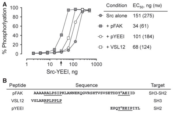
The c-Src tyrosine kinase co-operates with the focal adhesion kinase to regulate cell adhesion and motility. Focal adhesion kinase engages the regulatory SH3 and SH2 domains of c-Src, resulting in localized kinase activation that contributes to tumor cell metastasis. Using assay conditions where c-Src kinase activity required binding to a tyrosine phosphopeptide based on the focal adhesion kinase SH3-SH2 docking sequence, we screened a kinase-biased library for selective inhibitors of the Src/focal adhesion kinase peptide complex versus c-Src alone. This approach identified an aminopyrimidinyl carbamate compound, WH-4-124-2, with nanomolar inhibitory potency and fivefold selectivity for c-Src when bound to the phospho-focal adhesion kinase peptide. Molecular docking studies indicate that WH-4-124-2 may preferentially inhibit the 'DFG-out' conformation of the kinase active site. These findings suggest that interaction of c-Src with focal adhesion kinase induces a unique kinase domain conformation amenable to selective inhibition.

Keywords: SH2 domain; SH3 domain; Src kinase; cancer drug discovery; focal adhesion kinase; kinase inhibitors.


Figures

Figure 1
Figure 1
Structure and domain organization of c-Src and focal adhesion kinase (FAK). (A) Crystal structure of c-Src in the downregulated conformation (PDB ID: 2SRC) showing the SH3 domain, SH3-SH2 connector, the SH2 domain, the SH2-kinase linker, the kinase domain, and the C-terminal tail. Key kinase domain features include the N-lobe, activation loop, αC-helix, and C-lobe. The side chains of the conserved tyrosine residues in the activation loop (Y416) and the tail (pY527) are also shown. Intramolecular interactions between the SH3 domain and the SH2-kinase linker as well as the SH2 domain and the tyrosine-phosphorylated tail are required to maintain the inactive state. The modular domain organization of c-Src is shown below the structure, which includes a myristoylated N-terminal domain not present in the crystal structure. The organization of the recombinant Src-YEEI protein used for this study is also shown, in which the N-terminal unique domain is replaced with a His-tag and the C-terminal tail sequence is modified to encode YEEI. (B) FAK consists of an N-terminal FERM domain, central kinase domain, and a C-terminal FAT domain. The linker connecting the FERM and kinase domains encompasses the binding site for c-Src, consisting of a PxxP motif (SH3 binding) and an autophosphorylation site in the sequence context pYAEI (SH2 binding). The sequence of the pFAK peptide based on this region and used in the inhibitor screen is also shown, with the Src SH3 and SH2 binding sites underlined.
Figure 2
Figure 2
Activation of Src-YEEI by SH3- and SH2-binding peptides. The activity of Src-YEEI was measured in the Z’Lyte in vitro kinase assay in the absence or presence of peptides that bind to the SH3 domain alone (VSL12), the SH2 domain alone (pYEEI), or to both (pFAK). Recombinant Src-YEEI was titrated into the assay over the range of 2-500 ng as shown, and peptides were added at a 10-fold molar excess at each kinase concentration. The concentration of Src-YEEI required to reach 50% maximal activation (EC50) for each condition is shown on the right. The arrow indicates the Src-YEEI concentration used for the library screen, where Src-YEEI activity is dependent on the pFAK peptide (30 ng kinase input; 54.5 nM). The sequences of the peptides are shown below the graph with the docking sites for the Src SH3 and SH2 domains indicated.
Figure 3
Figure 3
Chemical library screen for selective inhibitors of pFAK-dependent Src-YEEI activity. A kinase-biased library of 586 compounds was screened for inhibitors of Src-YEEI activity in the presence of the pFAK peptide using the FRET-based Z’Lyte assay as described in the text. (A) Inhibitory activities of all 586 compounds are ranked by percent inhibition relative to the unphosphorylated substrate peptide control. Ninety-seven compounds inhibited kinase activity by at least 50% (dotted line). (B) A selectivity ratio was determined for compounds showing >50% inhibition of the Src-YEEI:pFAK complex in part A. This ratio was calculated as the percent inhibition of the Src-YEEI: pFAK complex divided by the percent inhibition observed with Src-YEEI alone. Twenty-four compounds exhibited a selectivity ratio >3 (dotted line). The hit compound WH-4-124-2 is indicated in each graph by a red dot, while the control compound WH-4-023 is indicated with a blue dot.
Figure 4
Figure 4
Selective inhibition of Src-YEEI by WH-4-124-2 in the presence of the pFAK peptide but not its structural analog, WH-4-023. WH-4-124-2 (A) and WH-4-023 (B) were assayed against Src-YEEI alone (gray circles) and the Src-YEEI:pFAK complex (black circles) over the range of inhibitor concentrations shown using the Z’Lyte end-point assay. All data points were measured in quadruplicate, and the values shown represent the mean ± SE. IC50 values were determined by nonlinear regression analysis and are shown in Table 1.
Figure 5
Figure 5
Selective inhibition of Src-YEEI by WH-4-124-2 by the pFAK peptide involves displacement of both SH2-tail and SH3-linker interactions. (A) Src-YEEI alone or together with the SH2 peptide ligand pYEEI, the SH3 peptide ligand VSL12, or the pFAK peptide was assayed over the range of inhibitor concentrations shown using the Z’Lyte end-point assay. All data points were measured in quadruplicate, and the values shown represent the mean ± SE. (B) IC50 values were determined by nonlinear regression analysis and are shown in Table 1.
Figure 6
Figure 6
WH-4-124-2 exhibits increased potency while maintaining selectivity in a kinetic kinase assay. (A) The activity of Src-YEEI was measured in the ADP Quest kinetic kinase assay in the absence (gray circles) or presence (black circles) of 30 μM pFAK peptide over the range of input kinase concentrations shown. Compounds WH-4-124-2 (B) and WH-4-023 (C) were assayed against Src-YEEI alone (gray circles) and the Src-YEEI: pFAK complex (black circles) in this assay over the range of inhibitor concentrations shown. Reaction rates at each compound concentration were measured in quadruplicate and are presented as the mean ± SE. IC50 values were determined by nonlinear regression analysis and are shown in Table 1.
Figure 7
Figure 7
Computational docking of WH-4-124-2 to the X-ray crystal structure of the Lck kinase domain bound to imatinib. (A) Comparison of the structures of WH-4-124-2 and imatinib reveals a shared methylpiperazinylmethyl-N-phenylbenzamide backbone (red). (B) Close-up view of the Lck active site with WH-4-124-2 (carbon atoms in cyan) aligned to imatinib (carbon atoms in yellow). WH-4-124-2 was readily accommodated by this imatinib-binding conformation of the Lck kinase domain. In this structure, the N-terminal portion of the activation loop (blue) adopts the DFG-out conformation, with the autophosphorylation site (Y416) extending outward. The side chains of the catalytic aspartate and the gatekeeper residue (T338) are shown for reference. Docking model was produced using the X-ray crystal structure of the Src-family kinase Lck bound to imatinib (PDB: 2PL0). Residue numbering is based on the crystal structure of c-Src (PDB: 2SRC).
Figure 8
Figure 8
Computational docking of WH-4-023 to the X-ray crystal structures of the DFG-in and DFG-out conformations of the Lck kinase domain. (A) View of the Lck active site with WH-4-023 (carbons in green) aligned to the related 2-aminopyrimidine carbamate (compound 43; carbons in yellow) in the crystal structure of the Lck kinase domain in the DFG-in conformation (PDB: 2OFU). In the DFG-in state, the activation loop tyrosine is phosphorylated (pY416). (B) Crystal structure of the Lck kinase domain in the DFG-out conformation (PDB: 2PL0) accommodates WH-4-023 in the same binding pocket as in panel A, with the ligand dimethoxyphenyl moiety rotated to accommodate the phenylalanine side chain of the DFG motif (arrow). In both models, the activation loop is colored in blue with the side chains of the DFG motif, the catalytic aspartate and the gatekeeper residue (T338) shown. Residue numbering is based on the crystal structure of c-Src (PDB: 2SRC).
Figure 9
Figure 9
Preincubation with ATP does not affect WH-4-124-2 potency for Src-YEEI. Src-YEEI was preincubated in the presence (top panel) or absence (bottom panel) of ATP (at the Km) for 3 h at 25 °C to induce autophosphorylation of Tyr416 in the activation loop as described previously (26). Each kinase preparation was then assayed for sensitivity to WH-4-124-2 in the presence or absence of the pFAK peptide using the Z’Lyte end-point assay. All data points were measured in quadruplicate, and the values shown represent the mean ± SE.
Figure 10
Figure 10
Inhibition of Src-YEEI by imatinib and dasatinib is influenced by pFAK peptide binding. Src-YEEI alone and the Src-YEEI:pFAK complex were assayed over the range of dasatinib and imatinib concentrations indicated using the Z’Lyte end-point assay. All data points were measured in quadruplicate, and the values shown represent the mean ± SE.

References

    1. Brown MT, Cooper JA. Regulation, substrates, and functions of Src. Biochim Biophys Acta. 1996;1287:121–149. - PubMed
    1. Resh MD. Myristylation and palmitylation of Src family members: the fats of the matter. Cell. 1994;76:411–413. - PubMed
    1. Boggon TJ, Eck MJ. Structure and regulation of Src family kinases. Oncogene. 2004;23:7918–7927. - PubMed
    1. Chong YP, Ia KK, Mulhern TD, Cheng HC. Endogenous and synthetic inhibitors of the Src-family protein tyrosine kinases. Biochim Biophys Acta. 2005;1754:210–220. - PubMed
    1. Chong YP, Chan AS, Chan KC, Williamson NA, Lerner EC, Smithgall TE, Bjorge JD, Fujita DJ, Purcell AW, Scholz G, Mulhern TD, Cheng HC. C-terminal Src kinase-homologous kinase (CHK), a unique inhibitor inactivating multiple active conformations of Src family tyrosine kinases. J Biol Chem. 2006;281:32988–32999. - PubMed

Publication types

MeSH terms