

Alternating magnetic field-induced hyperthermia increases iron oxide nanoparticle cell association/uptake and flux in blood-brain barrier models
- PMID: 25377069
- PMCID: PMC4803069
- DOI: 10.1007/s11095-014-1561-6
Alternating magnetic field-induced hyperthermia increases iron oxide nanoparticle cell association/uptake and flux in blood-brain barrier models
Abstract
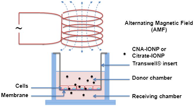
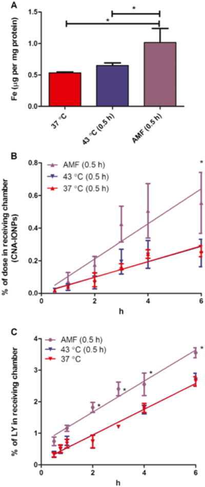
Purpose: Superparamagnetic iron oxide nanoparticles (IONPs) are being investigated for brain cancer therapy because alternating magnetic field (AMF) activates them to produce hyperthermia. For central nervous system applications, brain entry of diagnostic and therapeutic agents is usually essential. We hypothesized that AMF-induced hyperthermia significantly increases IONP blood-brain barrier (BBB) association/uptake and flux.
Methods: Cross-linked nanoassemblies loaded with IONPs (CNA-IONPs) and conventional citrate-coated IONPs (citrate-IONPs) were synthesized and characterized in house. CNA-IONP and citrate-IONP BBB cell association/uptake and flux were studied using two BBB Transwell(®) models (bEnd.3 and MDCKII cells) after conventional and AMF-induced hyperthermia exposure.
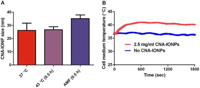
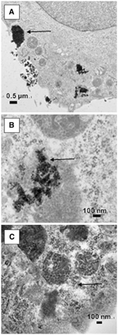
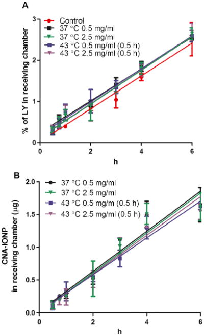
Results: AMF-induced hyperthermia for 0.5 h did not alter CNA-IONP size but accelerated citrate-IONP agglomeration. AMF-induced hyperthermia for 0.5 h enhanced CNA-IONP and citrate-IONP BBB cell association/uptake. It also enhanced the flux of CNA-IONPs across the two in vitro BBB models compared to conventional hyperthermia and normothermia, in the absence of cell death. Citrate-IONP flux was not observed under these conditions. AMF-induced hyperthermia also significantly enhanced paracellular pathway flux. The mechanism appears to involve more than the increased temperature surrounding the CNA-IONPs.
Conclusions: Hyperthermia induced by AMF activation of CNA-IONPs has potential to increase the BBB permeability of therapeutics for the diagnosis and therapy of various brain diseases.
Figures

References
-
- Bernacki J, Dobrowolska A, Nierwinska K, Malecki A. Physiology and pharmacological role of the blood–brain barrier. Pharmacol Rep. 2008;60(5):600–22. - PubMed
-
- Agarwal A, Lariya N, Saraogi G, Dubey N, Agrawal H, Agrawal GP. Nanoparticles as novel carrier for brain delivery: a review. Curr Pharm Des. 2009;15(8):917–25. - PubMed
-
- Peetla C, Labhasetwar V. Biophysical characterization of nanoparticle-endothelial model cell membrane interactions. Mol Pharm. 2008;5(3):418–29. - PubMed
-
- Dhanikula RS, Hammady T, Hildgen P. On the mechanism and dynamics of uptake and permeation of polyether-copolyester dendrimers across an in vitro blood–brain barrier model. J Pharm Sci. 2009;98(10):3748–60. - PubMed
-
- Boström M, Hellstroem Erkenstam N, Kaluza D, Jakobsson L, Kalm M, Blomgren K. The hippocampal neurovascular niche during normal development and after irradiation to the juvenile mouse brain. Int J Radiat Biol. 2014;90(9):778–89. - PubMed
Publication types
MeSH terms
Substances
Grants and funding
LinkOut - more resources
Full Text Sources
Other Literature Sources
