Review
. 2014 Nov 6;5(4):a019562.
doi: 10.1101/cshperspect.a019562.

Human Fungal Pathogens of Mucorales and Entomophthorales

Affiliations
Review

Human Fungal Pathogens of Mucorales and Entomophthorales

Leonel Mendoza et al. Cold Spring Harb Perspect Med. .

Abstract

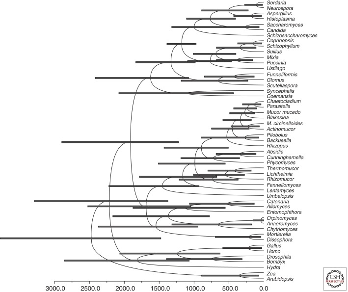
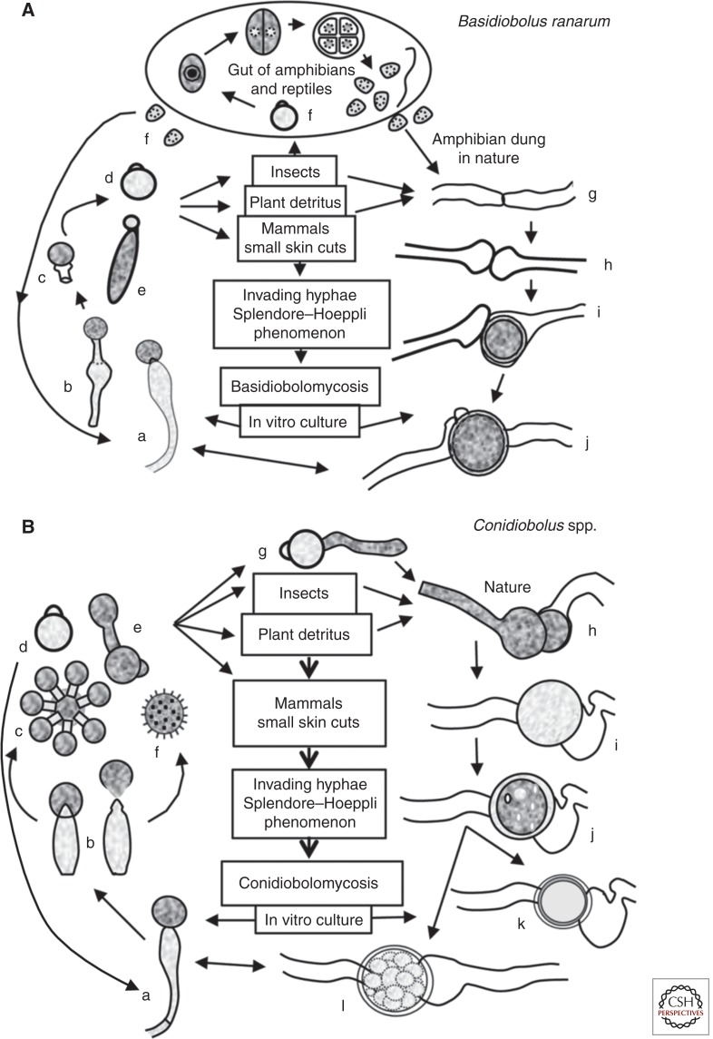
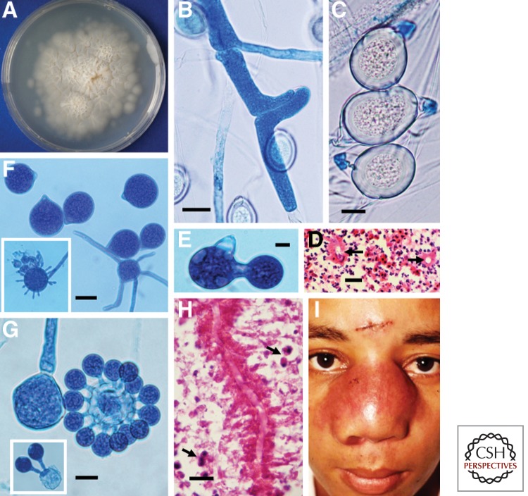
In recent years, we have seen an increase in the number of immunocompromised cohorts as a result of infections and/or medical conditions, which has resulted in an increased incidence of fungal infections. Although rare, the incidence of infections caused by fungi belonging to basal fungal lineages is also continuously increasing. Basal fungal lineages diverged at an early point during the evolution of the fungal lineage, in which, in a simplified four-phylum fungal kingdom, Zygomycota and Chytridiomycota belong to the basal fungi, distinguishing them from Ascomycota and Basidiomycota. Currently there are no known human infections caused by fungi in Chytridiomycota; only Zygomycotan fungi are known to infect humans. Hence, infections caused by zygomycetes have been called zygomycosis, and the term "zygomycosis" is often used as a synonym for "mucormycosis." In the four-phylum fungal kingdom system, Zygomycota is classified mainly based on morphology, including the ability to form coenocytic (aseptated) hyphae and zygospores (sexual spores). In the Zygomycota, there are 10 known orders, two of which, the Mucorales and Entomophthorales, contain species that can infect humans, and the infection has historically been known as zygomycosis. However, recent multilocus sequence typing analyses (the fungal tree of life [AFTOL] project) revealed that the Zygomycota forms not a monophyletic clade but instead a polyphyletic clade, whereas Ascomycota and Basidiomycota are monophyletic. Thus, the term "zygomycosis" needed to be further specified, resulting in the terms "mucormycosis" and "entomophthoramycosis." This review covers these two different types of fungal infections.


Figures

Figure 1.
Figure 1.
Morphology of Mucor circinelloides (left) and Rhizopus oryzae (right). (A) Sporangia form at the apices of sporangiophores and contain the asexual sporangiospores. (B) Zygospores formed during mating. (Figures adapted from Li et al. 2011 [left] and Gryganskyi et al. 2010 [right], respectively.) (C) Hyphae in the brains of infected mice. The tissue specimens were stained with Gomori’s methenamine silver at 48 h postinfection. Scale bars, 50 µm (A); 100 µm (B,C).
Figure 2.
Figure 2.
Timing of fungal divergences using BEAUTi and BEAST assuming a Basidiomycota–Ascomycota split 1200 ± 108 million years ago (Heckman et al. 2001). Node bars indicate 95% highest posterior density intervals of node heights. Calculation of diverging times was performed using BEAUTi v1.7.1 and BEAST v1.6.2 (Drummond et al. 2012). A reduced 18S rDNA alignment (51 taxa, 1195 characters) was used as input for BEAUTi. BEAUTi was run choosing the substitution model GTR with empirical base frequencies, GTR + I as site heterogeneity model, and six γ categories. The clock model was set to lognormal relaxed clock, estimate turned on. Calibration was performed by assuming a split of Ascomycota and Basidiomycota occurring 1208 ± 108 million years ago with normal distribution (Heckman et al. 2001). The generated xml file was run in BEAST over the CIPRES portal (www.phylo.org). Sampled reasonable trees were summarized with the TreeAnnotater from the BEAST package.
Figure 3.
Figure 3.
The life cycles of the representative species that cause entomophthoramycosis. (A) The life cycle of Basidiobolus ranarum. The life cycle starts after a single sporangiophore (a and b) forcibly ejects a conidium (c and d) or after the formation of an elongated sticky “capilliconidium” with a terminal sticky beak (e). Insects or other animals, including mammals, could come into contact with these sticky conidia. Some reptiles and amphibians feed from insects carrying these spores, and a new cycle begins inside their gut (f). Levisohn (1927) observed that a single conidium divided and could produce >50 small spores (meristospores), which could survive in dry environments. After being released into the environment, they develop short mycelia and produce single sporangiophores; alternatively, the nuclei in the same hyphae could exchange genetic material and develope zygospores (gj). They can also develop into structures called palmella, which resemble green algae. Mammals inhabiting areas of endemicity can contact B. ranarum propagules through trauma and develop zygomycosis (center). (B) The life cycle of the pathogenic Conidiobolus species. A single conidiophore develops terminal conidia (a and b), which are forcibly ejected (d) using a mechanism different from that in the genus Basidiobolus (see taxonomy) and geminate (g). Most species that are pathogenic to mammals can develop replicative conidia (c and e), but the “villose” conidia (f) can only be found in C. coronatus. These conidia could develop more sporangiophores or repeat the cycle, or the nuclei of the same coenocytic hyphae could form a septum and develop into a zygospore (hl). Drawings of k and l are examples of C. lampragues and C. incongruus zygospores, respectively.
Figure 4.
Figure 4.
Mycological and clinical aspects of Basidiobolus ranarum. (A) A culture of B. ranarum on 2% Sabouraud dextrose agar after 4 d incubation at 37°C. Note the small colonies formed by forcibly ejected conidia growing around the edges of the culture. (B) Coenocytic hypha of B. ranarum forming the first septum and the elongation across the plane in which the future zygospore will develop. (C) Three mature zygospores with thick walls and their prominent beaks. Note the Splendore–Hoeppli phenomenon around the empty, round hyphal structures in a histological section (D, arrows). (E) Single replicative conidium formation in Conidiobolus coronatus. (F) C. coronatus conidia with a typical basal papilla, which marks the residual section formed after ejection. The inset of panel F shows a villose conidium. (G) C. coronatus multiplicative secondary conidia around a single conidium. The left portion of panel G shows a sporangiophore before conidium discharge. The inset depicts two multiplicative conidia. (H) The eosinophilic inflammatory response and longitudinal sectioned hyphae of C. coronatus surrounded by an eosinophilic reaction. The presence of numerous eosinophils (arrows) is noted. (I) A young boy with bilateral rhinofacial infection caused by C. coronatus. Note the scar of the biopsy performed for diagnostic purposes. (Courtesy of Rafael Isa-Isa and Roberto Arenas.) Scale bars, 25 µm (B); 10 µm (C); 18 µm (D); 15 µm (E,G); 23 µm (F); 19 µm (H).
Figure 5.
Figure 5.
Mycological characteristics of Conidiobolus incongruus. (A) Three C. incongruus conidia with characteristic sharp papillae. (B) An example of multiple replicative conidia of C. incongruus. (C) A conidium with a sharp papilla and a single secondary replicative conidium. (D) Characteristic thick-walled zygospores of C. incongruus containing multiple hyaline globular structures in their cytoplasm. (E) Multiple zygospores of C. lampragues with homogeneous cytoplasm. (F and G) C. lampragues conidia with smooth papillae and the formation of a hypha from a primary conidium, respectively. Scale bars, 14 µm (A); 20 µm (B); 15 µm (C,F); 28 µm (D); 17 µm (E); 17 µm (G).

References

    1. Abe F, Inaba H, Katoh T, Hotchi M 1990. Effects of iron and desferrioxamine on Rhizopus infection. Mycopathologia 110: 87–91. - PubMed
    1. Abe A, Oda Y, Asano K, Sone T 2007. Rhizopus delemar is the proper name for Rhizopus oryzae fumaric-malic acid producers. Mycologia 99: 714–722. - PubMed
    1. Abzug MJ, Walsh TJ 2004. Interferon-γ and colony-stimulating factors as adjuvant therapy for refractory fungal infections in children. Pediatr Infect Dis J 23: 769–773. - PubMed
    1. Alastruey-Izquierdo A, Hoffmann K, de Hoog GS, Rodriguez-Tudela JL, Voigt K, Bibashi E, Walther G 2010. Species recognition and clinical relevance of the zygomycetous genus Lichtheimia (syn. Absidia Pro Parte, Mycocladus). J Clin Microbiol 48: 2154–2170. - PMC - PubMed
    1. Al-Hajjar S, Perfect J, Hashem F, Tufenkeji H, Kayes S 1996. Orbitofacial conidiobolomycosis in a child. Pediatr Infect Dis J 15: 1130–1132. - PubMed