

Concise reviews: Assisted reproductive technologies to prevent transmission of mitochondrial DNA disease
- PMID: 25377180
- PMCID: PMC4359624
- DOI: 10.1002/stem.1887
Concise reviews: Assisted reproductive technologies to prevent transmission of mitochondrial DNA disease
Abstract
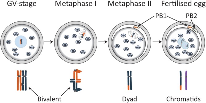
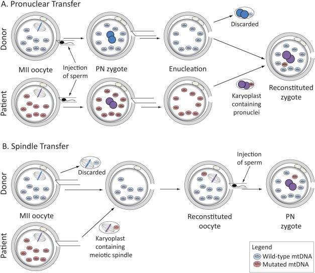
While the fertilized egg inherits its nuclear DNA from both parents, the mitochondrial DNA is strictly maternally inherited. Cells contain multiple copies of mtDNA, each of which encodes 37 genes, which are essential for energy production by oxidative phosphorylation. Mutations can be present in all, or only in some copies of mtDNA. If present above a certain threshold, pathogenic mtDNA mutations can cause a range of debilitating and fatal diseases. Here, we provide an update of currently available options and new techniques under development to reduce the risk of transmitting mtDNA disease from mother to child. Preimplantation genetic diagnosis (PGD), a commonly used technique to detect mutations in nuclear DNA, is currently being offered to determine the mutation load of embryos produced by women who carry mtDNA mutations. The available evidence indicates that cells removed from an eight-cell embryo are predictive of the mutation load in the entire embryo, indicating that PGD provides an effective risk reduction strategy for women who produce embryos with low mutation loads. For those who do not, research is now focused on meiotic nuclear transplantation techniques to uncouple the inheritance of nuclear and mtDNA. These approaches include transplantation of any one of the products or female meiosis (meiosis II spindle, or either of the polar bodies) between oocytes, or the transplantation of pronuclei between fertilized eggs. In all cases, the transferred genetic material arises from a normal meiosis and should therefore, not be confused with cloning. The scientific progress and associated regulatory issues are discussed.
Keywords: Mitochondria; Mitochondrial DNA; Preimplantation genetic diagnosis; Pronuclear transfer; Spindle transfer; Zygote.
© 2014 AlphaMed Press.
Figures
References
-
- Chen XJ, Butow RA. The organization and inheritance of the mitochondrial genome. Nat Rev Genet. 2005;6:815–825. - PubMed
-
- Sato M, Sato K. Maternal inheritance of mitochondrial DNA by diverse mechanisms to eliminate paternal mitochondrial DNA. Biochim. Biophys. Acta. 2013;1833:1979–1984. - PubMed
-
- Shoubridge EA, Wai T. Mitochondrial DNA and the Mammalian Oocyte. Curr Top Dev Biol. 2007;77:87–111. - PubMed
-
- Margulis L. Symbiotic theory of the origin of eukaryotic organelles; criteria for proof. Symp Soc Exp Biol. 1975;29:21–38. - PubMed
-
- Gray MW, Burger G, Lang BF. Mitochondrial evolution. Science. 1999;283:1476–1481. - PubMed
Publication types
MeSH terms
Substances
Grants and funding
LinkOut - more resources
Full Text Sources
Other Literature Sources
Medical
Miscellaneous
