. 2014 Dec;141(23):4628-39.
doi: 10.1242/dev.117804. Epub 2014 Nov 5.

TGFβ signaling inhibits goblet cell differentiation via SPDEF in conjunctival epithelium

Affiliations

TGFβ signaling inhibits goblet cell differentiation via SPDEF in conjunctival epithelium

Heather A McCauley et al. Development. 2014 Dec.

Abstract

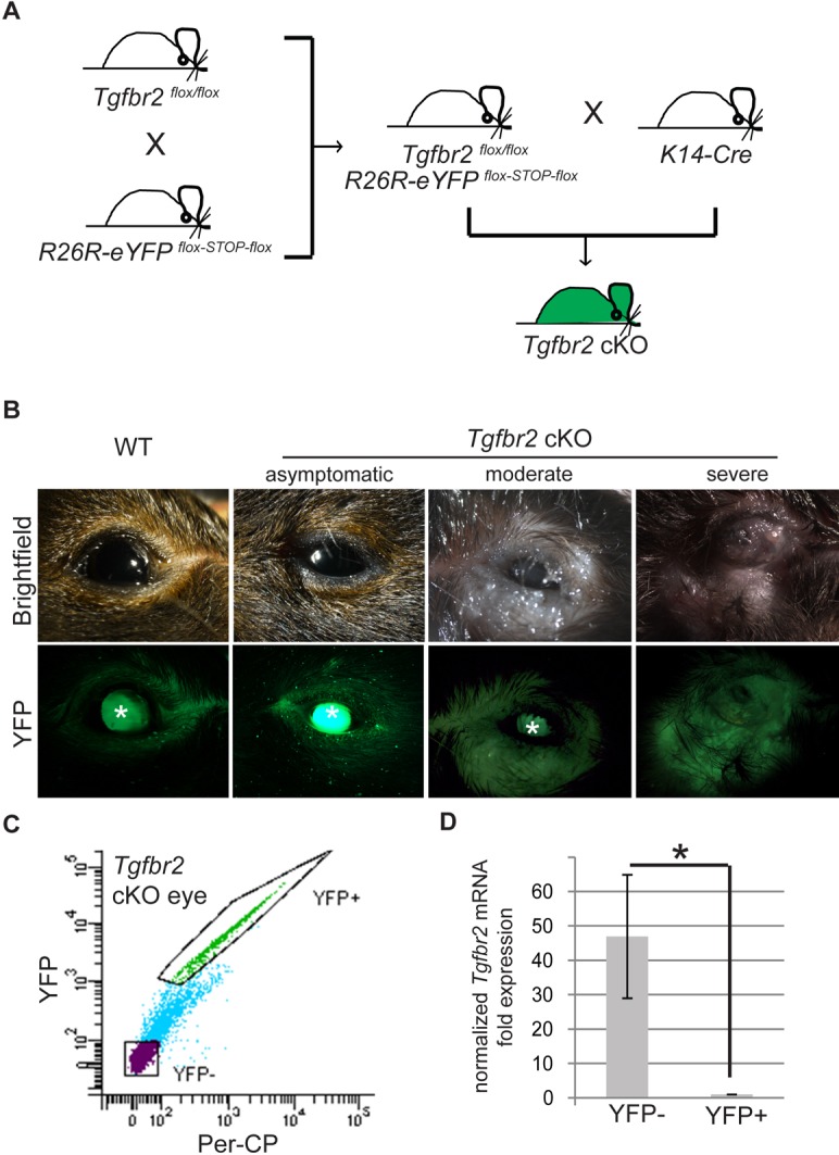
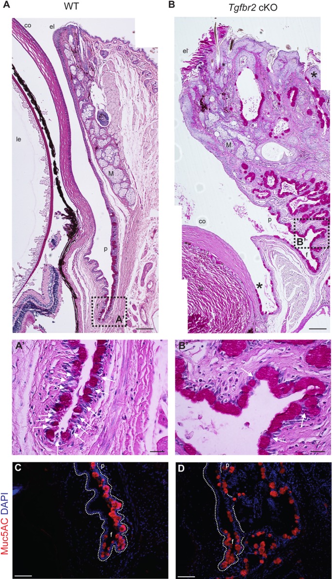
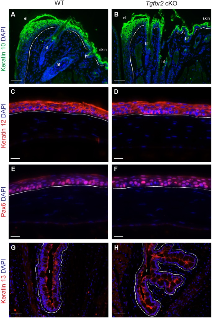
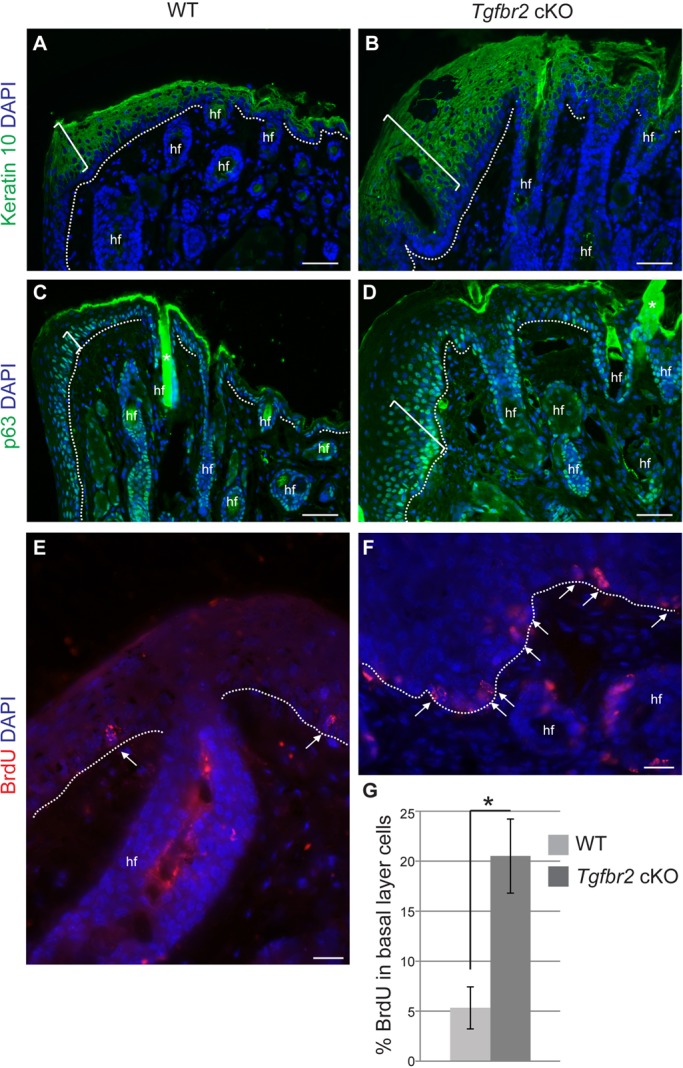
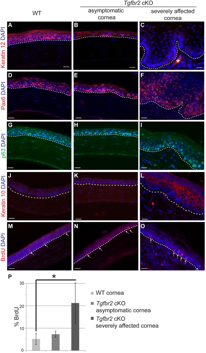
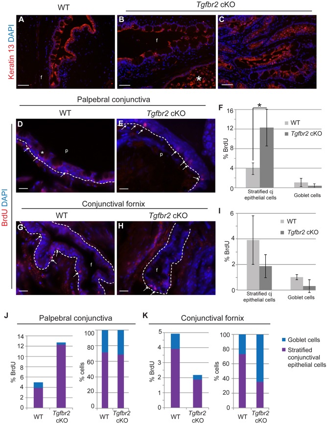
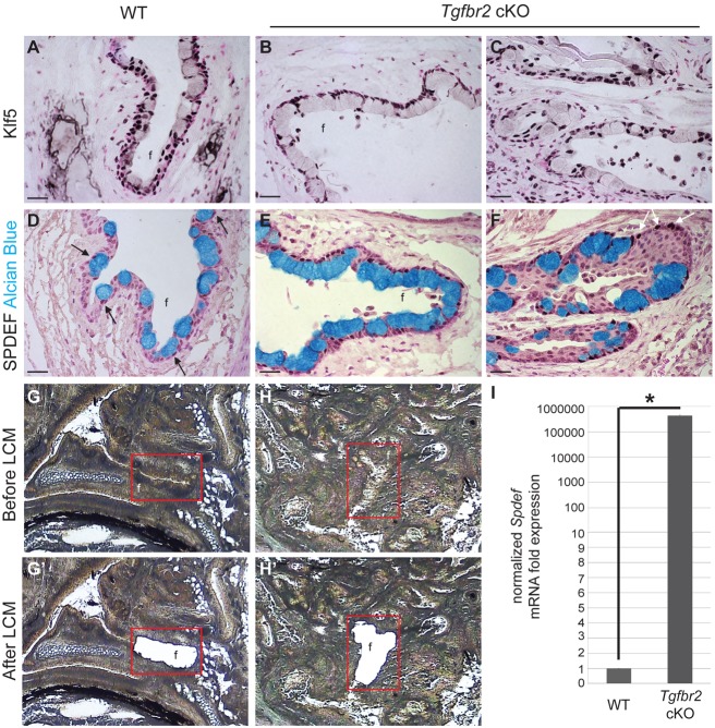
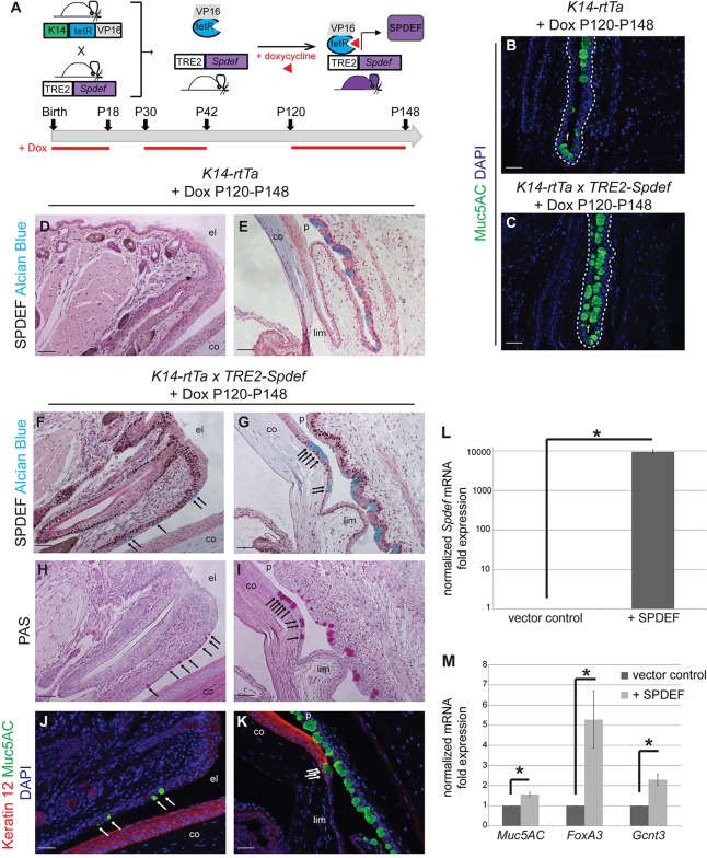
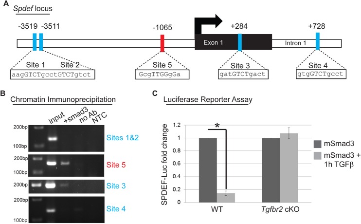
The ocular surface epithelia, including the stratified but non-keratinized corneal, limbal and conjunctival epithelium, in concert with the epidermal keratinized eyelid epithelium, function together to maintain eye health and vision. Abnormalities in cellular proliferation or differentiation in any of these surface epithelia are central in the pathogenesis of many ocular surface disorders. Goblet cells are important secretory cell components of various epithelia, including the conjunctiva; however, mechanisms that regulate goblet cell differentiation in the conjunctiva are not well understood. Herein, we report that conditional deletion of transforming growth factor β receptor II (Tgfbr2) in keratin 14-positive stratified epithelia causes ocular surface epithelial hyperplasia and conjunctival goblet cell expansion that invaginates into the subconjunctival stroma in the mouse eye. We found that, in the absence of an external phenotype, the ocular surface epithelium develops properly, but young mice displayed conjunctival goblet cell expansion, demonstrating that TGFβ signaling is required for normal restriction of goblet cells within the conjunctiva. We observed increased expression of SAM-pointed domain containing ETS transcription factor (SPDEF) in stratified conjunctival epithelial cells in Tgfbr2 cKO mice, suggesting that TGFβ restricted goblet cell differentiation directly by repressing Spdef transcription. Gain of function of Spdef in keratin 14-positive epithelia resulted in the ectopic formation of goblet cells in the eyelid and peripheral cornea in adult mice. We found that Smad3 bound two distinct sites on the Spdef promoter and that treatment of keratin 14-positive cells with TGFβ inhibited SPDEF activation, thereby identifying a novel mechanistic role for TGFβ in regulating goblet cell differentiation.

Keywords: Conjunctiva; Differentiation; Goblet cells; Mouse; SPDEF; TGFβ signaling.


Figures

Fig. 1.
Fig. 1.
Tgfbr2 conditional deletion in K14-expressing cells results in progressive periorbital tissue expansion with narrowing of the palpebral fissure. (A) Triple transgenic mice were obtained by crossing Tgfbr2flox/flox mice with R26R-eYFPflox-STOP-flox mice and K14-Cre mice. (B) External appearance of K14-Cre×Tgfbr2+/+×R26R-eYFPflox-STOP-flox wild-type and K14-Cre×Tgfbr2flox/flox×R26R-eYFPflox-STOP-flox (Tgfbr2 cKO) eyes showing representative examples of mice with an asymptomatic, a moderate and a severe phenotype. Asterisks indicate that the lens is autofluorescent. (C,D) YFP-positive and YFP-negative cells were isolated by FACS from dissected eyes of cKO mice and subjected to mRNA extraction and qPCR. Fluorescence in the PerCP channel was used to exclude autofluorescence. Data represent the mean±s.d.; Student's t-test, *P=0.007.
Fig. 2.
Fig. 2.
Tgfbr2-deficient mice develop eyelid, corneal and conjunctival epithelial hyperplasia. (A-B′) Combined PAS and Hematoxylin and Eosin staining demonstrated extensive squamous and mucous epithelial hyperplasia with invaginations into the underlying stroma, involving the palpebral conjunctiva, fornix and eyelids of symptomatic Tgfbr2 cKO mice compared with sections of comparable regions from age-matched wild-type mice. Ectopic goblet cells (magenta) were found within Tgfbr2 cKO hyperplastic eyelid epithelium and peripheral corneal epithelium (B, asterisks). Higher magnification of the boxed areas are shown in A′ and B′. White arrows indicate non-goblet cell stratified conjunctival epithelial cells interspersed between goblet cells. (C,D) Goblet cells within the expanded and invaginated Tgfbr2 cKO conjunctiva expressed Muc5AC. Dotted lines indicate the basal layer. DAPI counterstains nuclei in blue. co, cornea; le, lens; f, fornix; p, palpebral conjunctiva; M, Meibomian gland; el, eyelid; f, fornix. Scale bars: 100 µm in A,B,C,D; 20 µm in A′,B′.
Fig. 3.
Fig. 3.
Tgfbr2-deficient ocular surface epithelia differentiate properly. (A-F) Immunofluorescent staining with antibodies against keratin 10 (A,B), keratin 12 (C,D), Pax6 (E,F) and keratin 13 (G,H) on eyes dissected from 6-week-old mice indicated that the eyelid (A,B), cornea (C-F) and conjunctiva (G,H) developed normally in Tgfbr2 cKO mice. In the absence of external symptoms, the Tgfbr2 cKO conjunctiva was noted to invaginate into the subepithelial stroma by 6 weeks of age (H). Dotted lines indicate the basal layer. DAPI counterstains nuclei in blue. el, eyelid; hf, hair follicle; f, fornix. Scale bars: 50 µm in A,B,G,H; 20 µm in C-F.
Fig. 4.
Fig. 4.
The Tgfbr2-deficient eyelid becomes hyperplastic with age. Immunofluorescence staining with antibodies against the suprabasal marker keratin 10 (A,B), the basal marker p63 (C,D) and BrdU (E,F) indicated that the Tgfbr2 cKO eyelid epithelium (B) was hyperplastic with increased epithelial layers (brackets) compared with wild type. (E-G) Quantification (G) of BrdU immunofluorescence staining (E,F, arrows) indicated that the Tgfbr2 cKO eyelid epithelium was hyperproliferative. Data represent the mean number of BrdU-positive cells in relation to total basal epithelial cells±s.d.; Student's t-test; *P=0.003. hf, hair follicle. Asterisks in C,D indicate autofluorescence originating from the hair shaft. Scale bars: 50 µm in A-D; 20 µm in E,F.
Fig. 5.
Fig. 5.
The hyperplastic Tgfbr2-deficient adult cornea becomes keratinized. (A-C) Immunofluorescence staining with antibodies against keratin 12 (A-C), Pax6 (D-F), p63 (G-I), keratin 10 (J-L) and BrdU (M-O) indicated that the corneal epithelium of adult Tgfbr2 cKO mice becomes keratinized with increasing phenotypic severity. (M-P) Quantification (P) of BrdU immunofluorescence staining (M-O, arrows) indicated that the severely affected Tgfbr2 cKO corneal epithelium was hyperproliferative compared with wild-type and asymptomatic Tgfbr2 cKO adult corneal epithelium. Data represent the mean number of BrdU-positive cells in relation to total basal epithelial cells ±s.d.; Student's t-test; *P=0.04. Dotted lines indicate the basal layer. Asterisk in C denotes autofluorescence in the stroma. DAPI counterstains nuclei in blue. Scale bars: 20 µm.
Fig. 6.
Fig. 6.
Tgfbr2-deficient stratified conjunctival epithelial cells, but not goblet cells, are hyperproliferative. (A-I) Immunofluorescence staining with antibodies against keratin 13 (A-C) revealed positive keratin 13 expression in the Tgfbr2 cKO conjunctival epithelium (B), including the hyperplastic conjunctival epithelium (C). (D-I) Quantification (F,I) of BrdU immunofluorescence staining (D,E,G,H, arrows) indicated that the Tgfbr2 cKO palpebral conjunctiva, but not the fornix, was hyperproliferative compared with wild type. (J,K) Quantification of the ratio of BrdU-positive cells to total basal epithelial cells demonstrated that the stratified conjunctival epithelial cells, but not goblet cells, were hyperproliferative in Tgfbr2 cKO palpebral conjunctiva, whereas the ratio of stratified epithelial cells to goblet cells remained unchanged (J). Quantification of the ratio of BrdU-positive cells to total basal epithelial cells in the conjunctival fornix demonstrated no significant difference in proliferation between wild-type and Tgfbr2 cKO mice, whereas the ratio of stratified epithelial cells to goblet cells reflected an expansion of the goblet cell compartment (K). Asterisk in B denotes autofluorescence originating from inflammatory cells. Asterisk in D indicates a rare BrdU-positive goblet cell. Data represent the mean number of BrdU-positive cells in relation to total basal epithelial cells±s.d.; Student's t-test; *P=0.02. cj, conjunctival; p, palpebral conjunctiva; f, conjunctival fornix. Scale bars: 50 µm in A-C; 20 µm in D,E,G,H.
Fig. 7.
Fig. 7.
The hyperplastic Tgfbr2-deficient conjunctiva exhibits increased SPDEF expression associated with the goblet cell expansion. (A-C) Immunohistochemistry on the conjunctival epithelium in wild-type (A) and Tgfbr2 cKO mice (B), and the invaginated conjunctival epithelium within the subepithelial stroma of Tgfbr2 cKO eyes (C) showed similar expression of KLF5. (D-F) Alcian Blue-positive goblet cells within the conjunctiva of wild-type mice expressed SPDEF (D, black arrows). Alcian Blue-positive goblet cells (black arrows), as well as some stratified Alcian Blue-negative epithelial cells (white arrows) within the conjunctiva (E) and the epithelial invaginations (F) of Tgfbr2 cKO mice strongly expressed SPDEF. Nuclear Fast Red stains all nuclei in red. Scale bars: 20 µm. (G-H′) Conjunctival fornix (G,H) from wild-type (G,G′) and Tgfbr2 cKO (H,H′) mice were subjected to laser-capture microdissection and RNA extraction. (I) Tgfbr2 cKO conjunctival epithelium expressed Spdef over 400,000 fold more than in wild type. Data represent the mean±s.d.; Student's t-test, *P=0.00006. f, fornix.
Fig. 8.
Fig. 8.
Overexpression of SPDEF in K14-positive epithelium results in ectopic goblet cell formation in vivo and upregulation of goblet cell-associated genes in vitro. (A) K14-rtTA mice were crossed to TRE2-Spdef mice and fed doxycycline to induce SPDEF expression in K14-positive epithelium at three different timepoints (P0, P30 and P120), and analyzed at P18, P42 and P148, respectively. (B,C) Immunofluorescence staining with an antibody against Muc5AC revealed that adult K14-rtTA×TRE2-Spdef mice (C), which were induced with doxycycline from P120 to P148, exhibited increased goblet cell density in the conjunctival fornix, compared with K14-rtTA control littermates (B). Dotted lines indicate the basal layer. (D-K) Immunohistochemistry with an antibody against SPDEF revealed that adult K14-rtTA control mice expressed SPDEF exclusively in the nuclei of conjunctival goblet cells (D,E). However, adult K14-rtTA×TRE2-Spdef mice, induced at P120, expressed SPDEF abundantly throughout eyelid epithelium and conjunctival epithelium (F,G), and formed ectopic Alcian Blue-positive goblet cells in the eyelid epithelium (arrows, F) and peripheral cornea (arrows, G), which also expressed PAS (H,I, arrows) and Muc5AC (J,K, arrows). (J,K) Co-immunofluorescence staining with an antibody against keratin 12 revealed goblet cells in the keratin 12-positive peripheral corneal epithelium. (L,M) Lentiviral infection of K14-positive cells with the full-length Spdef gene resulted in a 10,000-fold enrichment of Spdef mRNA compared with cells infected with empty vector control (*P=0.00004) (L), and significantly upregulated genes associated with goblet cell differentiation, including Muc5aC (*P=0.00005), Foxa3 (*P=0.006) and Gcnt3 (*P=0.002) (M). Data represent the mean±s.d.; Student's t-test. Scale bars: 50 µm. f, fornix; el, eyelid; co, cornea; p, palpebral conjunctiva; lim, limbus.
Fig. 9.
Fig. 9.
TGFβ negatively regulates SPDEF in a Smad3-dependent manner. (A) Promoter analysis using MatInspector (Genomatrix) revealed five putative Smad-binding elements (SBEs) in the Spdef promoter, including four SBEs containing the consensus sequence GTCT (sites 1-4), and one repressive SBE containing the consensus sequence GNNTTGGNGN (site 5). All five sites are evolutionarily conserved and are not within repeat regions. Sites 1 and 2 are located 3511 bp upstream of the transcription start site (TSS, black arrow) of the Spdef gene. Site 3 is located within exon 1 of the Spdef gene and Site 4 is located within the first intron of the Spdef gene. The inhibitory Site 5 is located 1065 bp upstream of the TSS of Spdef. (B) Chromatin immunoprecipitation with an anti-Smad3 antibody was used to isolate DNA fragments that were amplified by primers designed to flank sites 3 and 5 in the Spdef promoter, but not sites 1 and 2 or site 4 after overexpressing Smad3 in NIH3T3 cells and treating with TGFβ1 (2 ng/ml) for 24 h. Non-template (NTC) and no-antibody (no Ab) controls were used to verify the specificity of binding. (C) SPDEF-Luciferase activity was reduced by 85% after overexpressing Smad3 in wild-type keratinocytes and treating with TGFβ1 (2 ng/ml) for 1 h compared with transfected cells without treatment. SPDEF-Luciferase activity was unchanged in Tgfbr2 cKO keratinocytes overexpressing Smad3 and treated with TGFβ1 (2 ng/ml) for 1 h compared with untreated Tgfbr2 cKO keratinocytes. Data represent the mean±s.d.; Student's t-test, *P=0.000002.

Similar articles

Cited by

References

    1. Aronson B. E., Stapleton K. A., Vissers L. A. T. M., Stokhuijzen E., Bruijnzeel H. and Krasinski S. D. (2014). Spdef deletion rescues the crypt cell proliferation defect in conditional Gata6 null mouse small intestine. BMC Mol. Biol. 15, 3 10.1186/1471-2199-15-3 - DOI - PMC - PubMed
    1. Barsky S., Siegal G., Jannotta F. and Liotta L. (1983). Loss of basement membrane components by invasive tumors but not by their benign counterparts. Lab. Invest. 49, 140-147. - PubMed
    1. Bian Y., Terse A., Du J., Hall B., Molinolo A., Zhang P., Chen W., Flanders K. C., Gutkind J. S., Wakefield L. M. et al. (2009). Progressive tumor formation in mice with conditional deletion of TGF-β signaling in head and neck epithelia is associated with activation of the PI3K/Akt pathway. Cancer Res. 69, 5918-5926 10.1158/0008-5472.CAN-08-4623 - DOI - PMC - PubMed
    1. Bleuming S. A., He X. C., Kodach L. L., Hardwick J. C., Koopman F. A., ten Kate F. J., van Deventer S. J. H., Hommes D. W., Peppelenbosch M. P., Offerhaus G. J. et al. (2007). Bone morphogenetic protein signaling suppresses tumorigenesis at gastric epithelial transition zones in mice. Cancer Res. 67, 8149-8155 10.1158/0008-5472.CAN-06-4659 - DOI - PubMed
    1. Chen G., Korfhagen T. R., Xu Y., Kitzmiller J., Wert S. E., Maeda Y., Gregorieff A., Clevers H. and Whitsett J. A. (2009). SPDEF is required for mouse pulmonary goblet cell differentiation and regulates a network of genes associated with mucus production. J. Clin. Invest. 119, 2914-2924. - PMC - PubMed

Publication types

MeSH terms

LinkOut - more resources