. 2014 Dec;141(23):4500-12.
doi: 10.1242/dev.113639. Epub 2014 Nov 5.

The sinus venosus contributes to coronary vasculature through VEGFC-stimulated angiogenesis

Affiliations

The sinus venosus contributes to coronary vasculature through VEGFC-stimulated angiogenesis

Heidi I Chen et al. Development. 2014 Dec.

Abstract

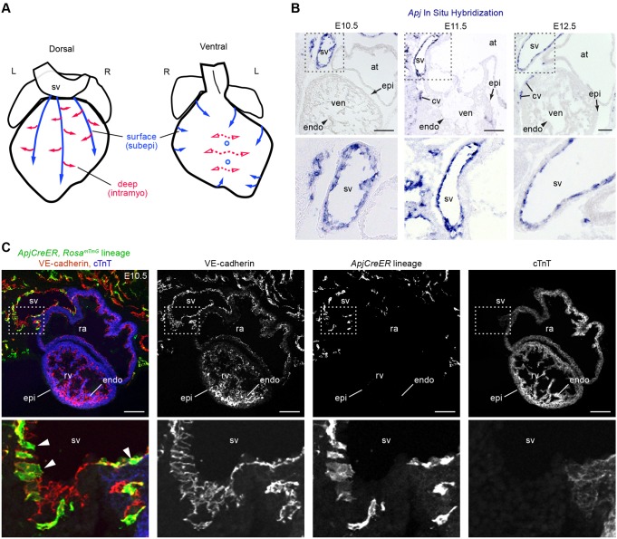
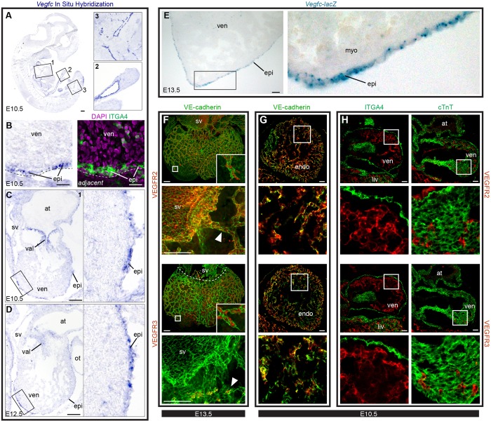
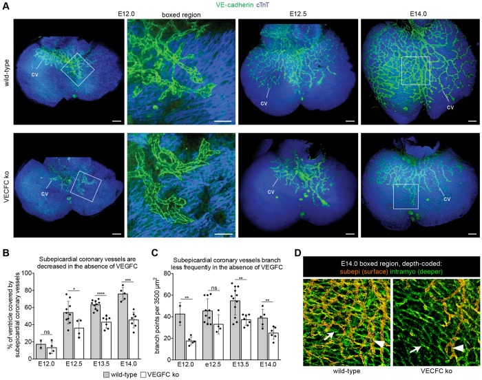
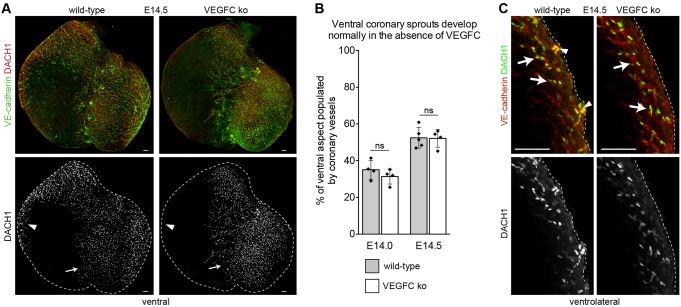
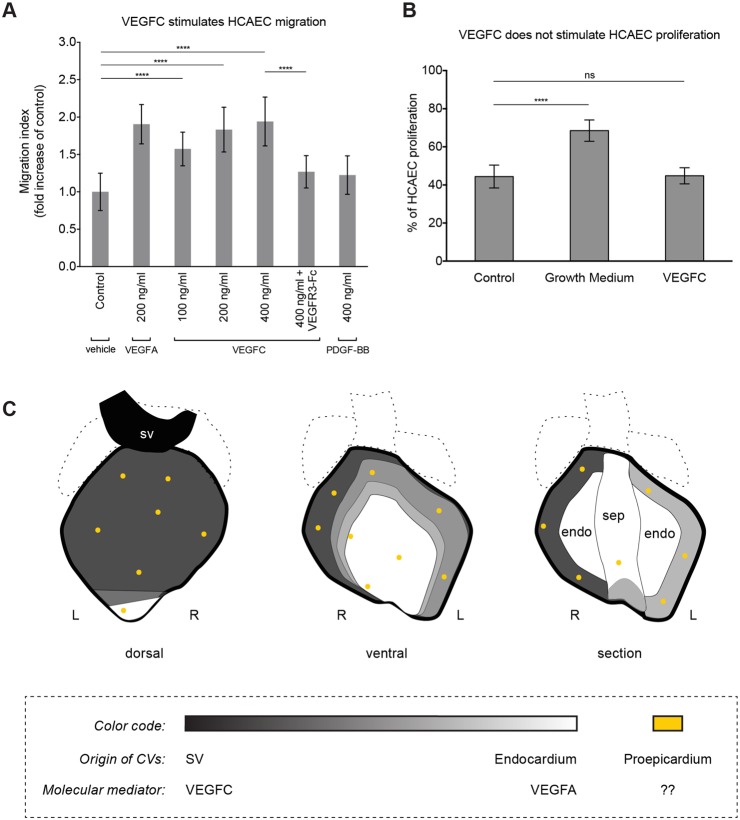
Identifying coronary artery progenitors and their developmental pathways could inspire novel regenerative treatments for heart disease. Multiple sources of coronary vessels have been proposed, including the sinus venosus (SV), endocardium and proepicardium, but their relative contributions to the coronary circulation and the molecular mechanisms regulating their development are poorly understood. We created an ApjCreER mouse line as a lineage-tracing tool to map SV-derived vessels onto the heart and compared the resulting lineage pattern with endocardial and proepicardial contributions to the coronary circulation. The data showed a striking compartmentalization to coronary development. ApjCreER-traced vessels contributed to a large number of arteries, capillaries and veins on the dorsal and lateral sides of the heart. By contrast, untraced vessels predominated in the midline of the ventral aspect and ventricular septum, which are vessel populations primarily derived from the endocardium. The proepicardium gave rise to a smaller fraction of vessels spaced relatively uniformly throughout the ventricular walls. Dorsal (SV-derived) and ventral (endocardial-derived) coronary vessels developed in response to different growth signals. The absence of VEGFC, which is expressed in the epicardium, dramatically inhibited dorsal and lateral coronary growth but left vessels on the ventral side unaffected. We propose that complementary SV-derived and endocardial-derived migratory routes unite to form the coronary vasculature and that the former requires VEGFC, revealing its role as a tissue-specific mediator of blood endothelial development.

Keywords: APLNR); Angiogenesis; Apelin receptor (APJ; Coronary vessel development; Endothelium; Sinus venosus; VEGF-C.


Figures

Fig. 1.
Fig. 1.
Apj and ApjCreER are expressed in the SV. (A) Schematics showing the direction of vessel migration (arrows) during coronary vascular plexus formation. Vessels take subepicardial (surface, subepi) and intramyocardial (deep, intramyo) routes as they populate the entire heart muscle. Solid and dashed lines represent proposed SV and endocardial migratory paths, respectively. (B) Apj mRNA is highly expressed in the SV endothelium but not at appreciable levels in the endocardium (arrowhead) or epicardium (arrow) from E10.5 to E12.5 as shown by in situ hybridization (ISH). Lower panels are magnifications of the boxed regions. (C) A tissue section from ApjCreER, RosamTmG embryos dosed with tamoxifen at E9.5 and analyzed at E10.5. Cre recombination is marked with GFP (green), endothelial cells with VE-cadherin (red) and myocardium with cTnT (blue). Recombination occurs in the SV (arrowheads), but not in the endocardium or epicardium. Lower panels are magnifications of the boxed regions. at, atrium; cv, coronary vessel; endo, endocardium; epi, epicardium; L, left; R, right; ra, right atrium; rv, right ventricle; sv, sinus venosus; ven, ventricle. Scale bars: 100 µm.
Fig. 2.
Fig. 2.
The ApjCreER lineage traces early SV sprouts. (A-F,H,I) Whole-mount confocal images immunostained for VE-cadherin (endothelium/endocardium) and cTnT (myocardium). Direct GFP fluorescence indicates lineage labeling. (A-C) Robust SV labeling at E10.5 (A) progressively spreads onto the heart at subsequent developmental stages (B,C). Little or no ventral labeling is detected at these stages. (D-F) High magnifications of the SV, endocardium (endo) and coronary vessels (CV) from the white-boxed regions in A-C. (G) Quantification of SV and coronary vessel labeling with a single E9.5 dose of tamoxifen. Error bars indicate one s.d. above and below the mean. Each dot represents the quantification for one heart. (H) Near complete labeling of the SV is achieved when tamoxifen is given once daily at E8.5, E9.5 and E10.5. (I) Depth-coded confocal images of red boxed regions in B and C showing that lineage-labeled cells first migrate subepicardially (red) at E11.5 and then into the myocardium (green) at E12.5. bi, blood island; cv, coronary vessels; la, left atrium; lv, left ventricle; ot, outflow tract; ra, right atrium; rv, right ventricle. Scale bars: 100 µm.
Fig. 3.
Fig. 3.
ApjCreER lineage-traced vessels give rise to coronary artery, capillaries and veins of the ventricular walls. Whole-mount confocal images (A,B,D,E) or tissue sections (C,F) of hearts from ApjCreER, RosamTmG crosses dosed with tamoxifen at E9.5 and isolated at the indicated ages. The lineage label is shown in green. (A,B) Many coronary vessels (DACH1+, red) on the dorsal and lateral sides of the heart are ApjCreER lineage traced (green), whereas parts of the apex and central region of the ventral side are not (asterisks). (C) Tissue sections through the left and right ventricular walls (lw and rw, respectively) and septum (sep, outlined by dotted line) show the paucity of ApjCreER lineage-traced vessels in the latter structure. (D) A series of optical sections through the ventricles (dorsal to ventral from left to right) shows lineage-traced cells on the surface (subepicardial) and within the myocardium (intramyocardial). (E) The boxed region from D showing a partially labeled coronary artery (ca) at the border between lineage-labeled (sv-derived) and non-labeled (non-sv-derived) vessels. (F) Immunostaining tissue sections for connexin 40 (Cx40) and COUP-TFII shows ApjCreER lineage contribution to arteries and veins, respectively. lv, left ventricle; rv, right ventricle. Scale bars: 100 µm.
Fig. 4.
Fig. 4.
ApjCreER-traced and Nfatc1Cre-traced coronary lineages populate complementary and overlapping regions of the heart. (A) Whole-mount confocal images of ApjCreER (left) and Nfatc1Cre (right) lineage traces immunostained with anti-DACH1 antibodies (red). Nfatc1Cre-labeled ventral vessels are dense on the ventral face, where ApjCreER-labeled vessels are sparse. High magnification of the boxed region is shown beneath. Arrowheads point to examples of GFP-positive ventral coronary vessels. (B) Representative tissue sections through the right lateral ventricular wall, left lateral ventricular wall, and septum of hearts from the indicated Cre lines highlight complementary contributions. Endothelial/endocardial cells are immunostained for VE-cadherin (top row) or PECAM1 (bottom row). (C) Quantification of ApjCreER-traced, Nfatc1Cre-traced and Sema3dCre-traced vessels shows that the latter population is at fairly consistent, low levels throughout the heart. ApjCreER and Nfatc1Cre lineages tend to contribute inversely proportional numbers to the cardiac compartments when considered from the dorsal to ventral aspect. Error bars represent one s.d. above and below the mean. cv, coronary vessels; endo, endocardium. Scale bars: 100 µm in A; 50 µm in B.
Fig. 5.
Fig. 5.
VEGFC is expressed in the epicardium, and its receptors are expressed in cardiac endothelial cells. (A-D) ISH for Vegfc mRNA. (A) Whole embryo and corresponding insets highlight probe specificity. (B) Adjacent sections subjected to either ISH (left) or immunostaining (right). Vegfc-positive cells (purple) overlap with α4-integrin (ITGA4)+ epicardial cells (epi). Dashed lines delineate the surface of the heart. (C) E10.5 heart (box 1 in A) showing high Vegfc expression in the epicardium and valves (val). (D) Epicardial expression at E12.5. (E) Tissue sections from Vegfc-lacZ mice in which X-Gal staining (blue) shows high Vegfc expression in the epicardium. (C-E) Boxed regions are shown at high magnification to the right. (F) Whole-mount confocal images of E13.5 wild-type hearts immunostained for VE-cadherin and either VEGFR2 or VEGFR3. Images with arrowheads show close-up views of receptor expression in the SV and in nearby coronary vessels. Both receptors are present in all coronary vessels, but VEGFR3 is absent from the SV. Insets are magnifications of the boxed regions showing receptor staining at the leading front of the growing coronary plexus. Arrowheads indicate coronary sprouts near the SV. (G,H) Tissue sections through E10.5 wild-type hearts showing colocalization of VEGFRs with VE-cadherin (G) but not ITGA4 (epicardium) or cTnT (myocardium) (H). Boxed regions (G,H) are magnified beneath. at, atrium; endo, endocardium; myo, myocardium; ot, outflow tract; ven, ventricle. Scale bars: 100 µm, except 20 µm in B.
Fig. 6.
Fig. 6.
VEGFC is required for SV-derived coronary vessel development. (A) Confocal images (transparent projections) showing that coronary vessels (cv, green) on the dorsal surface of the heart are stunted in VEGFC-deficient hearts. Myocardium is blue (cTnT). High magnification of E12.0 boxed regions shows less elaborate vessel branching in knockout (ko) hearts. (B,C) Ventricle coverage by subepicardial coronary vessels (B) and vessel branching (C) are decreased in Vegfc mutant hearts. Bar heights show mean coronary coverage and number of vessel branch points, respectively. Error bars represent one s.d. above and below the mean. Each dot represents quantification for one heart. ****P<0.0001; ***P=0.0001 to 0.001; **P=0.001 to 0.01; *P=0.01 to 0.05; ns, P≥0.05. (D) Depth-coded confocal images from boxed regions on E14.0 hearts in A, in which subepicardial (subepi) vessels appear orange (arrowheads) and intramyocardial vessels (intramyo) appear green (arrows). In mutants, intramyocardial vessels extend farther beyond subepicardial vessels than in wild type. Scale bars: 100 µm.
Fig. 7.
Fig. 7.
The migration of endocardial-derived ventral coronary vessels does not require VEGFC. (A) Ventral coronary vessels (DACH1+, red) corresponding to endocardial-derived regions of the vasculature are normal in Vegfc knockout (ko) hearts (arrows), as compared with wild type. By contrast, few coronary vessels are in areas that arise from the SV (arrowheads). (B) The area occupied by ventral intramyocardial coronary vessels is not significantly different (ns) between wild-type and Vegfc knockout embryos. (C) Subepicardial vessels (arrowheads) are present in wild-type but not in Vegfc knockout myocardium as shown in optical sections through the ventrolateral region of the heart. Intramyocardial vessels (arrows) are present in both genotypes. Scale bars: 100 µm in A; 25 µm in C.
Fig. 8.
Fig. 8.
VEGFC stimulates human coronary artery endothelial cell migration. (A) The addition of VEGFC stimulates HCAEC migration. (B) Proliferation is not increased by VEGFC. Error bars are one s.d. above and below the mean. ****P<0.0001; ns, P≥0.05. (C) Schematics showing the complementary, but overlapping, regions proposed to derive from the SV (black), endocardium (white) and proepicardium (yellow). endo, endocardium; L, left; R, right; sep, septum.

References

    1. Acharya A., Baek S. T., Huang G., Eskiocak B., Goetsch S., Sung C. Y., Banfi S., Sauer M. F., Olsen G. S., Duffield J. S. et al. (2012). The bHLH transcription factor Tcf21 is required for lineage-specific EMT of cardiac fibroblast progenitors. Development 139, 2139-2149 10.1242/dev.079970 - DOI - PMC - PubMed
    1. Arita Y., Nakaoka Y., Matsunaga T., Kidoya H., Yamamizu K., Arima Y., Kataoka-Hashimoto T., Ikeoka K., Yasui T., Masaki T. et al. (2014). Myocardium-derived angiopoietin-1 is essential for coronary vein formation in the developing heart. Nat. Commun. 5, 4552 10.1038/ncomms5552 - DOI - PMC - PubMed
    1. Benedito R., Rocha S., Woeste M., Zamykal M., Radtke F., Casanovas O., Duarte A., Pytowski B. and Adams R. (2012). Notch-dependent VEGFR3 upregulation allows angiogenesis without VEGF-VEGFR2 signalling. Nature 484, 110-114 10.1038/nature10908 - DOI - PubMed
    1. Cai C.-L., Martin J. C., Sun Y., Cui L., Wang L., Ouyang K., Yang L., Bu L., Liang X., Zhang X. et al. (2008). A myocardial lineage derives from Tbx18 epicardial cells. Nature 454, 104-108 10.1038/nature06969 - DOI - PMC - PubMed
    1. Cao Y., Linden P., Farnebo J., Cao R., Eriksson A., Kumar V., Qi J.-H., Claesson-Welsh L. and Alitalo K. (1998). Vascular endothelial growth factor C induces angiogenesis in vivo. Proc. Natl. Acad. Sci. USA 95, 14389-14394 10.1073/pnas.95.24.14389 - DOI - PMC - PubMed

Publication types

Substances