. 2014 Dec;141(23):4640-7.
doi: 10.1242/dev.115048. Epub 2014 Nov 5.

Repurposing an endogenous degradation system for rapid and targeted depletion of C. elegans proteins

Affiliations

Repurposing an endogenous degradation system for rapid and targeted depletion of C. elegans proteins

Stephen T Armenti et al. Development. 2014 Dec.

Abstract

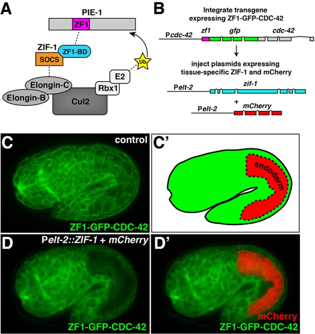
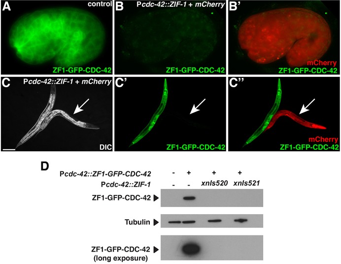
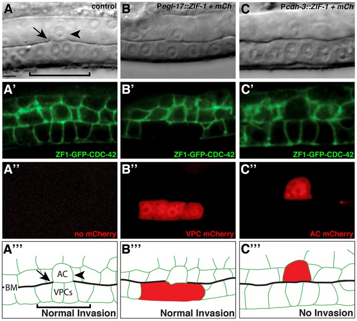
The capability to conditionally inactivate gene function is essential for understanding the molecular basis of development. In gene and mRNA targeting approaches, protein products can perdure, complicating genetic analysis. Current methods for selective protein degradation require drug treatment or take hours for protein removal, limiting their utility in studying rapid developmental processes in vivo. Here, we repurpose an endogenous protein degradation system to rapidly remove targeted C. elegans proteins. We show that upon expression of the E3 ubiquitin ligase substrate-recognition subunit ZIF-1, proteins tagged with the ZF1 zinc-finger domain can be quickly degraded in all somatic cell types examined with temporal and spatial control. We demonstrate that genes can be engineered to become conditional loss-of-function alleles by introducing sequences encoding the ZF1 tag into endogenous loci. Finally, we use ZF1 tagging to establish the site of cdc-42 gene function during a cell invasion event. ZF1 tagging provides a powerful new tool for the analysis of dynamic developmental events.

Keywords: C. elegans; Genetic tool; Mosaic; Protein degradation.


Figures

Fig. 1.
Fig. 1.
ZF1-tagged protein degradation in endodermal cells. (A) Schematic model of ZIF-1 recruiting the ZF1-containing protein PIE-1 to an ECS E3 ligase complex for ubiquitylation (Ub) and subsequent degradation. (B) Strategy for testing effectiveness of ZF1 tagging at later developmental stages. (C,C′) ZF1-GFP-CDC-42 expression in a 1.75-fold stage embryo; the position of the endodermal cells is indicated in C′; expression was detected in endodermal cells, in addition to all other somatic cells (n=14). (D,D′) ZF1-GFP-CDC-42 in a 1.5-fold embryo containing Pelt-2::ZIF-1+mCherry; ZF1-GFP-CDC-42 has degraded specifically in endodermal cells (n=22/22), where mCherry is expressed (D′). Whole embryos are ∼50 µm in length.
Fig. 2.
Fig. 2.
ZIF-1-mediated degradation in all somatic tissues. (A) Control 1.5-fold embryo expressing ZF1-GFP-CDC-42 in all somatic cells (n=22). (B,B′) ZF1-GFP-CDC-42 in an embryo containing Pcdc-42::ZIF-1+mCherry, which induces ZF1-GFP-CDC-42 degradation (n=41/41). (C-C″) DIC (C), GFP (C′) and GFP/mCherry (C″) channels showing larvae expressing ZF1-GFP-CDC-42. Control larvae express ZF1-GFP-CDC-42 in most or all somatic cells (n=37). In larvae also containing Pcdc-42::ZIF-1+mCherry (arrow), ZF1-GFP-CDC-42 degrades (n=14/14). (D) Western analysis of ZF1-GFP-CDC-42 levels in worms lacking (left two lanes) or expressing (right two lanes) ZIF-1 protein from two different Pcdc-42::ZIF-1+mCherry transgene insertions (xnIs520 and xnIs521). The top and bottom blots were probed with anti-GFP antibody. The leftmost lane is a wild-type (N2) control. Scale bar: 100 µm. Whole embryos are ∼50 µm in length.
Fig. 3.
Fig. 3.
Time-lapse analysis of induced ZF1-mediated degradation. (A-F) Frames from time-lapse movies of embryos expressing ZF1-GFP-CDC-42 and either lacking (A-C) or containing (D-F) Phsp::ZIF-1+mCherry. ZF1-GFP-CDC-42 expression persists in embryos lacking Phsp::ZIF-1+mCherry, whereas ZF1-GFP-CDC-42 degrades and mCherry is expressed in embryos containing Phsp::ZIF-1+mCherry. (G) GFP fluorescence levels from regions of interest (boxed region) in embryos containing Phsp::ZIF-1+mCherry relative to controls captured on the same slide. Dashed lines indicate 50% and 90% degradation. Error bars represent s.e.m. from three independent experiments. Whole embryos are ∼50 µm in length.
Fig. 4.
Fig. 4.
CDC-42 functions within the anchor cell to regulate invasion. (A-A‴) A DIC image (A), ZF1-GFP-CDC-42 expression in homozygous cdc-42(gk388) (A′), mCherry fluorescence of worm lacking mCherry construct (A″) and a schematic diagram (A‴). The anchor cell (arrowhead, labeled AC in A‴) has breached the basement membrane (arrow, interruption in phase-dense line in the DIC image; see black line labeled BM in schematic) and contacted the central primary vulval precursor cells (bracket) (n=50/50 normal invasion). (B-B‴) Expression of ZIF-1 in the primary vulval cells (B″, Pegl-17::ZIF-1+mCherry) led to loss of detectable ZF1-GFP-CDC-42 (B′), but did not affect anchor cell invasion (n=20/20 invaded normally). (C-C‴) Expression of ZIF-1 in the anchor cell (C″, Pcdh-3::ZIF-1+mCherry) led to loss of detectable ZF1-GFP-CDC-42 (C′, there is complete loss of fluorescence within the anchor cell; fluorescence outlining the anchor cell is from neighboring cell membranes) and resulted in an invasion defect (non-invaded basement membrane indicated by intact phase-dense line in DIC image; n=8/20 blocked invasion; P<0.005, Fisher's exact test). Scale bar: 5 µm.

References

    1. Abrams E. W. and Mullins M. C. (2009). Early zebrafish development: it's in the maternal genes. Curr. Opin. Genet. Dev. 19, 396-403 10.1016/j.gde.2009.06.002 - DOI - PMC - PubMed
    1. Achilleos A., Wehman A. M. and Nance J. (2010). PAR-3 mediates the initial clustering and apical localization of junction and polarity proteins during C. elegans intestinal epithelial cell polarization. Development 137, 1833-1842 10.1242/dev.047647 - DOI - PMC - PubMed
    1. Anderson D. C., Gill J. S., Cinalli R. M. and Nance J. (2008). Polarization of the C. elegans embryo by RhoGAP-mediated exclusion of PAR-6 from cell contacts. Science 320, 1771-1774 10.1126/science.1156063 - DOI - PMC - PubMed
    1. Armenti S. T., Chan E. and Nance J. (2014). Polarized exocyst-mediated vesicle fusion directs intracellular lumenogenesis within the C. elegans excretory cell. Dev. Biol. 394, 110-121 10.1016/j.ydbio.2014.07.019 - DOI - PMC - PubMed
    1. Asikainen S., Vartiainen S., Lakso M., Nass R. and Wong G. (2005). Selective sensitivity of Caenorhabditis elegans neurons to RNA interference. Neuroreport 16, 1995-1999 10.1097/00001756-200512190-00005 - DOI - PubMed

Publication types