. 2015 Jan 15;308(2):F92-F100.
doi: 10.1152/ajprenal.00510.2014. Epub 2014 Nov 5.

Mesenchymal stem cells and their secretome partially restore nerve and urethral function in a dual muscle and nerve injury stress urinary incontinence model

Affiliations

Mesenchymal stem cells and their secretome partially restore nerve and urethral function in a dual muscle and nerve injury stress urinary incontinence model

Kangli Deng et al. Am J Physiol Renal Physiol. .

Abstract

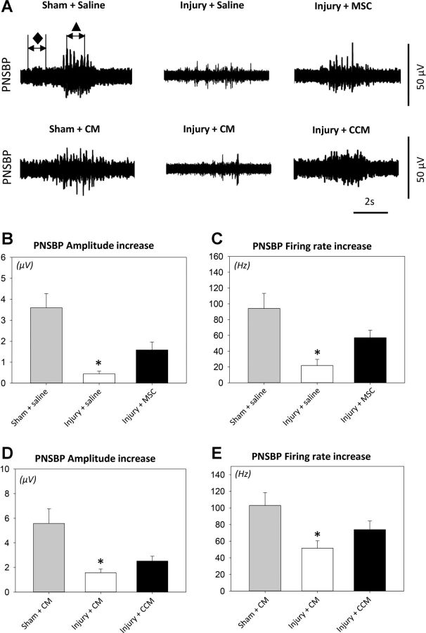
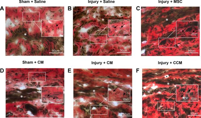
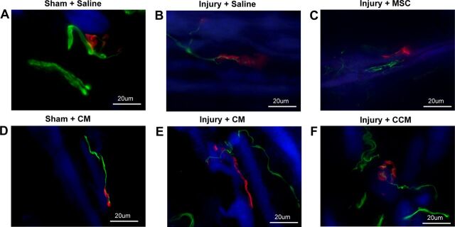
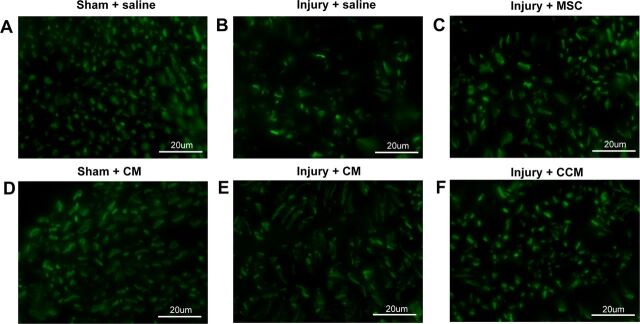
Childbirth injures muscles and nerves responsible for urinary continence. Mesenchymal stem cells (MSCs) or their secretome given systemically could provide therapeutic benefit for this complex multisite injury. We investigated whether MSCs or their secretome, as collected from cell culture, facilitate recovery from simulated childbirth injury. Age-matched female Sprague-Dawley rats received pudendal nerve crush and vaginal distension (PNC+VD) and a single intravenous (iv) injection of 2 million MSCs or saline. Controls received sham injury and iv saline. Additional rats received PNC+VD and a single intraperitoneal (ip) injection of concentrated media conditioned by MSCs (CCM) or concentrated control media (CM). Controls received a sham injury and ip CM. Urethral and nerve function were assessed with leak point pressure (LPP) and pudendal nerve sensory branch potential (PNSBP) recordings 3 wk after injury. Urethral and pudendal nerve anatomy were assessed qualitatively by blinded investigators. Quantitative data were analyzed using one-way ANOVA and Holm-Sidak post hoc tests with P < 0.05 indicating significant differences. Both LPP and PNSBP were significantly decreased 3 wk after PNC+VD with saline or CM compared with sham-injured rats, but not with MSC or CCM. Elastic fiber density in the urethra increased and changed in orientation after PNC+VD, with a greater increase in elastic fibers with MSC or CCM. Pudendal nerve fascicles were less dense and irregularly shaped after PNC+VD and had reduced pathology with MSC or CCM. MSC and CCM provide similar protective effects after PNC+VD, suggesting that MSCs act via their secretions in this dual muscle and nerve injury.

Keywords: elastin; external urethral sphincter; paracrine action; pudendal nerve; urinary incontinence.


Figures

Fig. 1.
Fig. 1.
Leak point pressure (LPP) with simultaneous external urethral sphincter electromyography (EUS EMG) results. Examples demonstrate the two 1-s segments selected from baseline (⧫) and peak activity (▲) that were used to calculate amplitude increase (μV) and firing rate increase (Hz) from baseline to peak pressure in EUS EMG recordings (A). LPP was recorded after pudendal nerve crush and vaginal distension (injury) or sham injury with mesenchymal stem cell (MSC) or saline treatment (B) as well as with concentrated conditioned media (CCM) or concentrated control media (CM) treatment (E). Also shown are EUS EMG amplitude increase after injury or sham injury with MSC or saline treatment (C) as well as with CCM or CM treatment (F) and firing rate increase after injury or sham injury with MSC or saline treatment (D) as well as with CCM or CM treatment (G). Values are means± SE of data from 8–14 animals. ⧫, Baseline EMG; ▲, LPP EMG.*Significant difference compared with corresponding sham-injured animals, P < 0.05.
Fig. 2.
Fig. 2.
Pudendal nerve sensory branch potential (PNSBP) results. Examples demonstrate the two 1-s segments selected from baseline (⧫) and brush activity (▲) that were used to calculate PNSBP amplitude difference (μV) and firing rate difference (Hz) during brush stimuli to the rat clitoris (A). Shown are PNSBP amplitude increase after pudendal nerve crush and vaginal distension (injury) or sham injury with MSC or saline treatment (B) as well as with CCM or CM treatment (D) and firing rate increase after injury or sham injury with MSC or saline treatment (C) as well as with CCM or CM treatment (E). Values are means± SE of data from 8–14 animals. ⧫, Baseline PNSBP; ▲, brush PNSBP. *Significant difference compared with corresponding sham-injured animals, P < 0.05.
Fig. 3.
Fig. 3.
Examples of transverse sections of the urethra stained with elastin von Giesson stain. Three weeks after pudendal nerve crush and vaginal distension (Injury) striated muscle fibers of the EUS (white *) had fewer striations and were atrophied (B, C, E, and F) compared with sham-injured animals (A and D). Elastin fibers (black arrows) were long and consecutive and mainly seen outside the EUS in sham-injured animals. Three weeks after injury, the elastin fibers closest to the urethral serosa were disrupted and twisted. Density of elastin fibers was increased in the EUS near urethral smooth muscle, and their orientation changed to be radial rather than circumferential after injury (B, C, E, and F). With MSC or CCM treatment (C and F), there was an additional increase in density of elastin fibers (black arrows) compared with saline or CM treatment (B and E).
Fig. 4.
Fig. 4.
Examples of immunofluorescence of the EUS showing neuromuscular junctions (red), innervating nerves (green), and striated muscle (blue). Sham-injured animals demonstrated discrete organized motor endplates innervated by straight thick innervating axons (A and D). Three weeks after pudendal nerve crush and vaginal distension (Injury) with saline or CM treatment, innervating axons were thinner and motor endplates were less organized and diffuse (B and E). With MSC or CCM treatment, innervating axons took a more torturous course and had multiple collaterals (C and F).
Fig. 5.
Fig. 5.
Examples of immunofluorescence of transections of the sensory branch of the pudendal nerve distal to the injury. Sham-injured animals demonstrated mostly circular nerve fascicles with small dense compact axons (A and D). Three weeks after pudendal nerve crush and vaginal distension (Injury) and treatment with saline or CM, nerve fascicles were less dense and axons had irregular shapes (B and E). With MSC or CCM treatment, the pudendal nerve demonstrated both normal and abnormal nerve fascicles, with a greater number of normal fascicles than when treated with saline or CM (C and F).

Comment in

  • Editorial Comment.
    Sievert KD. Sievert KD. J Urol. 2016 Dec;196(6):1815. doi: 10.1016/j.juro.2016.05.143. Epub 2016 Aug 30. J Urol. 2016. PMID: 27590989 No abstract available.

References

    1. Aggarwal S, Pittenger MF. Human mesenchymal stem cells modulate allogeneic immune cell responses. Blood 105: 1815–1822, 2005. - PubMed
    1. Bi B, Schmitt R, Israilova M, Nishio H, Cantley LG. Stromal cells protect against acute tubular injury via an endocrine effect. J Am Soc Nephrol 18: 2486–2496, 2007. - PubMed
    1. Cannon TW, Damaser MS. Effects of anesthesia on cystometry and leak point pressure of the female rat. Life Sci 69: 1193–1202, 2001. - PubMed
    1. Caplan AI, Correa D. The MSC: an injury drugstore. Cell Stem Cell 9: 11–15, 2011. - PMC - PubMed
    1. Carr LK, Robert M, Kultgen PL, Herschorn S, Birch C, Murphy M, Chancellor MB. Autologous muscle derived cell therapy for stress urinary incontinence: a prospective, dose ranging study. J Urol 189: 595–601, 2013. - PubMed

Publication types

Substances

LinkOut - more resources