. 2015 Apr;24(4):484-94.
doi: 10.1002/pro.2598. Epub 2014 Dec 11.

Severing of a hydrogen bond disrupts amino acid networks in the catalytically active state of the alpha subunit of tryptophan synthase

Affiliations

Severing of a hydrogen bond disrupts amino acid networks in the catalytically active state of the alpha subunit of tryptophan synthase

Jennifer M Axe et al. Protein Sci. 2015 Apr.

Abstract

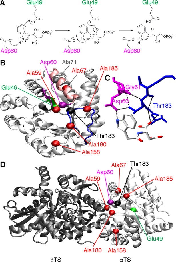
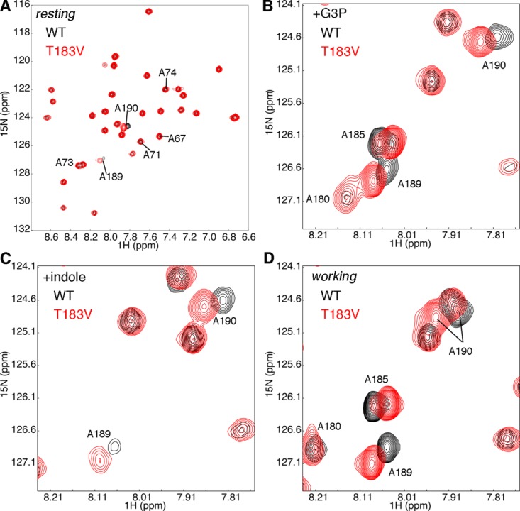
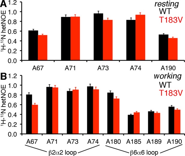
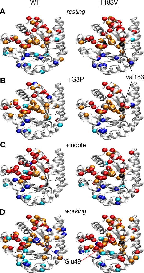
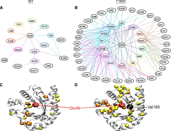
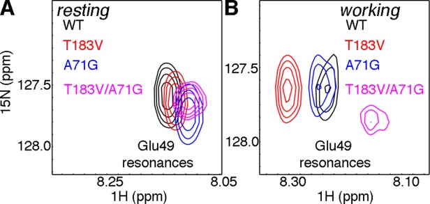
Conformational changes in the β2α2 and β6α6 loops in the alpha subunit of tryptophan synthase (αTS) are important for enzyme catalysis and coordinating substrate channeling with the beta subunit (βTS). It was previously shown that disrupting the hydrogen bond interactions between these loops through the T183V substitution on the β6α6 loop decreases catalytic efficiency and impairs substrate channeling. Results presented here also indicate that the T183V substitution decreases catalytic efficiency in Escherchia coli αTS in the absence of the βTS subunit. Nuclear magnetic resonance (NMR) experiments indicate that the T183V substitution leads to local changes in the structural dynamics of the β2α2 and β6α6 loops. We have also used NMR chemical shift covariance analyses (CHESCA) to map amino acid networks in the presence and absence of the T183V substitution. Under conditions of active catalytic turnover, the T183V substitution disrupts long-range networks connecting the catalytic residue Glu49 to the αTS-βTS binding interface, which might be important in the coordination of catalytic activities in the tryptophan synthase complex. The approach that we have developed here will likely find general utility in understanding long-range impacts on protein structure and dynamics of amino acid substitutions generated through protein engineering and directed evolution approaches, and provide insight into disease and drug-resistance mutations.

Keywords: amino acid networks; chemical shift covariance analysis; enzyme mechanisms; nuclear magnetic resonance; protein dynamics; tryptophan synthase.


Figures

Figure 1
Figure 1
Catalytic function and loop interactions in αTS. (A) Chemical mechanism for αTS, highlighting the roles of Glu49 and Asp60 according to the step-wise mechanism. (B) Structure of αTS (PDB: 1K3U), showing the location of important amino acid residues, including Glu49 on the β2 strand (green), Asp60 on the β2α2 loop (magenta), and Thr183 on the β6α6 loop (blue). Also shown are the sites of the perturbations used to characterize the amino acid networks in E. coli αTS, including Ala59, Ala67, Ala158, Ala180, and Ala185 (red). It should be pointed out that in the absence of βTS, the α2' helix (pink) does not form, and instead the β2α2 loop is extended. Ala71 (grey), which is on the same amino acid network as Glu49 under some conditions, is also shown. (C) Close-up of the hydrogen bond interactions between the β2α2 (magenta) and β6α6 (blue) loops involving Thr183. The ligand shown is N-2[1H-indol-3-YL-acetyl]aspartic acid. (D) The locations of important amino acid residues in the context of the α−β TS dimer (PDB: 1K3U). Importantly, all of the NMR and kinetic studies reported here were performed on the αTS subunit alone, in the absence of the βTS subunit.
Figure 2
Figure 2
The T183V substitution leads to local changes in the β2α2 and β6α6 loops of αTS. Shown are comparisons of the 1H-15N HSQC spectra for WT (black) and T183V (red) αTS enzymes (A) without ligand, (B) in the presence of 10 mM D-G3P, (C) in the presence of 10 mM indole and (D) under dynamic chemical equilibrium conditions representing a 4:1 ratio of E:IGP to E:indole:G3P forms; these working state conditions are initiated with the addition of 10 mM D-G3P and 10 mM indole. Note that only the Ala residues are 15N labeled. NMR data were collected at 298 K on samples containing 0.5 to 1 mM protein in 50 mM potassium phosphate, pH 7.8, 2 mM DTT, 0.2 mM Na2EDTA, and 10% 2H2O.
Figure 3
Figure 3
The T183V substitution alters structural dynamics in the β2α2 and β6α6 loops. Comparison of the 1H-15N heteronuclear Overhauser effects (hetNOE) for WT (black) and T183V (red) αTS enzymes for the (A) ligand-free resting state and the (B) working state conditions under dynamic chemical equilibrium. The working state conditions are initiated by the addition of 10 mM indole and 10 mM D-G3P to reach a ratio of E:IGP to E:indole:G3P of ∼4:1.
Figure 4
Figure 4
Amino acid networks in E. coli αTS are dependent upon the bound ligand and the presence of the T183V substitution. Agglomerative clustering is used to identify “nearest-neighbor” clusters in αTS (A) in the ligand-free resting state, (B) when bound to G3P, (C) when bound to indole, and (D) in the working state under dynamic chemical equilibrium conditions (i.e. ∼4:1 ratio of E:IGP to E:indole:G3P). To generate the WT networks (left), the CHESCA approach utilized NMR data from WT, A59G, A67G, A158G, A180G, and A185G proteins. To generate the T183V networks (right), the CHESCA approach utilized NMR data from T183V, T183V/A59G, T183V/A67G, T183V/A158G, T183V/A180G, and T183V/A185G proteins. Residues in cluster 1 and cluster 2 are plotted as dark blue/light blue and red/orange spheres onto the αTS structure (PDB: 1K3U). Colors also correspond to the level of statistical significance that these residues are found in their appropriate clusters (P < 0.01, dark blue/red; P < 0.05, light blue/orange). Chemical shift covariance matrices and the associated dendrograms are presented in Supporting Information Figures S1 and S2 respectively.
Figure 5
Figure 5
Network associations with Glu49 are different in the presence/absence of the T183V substitution. Shown are the dendrograms for (A) WT and (B) T183V networks under the working state conditions. The WT network utilized NMR data from WT, A59G, A67G, A158G, A180G, and A185G proteins, whereas the T183V network utilized NMR data from T183V, T183V/A59G, T183V/A67G, T183V/A158G, T183V/A180G, and T183V/A185G proteins. Dendrograms are drawn based on the chemical shift covariance matrices in Supporting Information Figure S1. Lines are drawn between residues showing significant (i.e. P < 0.05) linear chemical shift correlations, starting with Glu49 and then to other residues. For improved clarity, connecting lines are not shown between residues within the same “circle” of residues. Residues from these dendrograms are plotted as spheres on the αTS structure for the (C) WT and (D) T183V networks (PDB: 1K3U). Glu49 is plotted as a larger red sphere, the residues showing significant linear chemical shift correlations with Glu49 are plotted as orange spheres, and the residues showing significant linear chemical shift correlations with the first set of residues are plotted as yellow spheres. Analysis of other states is presented in Supporting Information Figure S4.
Figure 6
Figure 6
Perturbing unique pathways in the WT and T183V networks of E. coli αTS. Zoomed in pictures of the 1H-15N HSQC spectra showing the Glu49 resonance for WT (black), A71G (blue), T183V (red), and T183V/A71G (magenta) αTS enzymes for the (A) ligand-free resting state and (B) working state under dynamic chemical equilibrium conditions (i.e. a ∼4:1 ratio of E:IGP to E:indole:G3P). The A71G substitution induces a more substantial chemical shift change for the Glu49 resonance in the working state in the presence of the T183V substitution, consistent with Ala71 being in the same cluster as Glu49 in the T183V but not the WT network.
Figure 7
Figure 7
Potential pathway of communication between the active site of αTS and the βTS binding interface in the (A) WT enzyme that is disrupted in the (B) T183V variant. Working state clusters from Figure 4 are plotted onto αβTS dimer (PDB: 1K3U). αTS and βTS subunits are indicated by white and dark grey ribbons, respectively. Cluster residues are plotted as colored spheres (cluster 1, blue; cluster 2, orange). Green indicates a potential pathway from Glu49 to the αTS-βTS binding interface identified from the cluster analysis in Figure 4, where Phe107 from αTS interacts with β-strands from βTS forming part of the indole channel. These interactions might influence the conformation of Phe280 in βTS, which has been shown to block the indole channel in some TS crystal structures. The T183V substitution also likely disrupts structural dynamics of the β2α2 and β6α6 loops in αTS that are important for TS catalysis and indole channeling.

References

    1. Amitai G, Shemesh A, Sitbon E, Shklar M, Netanely D, Venger I, Pietrokovski S. Network analysis of protein structures identifies functional residues. J Mol Biol. 2004;344:1135–1146. - PubMed
    1. Bode C, Kovacs IA, Szalay MS, Palotai R, Korcsmaros T, Csermely P. Network analysis of protein dynamics. FEBS Lett. 2007;581:2776–2782. - PubMed
    1. Csermely P, Sandhu KS, Hazai E, Hoksza Z, Kiss HJ, Miozzo F, Veres DV, Piazza F, Nussinov R. Disordered proteins and network disorder in network descriptions of protein structure, dynamics and function: hypotheses and a comprehensive review. Curr Protein Pept Sci. 2012;13:19–33. - PubMed
    1. Lee J, Goodey NM. Catalytic contributions from remote regions of enzyme structure. Chem Rev. 2011;111:7595–7624. - PubMed
    1. Reynolds KA, Russ WP, Socolich M, Ranganathan R. Evolution-based design of proteins. Methods Enzymol. 2013;523:213–235. - PubMed

Publication types

MeSH terms

LinkOut - more resources