. 2014 Nov 5;34(45):14820-6.
doi: 10.1523/JNEUROSCI.3369-14.2014.

Caspr and caspr2 are required for both radial and longitudinal organization of myelinated axons

Affiliations

Caspr and caspr2 are required for both radial and longitudinal organization of myelinated axons

Aaron Gordon et al. J Neurosci. .

Abstract

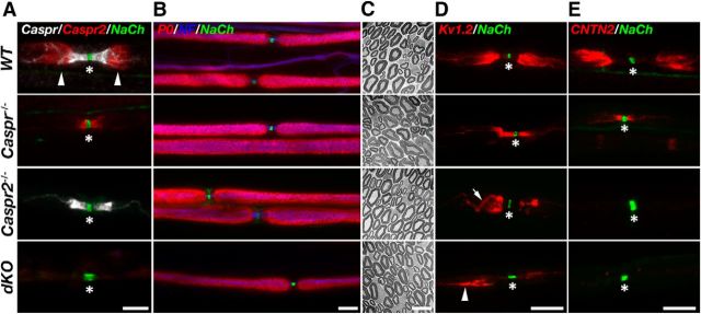
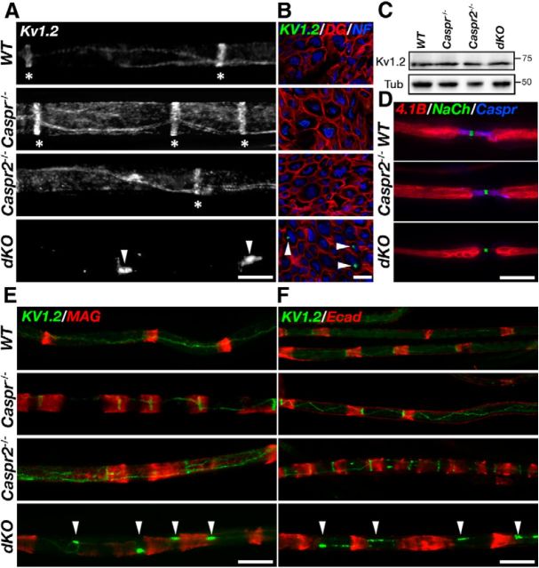
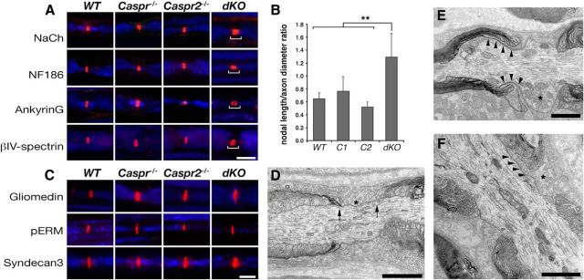
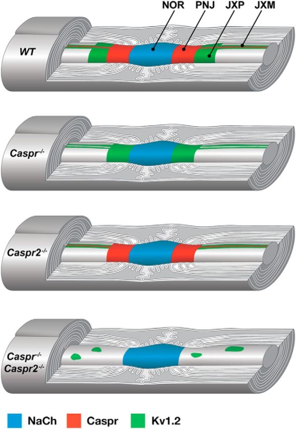
In myelinated peripheral axons, Kv1 potassium channels are clustered at the juxtaparanodal region and at an internodal line located along the mesaxon and below the Schmidt-Lanterman incisures. This polarized distribution is controlled by Schwann cells and requires specific cell adhesion molecules (CAMs). The accumulation of Kv1 channels at the juxtaparanodal region depends on the presence of Caspr2 at this site, as well as on the presence of Caspr at the adjacent paranodal junction. However, the localization of these channels along the mesaxonal internodal line still persists in the absence of each one of these CAMs. By generating mice lacking both Caspr and Caspr2 (caspr(-/-)/caspr2(-/-)), we now reveal compensatory functions of the two proteins in the organization of the axolemma. Although Kv1 channels are clustered along the inner mesaxon and in a circumferential ring below the incisures in the single mutants, in sciatic nerves of caspr(-/-)/caspr2(-/-) mice, these channels formed large aggregates that were dispersed along the axolemma, demonstrating that internodal localization of Kv1 channels requires either Caspr or Caspr2. Furthermore, deletion of both Caspr and Caspr2 also resulted in widening of the nodes of Ranvier, suggesting that Caspr2 (which is present at paranodes in the absence of Caspr) can partially compensate for the barrier function of Caspr at this site even without the formation of a distinct paranodal junction. Our results indicate that Caspr and Caspr2 are required for the organization of the axolemma both radially, manifested as the mesaxonal line, and longitudinally, demarcated by the nodal domains.

Keywords: Caspr; adhesion molecule; axon–glia interaction; myelin; node of Ranvier; peripheral nerve.


Figures

Figure 1.
Figure 1.
Caspr and Caspr2, respectively, dictate the location and membrane accumulation of Kv1 channels at the nodal environ. A, Caspr and Caspr2 are not detectable in dKO mice. Immunofluorescence of teased sciatic nerves isolated from WT, Caspr-null (Caspr−/−), Caspr2-null (Caspr2−/−), and dKO mice using antibodies to Caspr, Caspr2, and Na+ channels (NaCh). The location of the nodes and the juxtaparanodal region are labeled with asterisks and arrowheads, respectively. Note that Caspr2 is mislocalized at the paranodes in the absence of Caspr, whereas the distribution of Caspr is unchanged in the absence of Caspr2. B, C, Absence of both Caspr and Caspr2 does not lead to myelin abnormalities. Immunofluorescence analysis using antibodies to P0, neurofilament (NF) and NaCh (B), as well as electron microscopy analysis of cross-sections (C) of sciatic nerves isolated from the indicated genotypes. D, E, Absence of Caspr or Caspr2 results in altered distribution of juxtaparanodal components. Shown is immunofluorescence labeling of teased sciatic nerves isolated from the indicated genotypes using antibodies to NaCh and Kv1.2 potassium channels (D) or CNTN2 (E). The location of the nodes of Ranvier is marked with an asterisk in all panels. In the absence of Caspr2, Kv1.2 channels are not accumulated at the juxtaparanodal region, but are occasionally detected at the double mesaxonal lines that passed through this region (arrow). Arrowhead marks the presence of abnormal patches of Kv1 channels at the edge of the juxtaparanodal region in the dKO nerves. Scale bars: A, B, D, E, 10 μm; C, 5 μm.
Figure 2.
Figure 2.
Absence of Caspr and Caspr2 results in the disruption of Kv1.2 localization along the mesaxonal line. A, Immunofluorescence of teased sciatic nerves isolated from WT, Caspr-null (Caspr−/−), Caspr2-null (Caspr2−/−), and dKO mice using antibodies to Kv1.2 channels. In WT mice and the single mutants, these channels are present at the juxtamesaxonal line, as well as in radial rings (asterisks) located beneath the Schmidt Lanterman incisures. In contrast, Kv1.2 channels are found in large aggregates (arrowheads) that are distributed along the internodes. B, Cross-sections of sciatic nerves isolated from the indicated genotypes labeled using antibodies to Kv1.2, dystroglycan (DG), and neurofilament (NF). Kv1.2 aggregates (arrowheads) are present in proximity to the axolemma. C, Western blot analysis of sciatic nerves reveals similar levels of Kv1.2 in WT, Caspr−/−, Caspr2−/−, and dKO mice. D, Absence of Caspr and Caspr2 does not affect the distribution of protein 4.1B. E, F, Distribution of Kv1.2 aggregates in relation to Schmidt-Lanterman incisures. Teased sciatic nerves isolated from the four different genotypes were labeled using antibodies to Kv1.2 and MAG (E) or E-cadherin (Ecad; F). Arrowheads mark the location of Kv1.2 aggregates. Scale bars, 10 μm.
Figure 3.
Figure 3.
Deletion of both Caspr and Caspr2 results in widening of the nodes. A, Double immunofluorescence labeling of sciatic nerves isolated from the indicated genotypes using antibodies to neurofilament (blue) and axonodal components: Na+ channels (NaCh), NF186, ankyrin G, or βIV spectrin. Bars highlight the wider nodal region in the dKO nerves. B, Quantification of axonodal width-to-height ratio showed a widening of the dKO node compared with WT mice and the single mutants. Error bars indicate SEM of n = 4 mice for each genotype (**p < 0.05). C, Double immunofluorescence labeling of sciatic nerves isolated from the indicated genotypes using antibodies to neurofilament (blue) and glial microvilli proteins: gliomedin, phospho-ERM (pERM), or Syndecan-3. DF, Electron microscopy images showing a longitudinal view of the nodes of Ranvier in sciatic nerves of WT (D) and dKO (E,F) mice. In WT nerves, the node is bordered by the PNJ and Schwann cell microvilli (asterisk) are confined to the nodal gap (arrows). In dKO nerves, Schwann cell microvilli processes are detected between the axolemma and the detached paranodal loops (E,F, arrowheads). Asterisks in E and F mark microvilli and the accumulation of mitochondria at the nodes, respectively. Scale bars: A, C, 10 μm; DF, 1 μm.
Figure 4.
Figure 4.
Caspr proteins organize myelinated axons. A schematic drawing illustrating the distribution of Na+ channels (NaCh), Caspr, and Kv1.2 channels along myelinated PNS axons in WT, Caspr−/−, Caspr2−/−, and double Caspr−/−/Caspr2−/− mutant mice. Whereas in Caspr−/− or Caspr2−/− nerves, Kv1.2 channels accumulate at the JXM, in the absence of both Caspr proteins, these channels form large aggregates along the internodal axolemma. NOR indicates node of Ranvier; PNJ, paranodal junction; and JXP, juxtaparanodal region.

References

    1. Amor V, Feinberg K, Eshed-Eisenbach Y, Vainshtein A, Frechter S, Grumet M, Rosenbluth J, Peles E. Long-term maintenance of Na+ channels at nodes of Ranvier depends on glial contact mediated by gliomedin and NrCAM. J Neurosci. 2014;34:5089–5098. doi: 10.1523/JNEUROSCI.4752-13.2014. - DOI - PMC - PubMed
    1. Arancibia-Carcamo IL, Attwell D. The node of Ranvier in CNS pathology. Acta Neuropathol. 2014;128:161–175. doi: 10.1007/s00401-014-1305-z. - DOI - PMC - PubMed
    1. Arroyo EJ, Xu YT, Zhou L, Messing A, Peles E, Chiu SY, Scherer SS. Myelinating Schwann cells determine the internodal localization of Kv1.1, Kv1.2, Kvbeta2, and Caspr. J Neurocytol. 1999;28:333–347. doi: 10.1023/A:1007009613484. - DOI - PubMed
    1. Babbs CF, Shi R. Subtle paranodal injury slows impulse conduction in a mathematical model of myelinated axons. PLoS One. 2013;8:e67767. doi: 10.1371/journal.pone.0067767. - DOI - PMC - PubMed
    1. Bhat MA, Rios JC, Lu Y, Garcia-Fresco GP, Ching W, St Martin M, Li J, Einheber S, Chesler M, Rosenbluth J, Salzer JL, Bellen HJ. Axon-glia interactions and the domain organization of myelinated axons requires neurexin IV/Caspr/Paranodin. Neuron. 2001;30:369–383. doi: 10.1016/S0896-6273(01)00294-X. - DOI - PubMed

Publication types

MeSH terms