. 2014 Dec 1;42(21):13353-69.
doi: 10.1093/nar/gku1077. Epub 2014 Nov 5.

Functional characterization of C. elegans Y-box-binding proteins reveals tissue-specific functions and a critical role in the formation of polysomes

Affiliations

Functional characterization of C. elegans Y-box-binding proteins reveals tissue-specific functions and a critical role in the formation of polysomes

Andreas Arnold et al. Nucleic Acids Res. .

Abstract

The cold shock domain is one of the most highly conserved motifs between bacteria and higher eukaryotes. Y-box-binding proteins represent a subfamily of cold shock domain proteins with pleiotropic functions, ranging from transcription in the nucleus to translation in the cytoplasm. These proteins have been investigated in all major model organisms except Caenorhabditis elegans. In this study, we set out to fill this gap and present a functional characterization of CEYs, the C. elegans Y-box-binding proteins. We find that, similar to other organisms, CEYs are essential for proper gametogenesis. However, we also report a novel function of these proteins in the formation of large polysomes in the soma. In the absence of the somatic CEYs, polysomes are dramatically reduced with a simultaneous increase in monosomes and disomes, which, unexpectedly, has no obvious impact on animal biology. Because transcripts that are enriched in polysomes in wild-type animals tend to be less abundant in the absence of CEYs, our findings suggest that large polysomes might depend on transcript stabilization mediated by CEY proteins.


Figures

Figure 1.
Figure 1.
Characteristics of CEY proteins. (A) Phylogenetic tree showing a relation between CEYs and Y-box-binding proteins in other organisms, the closest being Yps in Drosophila melanogaster. The distance values show the number of amino acid substitutions as a proportion of the length of the alignment. (B) Besides the CSD, CEY-1 and CEY-4 contain RG/RGG repeats, which are absent in CEY-2 and CEY-3. (C) A schematic gonad with embryos. Self-renewing germ cells are located in the most distal part of the gonad. More proximally, germ cells enter meiosis via a so-called transition zone and, in adults, eventually differentiate into oocytes. Ovulated oocytes become fertilized by sperm stored in the spermatheca (in gray). Embryogenesis follows. (D) Fluorescent micrographs from live animals expressing reporter constructs for each of the four cey genes. The indicated cey promoters and the corresponding 3′UTRs drive expression of GFP fused to histone H2B (localizing GFP to the nucleus). The gonads are outlined by dotted lines and the animals by solid lines. Asterisks indicate the distal ends of the gonads. In the germline, cey-1 and cey-4 begin to be expressed in the distal most region of the gonad. In contrast, cey-2 and cey-3 are very lowly expressed distally but become strongly upregulated more proximally, when germ cells enter meiosis. In the soma, cey-1 and cey-4 are expressed in all tissues, albeit at different levels. Upper panel: cey-1 reporter is expressed in neurons and muscles (yellow arrows point to exemplary neurons and yellow arrowheads to muscles). Lower panel: cey-4 reporter is strongly expressed in the intestinal cells (yellow arrows). Scale bars = 50 μm. (E) Fluorescent micrographs from live animals expressing GFP-tagged CEY-2 and CEY-3. Gonad and embryos are outlined by dotted lines. Consistently with the reporters shown above, both GFP-CEY-2 and CEY-3-GFP are upregulated upon the meiotic entry. The GFP signal starts to decrease in most proximal oocytes and disappears in early embryos. Scale bar = 50 μm.
Figure 2.
Figure 2.
CEY proteins are essential for fertility. Light micrographs from live wild-type and mutant animals grown at the indicated temperatures. Gonads and embryos are outlined by white dotted lines, and oocyte nuclei by yellow dotted circles. Asterisk indicates the distal end of the gonad. Wild type looking oocytes form in the cey-1,-2,-3,-4 quadruple mutant at 20°C, but all embryos fail to develop. A double row of smaller oocytes form in the cey-1,-2,-3,-4 quadruple mutant at 25°C. Scale bar = 50 μm.
Figure 3.
Figure 3.
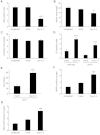
Germline defects in the absence of CEY proteins. Gonads were either not injected (uninjected), control injected (mock RNAi) or injected with RNAi clones targeting both cey-2 and cey-3 (cey-2,-3 RNAi). (A–G) Error bars represent SEM. Asterisks denote P-values <0.01 by t-test. (A) Proliferative germ cells were stained with anti-pH3 antibody and the number of positive cells was subsequently quantified by fluorescence microscopy. (B) The proliferative zone was shorter in cey-2,-3 RNAi gonads compared to controls, while the length of the transition zone remained constant (C). (D) Depletion of CEY-2,-3 caused an increase of acridine orange (AO) stained apoptotic cells in both wild-type and ced-9(n1950) animals. (E) RNAi of cey-2,-3 in the cell corpse engulfment-defective strain, ced-1(e1735), resulted in a significant increase in the number of germline corpses. (F and G) RNAi of cey-2,-3 resulted in premature appearance of activated MAPK in proximal oocytes and AIR-2-GFP on the chromosomes of oocytes in diakinesis.
Figure 4.
Figure 4.
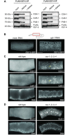
CEYs are required for the integrity of germline mRNPs. (A) FLAG IPs performed in the presence or absence of RNAse on extracts from transgenic animals expressing either FLAG-CEY-2 or FLAG-CEY-4. In both cases, CGH-1, CAR-1 and PAB-1 were no longer co-IPed upon RNAse treatment. (B–D) All shown images come from the medial gonadal region, boxed in red on the schematic gonad. All partial gonads are outlined by dotted lines. Asterisk indicates the distal end of the gonad. (B) Fluorescent micrographs of medial gonads from live animals expressing CEY-3-GFP. In the cytoplasm of wild-type gonads, the CEY-3-GFP was distributed evenly. Upon cgh-1 RNAi, CEY-3-GFP localized to sheet-like structures. Scale bar = 20 μm. (C) Fluorescent micrographs of wild-type and cey-1,-2,-3,-4 mutant gonads co-immunostained for CAR-1 and CGH-1, and additionally stained with DAPI to visualize DNA. Both proteins localized to aberrant RNP granules in cey mutant germlines (yellow arrow points to an exemplary RNP granule). Scale bar = 20 μm. (D) Fluorescent micrographs of wild-type and cey-1,-2,-3,-4 mutant gonads immunostained for GLD-1 and stained with DAPI. GLD-1 also localized to aberrant granules in the absence of CEY proteins. Scale bar = 20 μm.
Figure 5.
Figure 5.
CEY-1 and CEY-4 are essential for the assembly of large polysomes. (A) Polysome profiles from wild-type and cey-2,-3 mutants were indistinguishable. Indicated are the positions of mono-, di- and polysomes. (B) The depletion of CEY-1 and CEY-4 caused a strong decrease of large polysomes with a concomitant increase of mono- and disomes. (C) The loss of polysomes observed in cey-1,-4 mutants was also observed in the germline-less glp-1(e2144) background. Red asterisks indicate the positions of additional small peaks present to the lighter side of normal di- and trisome peaks. (D) A FLAG-tagged CEY-4 transgene (FLAG-CEY-4) expressed specifically in the germline from the mex-5 promoter could not restore polysomes in the cey-1,-4 mutant. The same fusion protein when expressed ubiquitously, partially restored polysomes (see Supplementary Figure S8F). (E) Expressing a FLAG-tagged CEY-2 transgene (FLAG-CEY-2) from the cey-1 promoter also did not restore polysomes in the cey-1,-4 mutant. Expression of a FLAG-tagged CEY-1 transgene (FLAG-CEY-1) from the cey-1 promoter partially restored polysomes (see Supplementary Figure S8G). (F) Proteins were extracted from each of the 12 fractions from a polysome profiling experiment and analysed by western blot. CEY-1 (FLAG-tagged) was mainly present in subpolysomal fractions, while a significant part of CEY-4 was additionally found in ribosomal fractions. PAB-1 was, as expected, present in both submonosomal and ribosomal (mono- and polysomal) fractions and served here as a control.
Figure 6.
Figure 6.
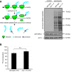
Global protein synthesis rates are similar between wild-type and cey-1,-4 mutants. (A) SUrface SEnsing of Translation (SUnSET) was adapted for Caenorhabditis elegans. Animals were grown with or without puromycin. A 4-h treatment was sufficient to detect puromycin incorporation into nascent proteins on a western blot, but, importantly, did not yet affect global translation (data not shown). Actin (ACT-1) was used as a loading control. Additionally, as an external loading control, extracts were spiked with an identical amount of extract from animals expressing GFP-tagged RPS-1, which were grown without puromycin. (B) Animals were grown in 35S-methionine-labeled OP50 bacteria for 3 h, and the amount of radioactivity incorporated into newly synthesized proteins was measured for wild-type and cey-1,-4 mutants. The wild-type value was set to 100.
Figure 7.
Figure 7.
CEY-1 and CEY-4 regulate mRNA translation and abundance. Both ribosome profiling and total RNA sequencing were performed in duplicates. The mean values were calculated and the wild-type values were subtracted from the cey-1,-4 values. The changes in mRNA levels were then plotted against the changes in RPF levels (indicating translation). The same plot is shown in A–D. The ‘germline-specific mRNAs’, ‘ribosome-depleted mRNAs’ and ‘ribosome-associated mRNAs’ (marked in B, C and D, respectively) were selected as shown in Supplementary Figure S12A–C and G and H. (A) Gray dotted lines demarcate 1.5-fold changes. As expected, cey-1 and cey-4 reads were strongly depleted in the mutants. We found that a subpopulation of transcripts (red arrow) displayed little or no change in mRNA levels but showed reduced association with ribosomes. (B) In green are marked mRNAs expressed in gonads (mostly germline mRNAs) but not in the soma (see Supplementary Figure S12A and B). (C) In red are marked mRNAs that are depleted from mono- and polysomes (i.e. are either poorly translated or repressed) in wild-type animals (see Supplementary Figure S12C). (D) In blue are marked mRNAs enriched in mono- and polysomes in wild-type animals (see Supplementary Figure S12G and H). The vertical dotted line marks no change at the mRNA level in wild-type and mutant. The majority of ‘ribosome-associated mRNAs’ (77%) appear to the left of the dotted line. Therefore, compared to all mRNAs, genes in this subset have a higher chance to be lower in abundance in the mutant (P-value <2.2 × 10⁻¹⁶ by t-test).
Figure 8.
Figure 8.
CEYs promote mRNA abundance in the soma and the germline. (A and B) Polysome profiling was performed for wild-type, cey-1,-4, cey-2,-3, and cey-1,-2,-3,-4 animals. qRT-PCR analysis was performed on RNA extracted from pooled sucrose fractions 1–12 (total RNA) (see Supplementary Figure S13A). The data were normalized to a mouse mRNA, cytc (cytochrome c), to correct for any discrepancies during RNA extraction and cDNA synthesis. One asterisk denotes P-value < 0.05 by t-test. Two asterisks denote P-value < 0.01 by t-test. Three asterisks denote P-value < 0.005 by t-test. Error bars represent SEM. (A) The abundance of tested ubiquitous (expressed in germline and soma) or soma-specific mRNAs was reduced in the cey-1,-4 mutant but not in the absence of the germline-specific CEY-2 and -3. (B) The abundance of germline-specific transcripts was more strongly affected in the cey-2,-3 double mutant compared to cey-1,-4 mutant animals. The mRNA levels dropped even further in the cey-1,-2,-3,-4 quadruple mutant. (C) Changes in RME-2 protein levels mirrored the changes of mRNA levels (B), showing the strongest decrease in the cey-1,-2,-3,-4 mutant.

References

    1. Sommerville J. Activities of cold-shock domain proteins in translation control. Bioessays. 1999;21:319–325. - PubMed
    1. Landsman D. RNP-1, an RNA-binding motif is conserved in the DNA-binding cold shock domain. Nucleic Acids Res. 1992;20:2861–2864. - PMC - PubMed
    1. Sasaki K., Imai R. Pleiotropic roles of cold shock domain proteins in plants. Front. Plant. Sci. 2012;2:116. - PMC - PubMed
    1. Eliseeva I.A., Kim E.R., Guryanov S.G., Ovchinnikov L.P., Lyabin D.N. Y-box-binding protein 1 (YB-1) and its functions. Biochemistry (Moscow) 2011;76:1402–1433. - PubMed
    1. Ruzanov P.V., Evdokimova V.M., Korneeva N.L., Hershey J.W., Ovchinnikov L.P. Interaction of the universal mRNA-binding protein, p50, with actin: a possible link between mRNA and microfilaments. J. Cell Sci. 1999;112:3487–3496. - PubMed

Publication types

MeSH terms

Associated data