. 2016 Jan;75(1):295-302.
doi: 10.1136/annrheumdis-2014-205790. Epub 2014 Nov 6.

TGFβ responsive tyrosine phosphatase promotes rheumatoid synovial fibroblast invasiveness

Affiliations

TGFβ responsive tyrosine phosphatase promotes rheumatoid synovial fibroblast invasiveness

Stephanie M Stanford et al. Ann Rheum Dis. 2016 Jan.

Abstract

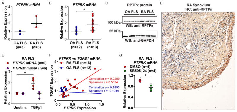
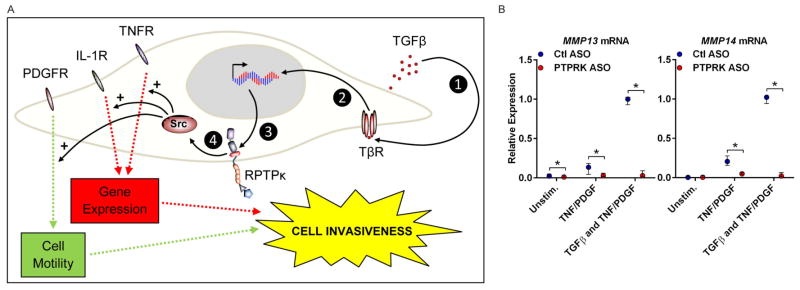
Objective: In rheumatoid arthritis (RA), fibroblast-like synoviocytes (FLS) that line joint synovial membranes aggressively invade the extracellular matrix, destroying cartilage and bone. As signal transduction in FLS is mediated through multiple pathways involving protein tyrosine phosphorylation, we sought to identify protein tyrosine phosphatases (PTPs) regulating the invasiveness of RA FLS. We describe that the transmembrane receptor PTPκ (RPTPκ), encoded by the transforming growth factor (TGF) β-target gene, PTPRK, promotes RA FLS invasiveness.

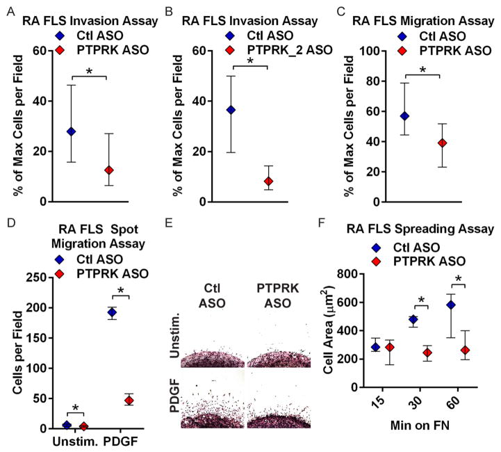
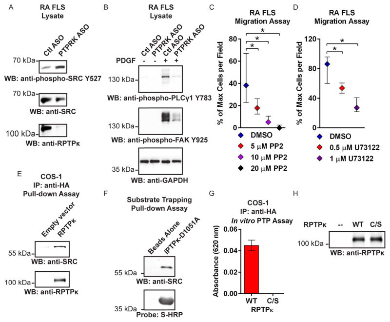
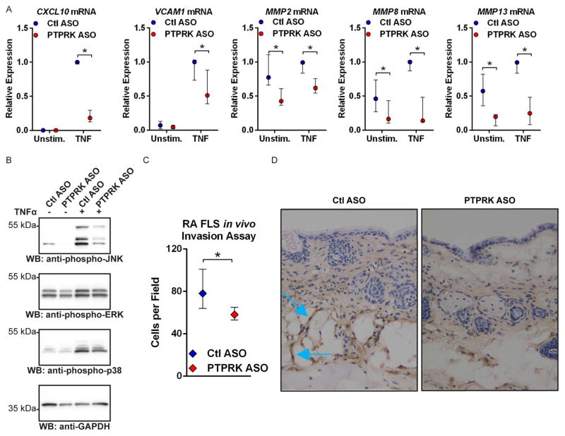
Methods: Gene expression was quantified by quantitative PCR. PTP knockdown was achieved using antisense oligonucleotides. FLS invasion and migration were assessed in transwell or spot assays. FLS spreading was assessed by immunofluorescence microscopy. Activation of signalling pathways was analysed by Western blotting of FLS lysates using phosphospecific antibodies. In vivo FLS invasiveness was assessed by intradermal implantation of FLS into nude mice. The RPTPκ substrate was identified by pull-down assays.

Results: PTPRK expression was higher in FLS from patients with RA versus patients with osteoarthritis, resulting from increased TGFB1 expression in RA FLS. RPTPκ knockdown impaired RA FLS spreading, migration, invasiveness and responsiveness to platelet-derived growth factor, tumour necrosis factor and interleukin 1 stimulation. Furthermore, RPTPκ deficiency impaired the in vivo invasiveness of RA FLS. Molecular analysis revealed that RPTPκ promoted RA FLS migration by dephosphorylation of the inhibitory residue Y527 of SRC.

Conclusions: By regulating phosphorylation of SRC, RPTPκ promotes the pathogenic action of RA FLS, mediating cross-activation of growth factor and inflammatory cytokine signalling by TGFβ in RA FLS.

Keywords: Fibroblasts; Inflammation; Rheumatoid Arthritis.


Conflict of interest statement

Competing interests None.

Figures

Figure 1
Figure 1
Transforming growth factor (TGF)-β1-responsive PTPRK is overexpressed in rheumatoid arthritis (RA) fibroblast-like synoviocytes (FLS). (A and B) PTPRK mRNA expression in FLS was measured by quantitative PCR. (A) Median±range is shown. (B) Median±IQR is shown. *p<0.05, Mann–Whitney test. (C) Western blotting of lysates of RA and osteoarthritis (OA) FLS. (D) Immunohistochemical (IHC) staining of RA synovial section using antireceptor protein tyrosine phosphatase κ (RPTP κ) antibody. (E) PTPRK and PTPRM mRNA expression in RA FLS was measured following cell stimulation with 50 ng/mL TGFβ1 for 24 h. Median±IQR is shown. *p<0.05, Mann–Whitney test. (F) PTPRK and TGFB1 mRNA expression in RA and OA FLS was measured. Graph shows PTPRK vs TGFB1 expression for each line. (G) RA FLS were treated with 25 μM SB505124 or dimethylsulfoxide (DMSO) for 24 h. Median±IQR PTPRK expression is shown. *p<0.05, Mann–Whitney test.
Figure 2
Figure 2
Receptor protein tyrosine phosphatase κ (RPTPκ) is required for rheumatoid arthritis (RA) fibroblast-like synoviocyte (FLS) invasiveness. (A and B) Following treatment with control (Ctl) or PTPRK (A) or PTPRK_2 (B) antisense oligonucleotide (ASO) for 7 days, RA FLS invaded through Matrigel-coated transwell chambers in response to 50 ng/mL platelet-derived growth factor BB (PDGF-BB) for 24 h. Graphs show median±IQR % maximum number of cells per field. Data from four (A) or three (B) independent experiments in different FLS lines are shown. (C) ASO-treated RA FLS migrated through uncoated transwell chambers in response to 5% fetal bovine serum (FBS) for 24 h. Graph shows median±IQR % maximum number of cells per field. Data from five independent experiments in different FLS lines are shown. (A–C) *p<0.05, Mann–Whitney test. (D and E) ASO-treated RA FLS migrated out of a Matrigel spot for 2 days in response to 10 ng/mL PDGF or media alone. (D) Median±IQR cells per field. Data from three independent experiments in different FLS lines are shown. *p<0.05, Wilcoxon-matched pairs signed-rank test. (E) Representative image of cells from (D). (F) ASO-treated RA FLS were plated on fibronectin (FN)-coated coverslips in the presence of 5% FBS. Graph shows median±IQR cell area after 15, 30 and 60 min. Data from three independent experiments in different FLS lines are shown. *p<0.05, Wilcoxon-matched pairs signed-rank test.
Figure 3
Figure 3
RPTPκ promotes RA FLS migration through dephosphorylation of SRC. (A) Western blotting of ASO-treated RA FLS lysates. Data are representative of three independent experiments in different FLS lines. (B) Western blotting of lysates of ASO-treated RA FLS stimulated with 50 ng/mL PDGF-BB for 30 min or left unstimulated. Data are representative of two independent experiments in different FLS lines. (C and D) RA FLS migrated through uncoated transwell chambers in response to 5% FBS in the presence of PP2 (C) or U73122 (D). Graphs show median±IQR % maximum number of cells per field. Data from two independent experiments in different FLS lines are shown. *p<0.05, Mann–Whitney test. (E) HA-tagged RPTPκ was immunoprecipitated from COS-1 cells and incubated in vitro with RA FLS lysates, and pull-down was subjected to Western blotting. Data are representative of two independent experiments. (F) Agarose-bound S-tagged-substrate trapping mutant iPTPκ-D1051A was incubated in vitro with RA FLS lysates, and pull-down was subjected to Western blotting and probed using HRP-conjugated S-protein. A similar result was obtained when RA FLS were stimulated with 100 μM pervanadate for 15 min immediately prior to lysis (data not shown). (G and H) Immunoprecipitated wild type (WT) or catalytically inactive C1100S (C/S) HA-tagged RPTPκ was incubated in vitro with SRC pY527 phosphopeptide for 30 min. The reaction was stopped by addition of Biomol Green. (G) Absorbance following subtraction of the blank reaction. (H) Western blotting of a fraction of the immunoprecipitation reaction. (G and H) Data are representative of two independent experiments. ASO, antisense oligonucleotide; COS, cells Simian CV-1 in origin and carrying SV40 genetic material; DMSO, dimethylsulfoxide; FLS, fibroblast-like synoviocytes; FBS, fetal bovine serum; GADPH, glyceraldehyde 3-phosphate dehydrogenase; HA, haemagglutinin tag; HRP, horseradish peroxidase; IP, immunoprecipitation; PDGF-BB, platelet-derived growth factor BB; RA, rheumatoid arthritis; RPTPκ, receptor protein tyrosine phosphatase κ; WB, Western blotting.
Figure 4
Figure 4
RPTPκ is required for the pathogenic action of RA FLS. (A) ASO-treated RA FLS were stimulated with 50 ng/mL TNFα for 24 h or left unstimulated. Graph shows median±IQR mRNA expression levels relative to the Ctl ASO-treated, TNFα-stimulated samples from the same FLS line. Data from four (MMP8 and MMP13) or five (CXCL10, VCAM1, MMP2) independent experiments in different FLS lines are shown. *p<0.05, Mann–Whitney test. (B) Western blotting of lysates from ASO-treated RA FLS stimulated with 50 ng/mL TNFα for 15 min or left unstimulated. Data are representative of four independent experiments in different FLS lines. (C and D) ASO-treated RA FLS were intradermally implanted into nude mice following subcutaneous injection of CFA. After 5 days, FLS invasion towards the inflammation site was measured by immunohistochemical staining of FLS in skin immediately adjacent the CFA injection site with an anti-human class I HLA antibody. (C) Graph shows median±IQR cells per field. Data from three independent experiments in different FLS lines are shown. *p<0.05, Wilcoxon-matched pairs signed-rank test. (D) Representative 40× images of mouse skin samples. Blue arrows indicate invading FLS, identified by anti-human class I HLA antibody positivity. RPTPκ, receptor protein tyrosine phosphatase κ; RA, rheumatoid arthritis; FLS, fibroblast-like synoviocytes; ASO, antisense oligonucleotide; GADPH, glyceraldehyde 3-phosphate dehydrogenase; JNK, Jun N-terminal kinase; TNF, tumour necrosis factor; CFA, complete Freunds adjuvant; HLA, human leucocyte antigen; ERK, extracellular-signal-regulated kinases; WB, Western blotting.
Figure 5
Figure 5
Receptor protein tyrosine phosphatase κ (RPTPκ) mediates cross-activation of platelet-derived growth factor (PDGF) and tumour necrosis factor (TNF) signalling by transforming growth factor (TGF)-β. (A) Model depicting the role of TGFβ-dependent RPTPκ in rheumatoid arthritis (RA) pathogenesis. Autocrine TGFβ binds to the TGFβ receptor complex (TβR) (1), inducing SMAD activation (2) and transcription of PTPRK (3). RPTPκ activates SRC (4), which promotes proinvasive signalling through the PDGF receptor (PDGFR), and the TNF and interleukin (IL)-1 receptors (TNFR and IL-1R). (B) Antisense oligonucleotide (ASO)-treated RA fibroblast-like synoviocytes (FLS) were prestimulated with 50 ng/mL TGFβ1 for 24 h (or left unstimulated) in the presence of ASO. Cells were then stimulated with 50 ng/mL PDGF/TNFα, or 50 ng/mL TGFβ1/PDGF/TNFα, or left unstimulated for 24 h. Graph shows median±IQR mRNA expression levels relative to the Ctl ASO-treated, TGFβ1/PDGF/TNFα-stimulated samples from the same FLS line. Data from three independent experiments in different FLS lines are shown. *p<0.05, Mann–Whitney test.

References

    1. Noss EH, Brenner MB. The role and therapeutic implications of fibroblast-like synoviocytes in inflammation and cartilage erosion in rheumatoid arthritis. Immunol Rev. 2008;223:252–70. - PubMed
    1. Bottini N, Firestein GS. Duality of fibroblast-like synoviocytes in RA: passive responders and imprinted aggressors. Nat Rev Rheumatol. 2013;9:24–33. - PMC - PubMed
    1. Ospelt C, Gay S. The role of resident synovial cells in destructive arthritis. Best Pract Res Clin Rheumatol. 2008;22:239–52. - PubMed
    1. Lefevre S, Knedla A, Tennie C, et al. Synovial fibroblasts spread rheumatoid arthritis to unaffected joints. Nat Med. 2009;15:1414–20. - PMC - PubMed
    1. Tolboom TC, van der Helm-Van Mil AH, Nelissen RG, et al. Invasiveness of fibroblast-like synoviocytes is an individual patient characteristic associated with the rate of joint destruction in patients with rheumatoid arthritis. Arthritis Rheum. 2005;52:1999–2002. - PubMed

Publication types

MeSH terms

Substances