. 2015 Jan 15;89(2):1094-104.
doi: 10.1128/JVI.02005-14. Epub 2014 Nov 5.

The influenza virus neuraminidase protein transmembrane and head domains have coevolved

Affiliations

The influenza virus neuraminidase protein transmembrane and head domains have coevolved

Diogo V da Silva et al. J Virol. .

Abstract

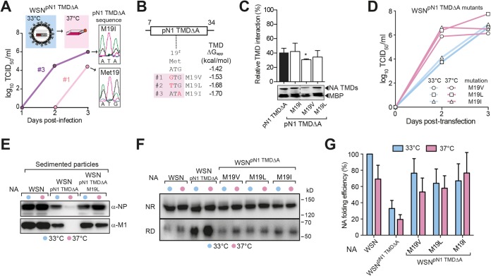
Transmembrane domains (TMDs) from single-spanning membrane proteins are commonly viewed as membrane anchors for functional domains. Influenza virus neuraminidase (NA) exemplifies this concept, as it retains enzymatic function upon proteolytic release from the membrane. However, the subtype 1 NA TMDs have become increasingly more polar in human strains since 1918, which suggests that selection pressure exists on this domain. Here, we investigated the N1 TMD-head domain relationship by exchanging a prototypical "old" TMD (1933) with a "recent" (2009), more polar TMD and an engineered hydrophobic TMD. Each exchange altered the TMD association, decreased the NA folding efficiency, and significantly reduced viral budding and replication at 37°C compared to at 33°C, at which NA folds more efficiently. Passaging the chimera viruses at 37°C restored the NA folding efficiency, viral budding, and infectivity by selecting for NA TMD mutations that correspond with their polar or hydrophobic assembly properties. These results demonstrate that single-spanning membrane protein TMDs can influence distal domain folding, as well as membrane-related processes, and suggest the NA TMD in H1N1 viruses has become more polar to maintain compatibility with the evolving enzymatic head domain.

Importance: The neuraminidase (NA) protein from influenza A viruses (IAVs) functions to promote viral release and is one of the major surface antigens. The receptor-destroying activity in NA resides in the distal head domain that is linked to the viral membrane by an N-terminal hydrophobic transmembrane domain (TMD). Over the last century, the subtype 1 NA TMDs (N1) in human H1N1 viruses have become increasingly more polar, and the head domains have changed to alter their antigenicity. Here, we provide the first evidence that an "old" N1 head domain from 1933 is incompatible with a "recent" (2009), more polar N1 TMD sequence and that, during viral replication, the head domain drives the selection of TMD mutations. These mutations modify the intrinsic TMD assembly to restore the head domain folding compatibility and the resultant budding deficiency. This likely explains why the N1 TMDs have become more polar and suggests the N1 TMD and head domain have coevolved.


Figures

FIG 1
FIG 1
A recent N1 TMD impairs the replication of an old H1N1 virus at 37°C. (A) The three domains of an NA tetramer are shown with the 7-face TMD model for its amphipathic assembly via the polar faces a and e (21). (B) The timeline depicts the different human IAV lineages by their hemagglutinin (H) and neuraminidase (N) subtypes with the predicted hydrophobicity values (ΔGapp values for membrane insertion) of the consensus N1 TMD sequences for the H1N1 lineages. (C) Split-transfection method for comparative viral production by reverse genetics. Trypsinized 293T cells are transfected with WSN plasmids encoding its old N1 TMD or the recent pN1 TMD, divided into flasks containing MDCK cells at 33°C and 37°C, and viral titers are assayed by TCID50 values. (D) The recent pN1 TMD significantly decreases the replication of the 1933 H1N1 virus WSN at 37°C compared to at 33°C. The viral titers are displayed as means from three independent experiments ± standard errors of the means (SEM). (E) WSN and WSNpN1-TMD viral particles created by reverse genetics were isolated from the culture medium 3 days after recovery by ultracentrifugation. The M1 and NP contents were assayed by immunoblotting and the NA content by activity, which is displayed as a ratio to the WSN sedimented particles from 33°C. Cells that did not receive the NA plasmid were used as a control.
FIG 2
FIG 2
Nonoptimal N1 TMDs are under temperature-dependent selection pressure. (A) Viral passaging and sequencing. MDCK cells at 33°C and 37°C were infected at an MOI of ∼0.001 with the WSN and WSNpN1-TMD viruses generated at 33°C or 37°C for 3 days and repeated for the indicated number of passages (P). For each passage, viruses were isolated 3 days postinfection, and genomes were extracted and sequenced. (B) pN1 TMD mutations were selected for in the WSNpN1-TMD viruses produced and passaged twice at 37°C (pink box, 37°C P = 2) and when it was produced at 33°C and passaged twice at 37°C (pink box, 33°C to 37°C P = 2). Nonsynonymous (red) and synonymous (green) mutations are displayed with the amino acid number and the TMD face (a to g). (C) The replication efficiency of WSNpN1-TMD virus at 37°C was rescued by the virus-selected pN1 TMD mutations (M19V, I32T, and I29T/I32T). Graphs display the mean titers ± SEM at 33°C (blue lines) and 37°C (pink lines) from split transfections of the indicated WSNpN1-TMD mutants created by reverse genetics. WSN and WSNpN1-TMD titers from Fig. 1D were included for comparison. (D) Following reverse genetics recovery for 3 days, the indicated viral particles were isolated from the culture medium, and the NP and M1 contents were determined by immunoblotting.
FIG 3
FIG 3
Head domain folding drives the selection of the polar pN1 TMD mutations. (A) The adaptive mutations localize to different faces of the amphipathic pN1 TMD and are mainly polar residues that decrease the face and TMD hydrophobicity (increase the ΔGapp values). Predicted ΔG values of the heterogeneous TMD sequences are also displayed independently. (B) The mutations decrease the strong pN1 TMD association to the moderate levels of the N1 TMD from WSN. TMD interaction strengths were measured in the GALLEX system, where the association is proportional to the decrease in β-galactosidase activity. The relative TMD interaction strength was determined with respect to an empty-vector control. Error bars represent the standard deviation (SD) from three experiments, and the Student t test was used to calculate the significant differences from the WSN TMD (**, P ≤ 0.01). Equal expression of the different NA TMD constructs was confirmed by immunoblotting. (C) The procedure for determining the NA folding efficiency at different temperatures based on the presence of the same enzymatic head domain. (D) Nonreducing (NR) and reducing (RD) immunoblots of the indicated NA constructs that were expressed in 293T cells at 33°C and 37°C for 48 h and standardized to their activity levels are shown. The resulting NA folding efficiencies displayed in panel E were determined by normalizing the activity levels in each sample to their total protein levels from the RD immunoblots. The values for NAWSN expressed at 33°C were set to 100%, and the error bars represent the SD from three independent experiments.
FIG 4
FIG 4
Disrupting the conserved amphipathic N1 TMD restricts viral replication to 33°C. (A) Graph displaying the conservation of the amphipathic characteristic (adjacent polar a and e faces with positive ΔGapp-face values) in 100% of the N1 TMDs from human, avian, and swine IAVs. (B) Proposed model of how mutating the polar a face residues (Asn 21 and Asn 28) in the pN1-TMDΔA to Ala alter its amphipathic assembly (white faces are polar). (C) Replication of WSNpN1-TMDΔA is restricted to 33°C. The graph displays mean viral titers ± SEM from three independent split transfections of WSNpN1-TMDΔA virus at 33°C and 37°C. (D) The indicated viral particles were isolated from the culture medium after 3 days of recovery at the indicated temperatures. The NP and M1 contents were determined by immunoblotting.
FIG 5
FIG 5
Hydrophobic N1 TMD mutations restore WSNpN1-TMDΔA replication at 37°C. (A) The pN1 TMDΔA mutations rescue WSNpN1-TMDΔA replication at 37°C. The graph displays the viral titers from two independent experiments where WSNpN1-TMDΔA virus that had been generated at 33°C was used to infect MDCK cells at 37°C using an MOI of ∼0.001 that was determined at 33°C. Chromatograms show the mutations at Met 19 in the TMD at day 3. (B) The pN1 TMDΔA mutations at 37°C are all hydrophobic (ΔGapp values are more negative) and positionally conserved (Met 19). (C) The relative interaction strengths of the pN1 TMDΔA and the rescue mutations were determined as described for Fig. 3B. (D) The M19V, M19L, and M19I pN1 TMDΔA mutations were introduced into the WSNpN1-TMDΔA virus by reverse genetics, and each one rescued the replication at 37°C based on their viral titers at 33°C and 37°C. (E) The M1 and NP contents within the indicated viral particles that were isolated 3 days postrecovery are shown by the representative immunoblots. (F) Altering the N1 TMD amphipathicity significantly impairs the head domain folding, which is restored by the adaptive TMD mutations. Representative NR and RD immunoblots are shown of the indicated NA constructs expressed in 293T cells at 33°C and 37°C for 48 h and standardized to their activity levels. The resulting NA folding efficiencies displayed in panel G were performed as described for Fig. 3E.
FIG 6
FIG 6
The Eurasian swine origin of the pN1 head domain can be traced by its low TMD hydrophobicity. (A) Model displaying how N1 head domain changes disrupt the folding compatibility with the N1 TMD. Head domain changes create selection pressure for residues (mainly polar) in the TMD that alter its amphipathic assembly and restore the compatibility. (B) Diagram showing the reassortment of the NA (N1 and N2) and M segments from geographically separated North American swine H1N2 viruses and the Eurasian swine H1N1 viruses that created the 2009 human pandemic pH1N1 virus. (C) Prior to 2009, the low N1 TMD hydrophobicity found in the pH1N1 viruses globally was present only in the swine H1N1 viruses from Eurasia (see “Swine ≤2008”). All the predicted N1 and N2 TMD hydrophobicities from the available swine (H1N1 and H1N2) and human pH1N1 viruses are displayed with respect to their geographic location and date of isolation (2009 and ≤2008).

References

    1. Li E, Wimley WC, Hristova K. 2012. Transmembrane helix dimerization: beyond the search for sequence motifs. Biochim Biophys Acta 1818:183–193. doi:10.1016/j.bbamem.2011.08.031. - DOI - PMC - PubMed
    1. Langosch D, Arkin IT. 2009. Interaction and conformational dynamics of membrane-spanning protein helices. Protein Sci 18:1343–1358. doi:10.1002/pro.154. - DOI - PMC - PubMed
    1. Moore DT, Berger BW, DeGrado WF. 2008. Protein-protein interactions in the membrane: sequence, structural, and biological motifs. Structure 16:991–1001. doi:10.1016/j.str.2008.05.007. - DOI - PMC - PubMed
    1. Shaw AC, Mitchell RN, Weaver YK, Campos-Torres J, Abbas AK, Leder P. 1990. Mutations of immunoglobulin transmembrane and cytoplasmic domains: effects on intracellular signaling and antigen presentation. Cell 63:381–392. doi:10.1016/0092-8674(90)90171-A. - DOI - PubMed
    1. Call ME, Pyrdol J, Wiedmann M, Wucherpfennig KW. 2002. The organizing principle in the formation of the T cell receptor-CD3 complex. Cell 111:967–979. doi:10.1016/S0092-8674(02)01194-7. - DOI - PMC - PubMed

Publication types