

Borrelia burgdorferi arthritis-associated locus Bbaa1 regulates Lyme arthritis and K/B×N serum transfer arthritis through intrinsic control of type I IFN production
- PMID: 25378596
- PMCID: PMC4258437
- DOI: 10.4049/jimmunol.1401746
Borrelia burgdorferi arthritis-associated locus Bbaa1 regulates Lyme arthritis and K/B×N serum transfer arthritis through intrinsic control of type I IFN production
Abstract
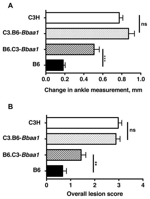
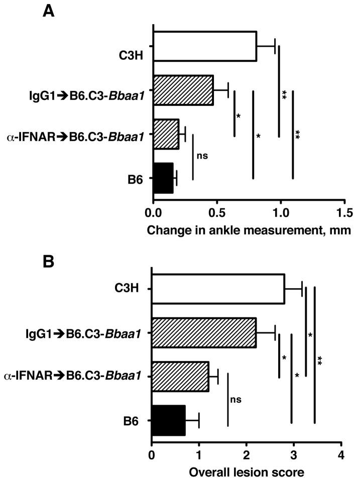
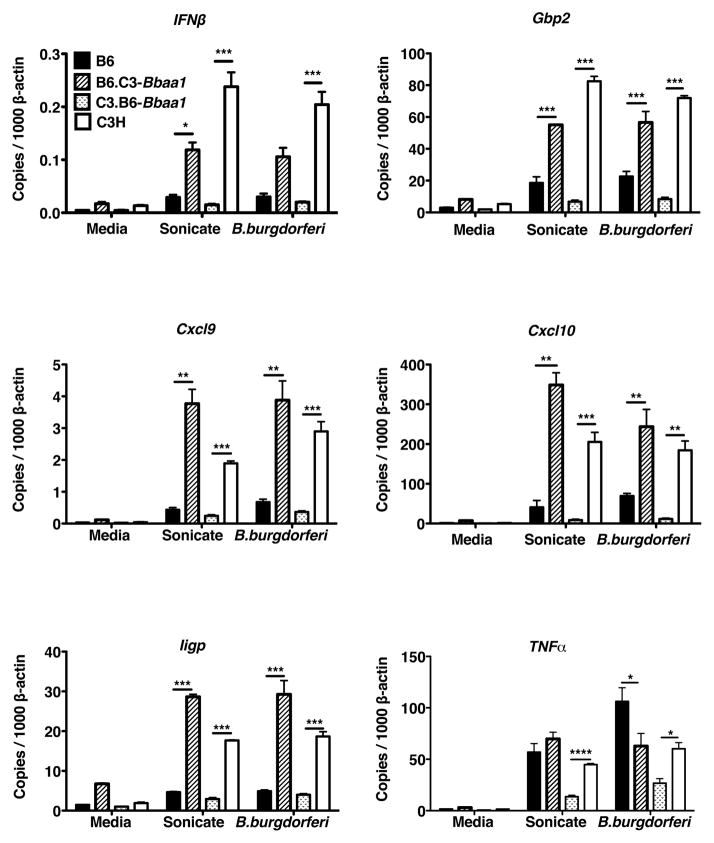
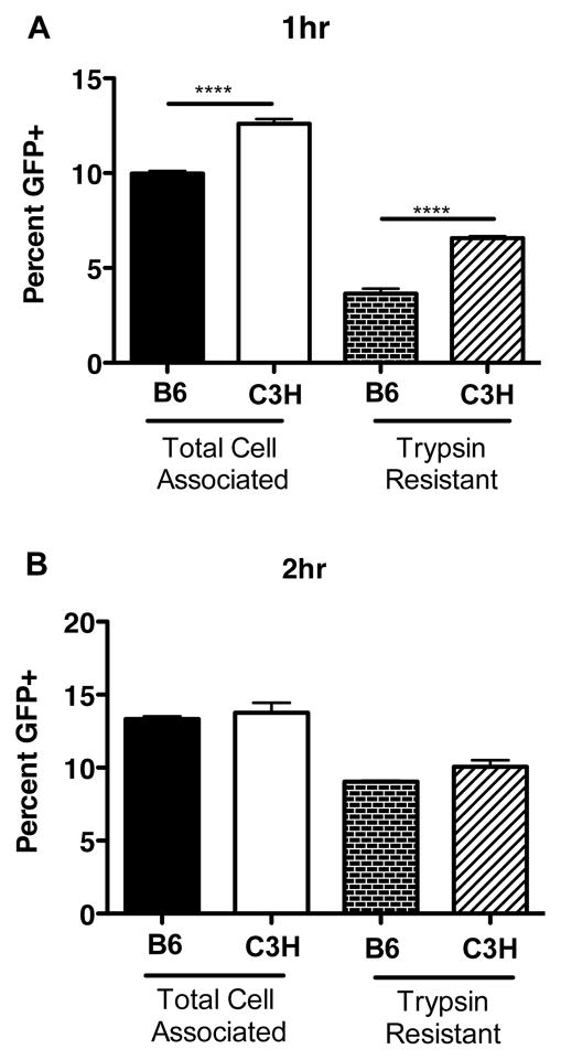
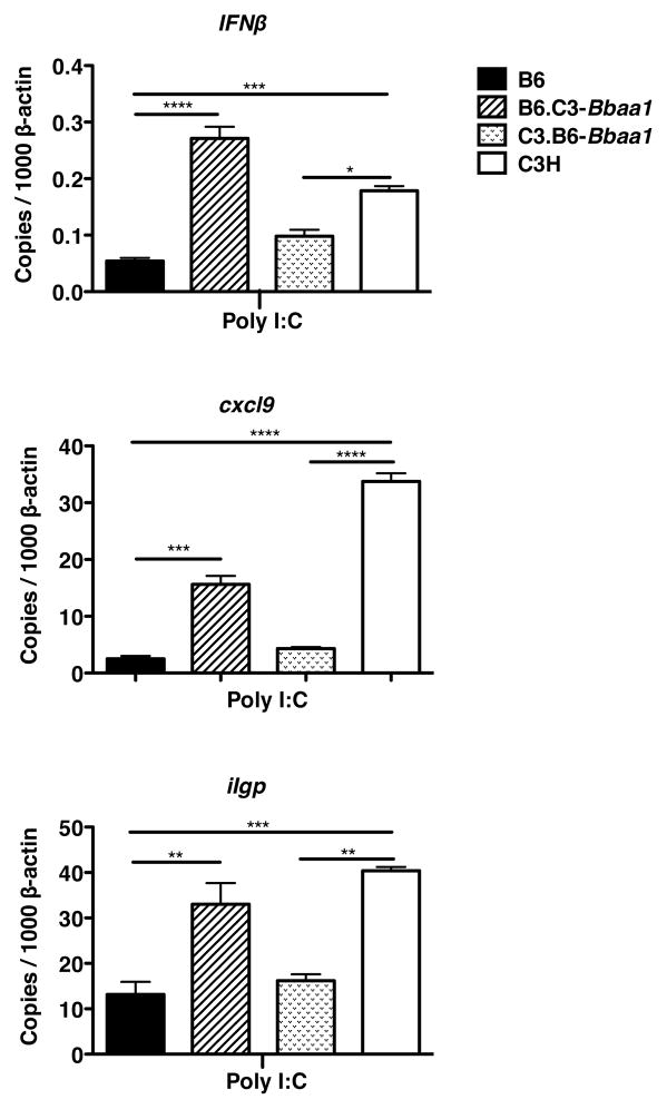
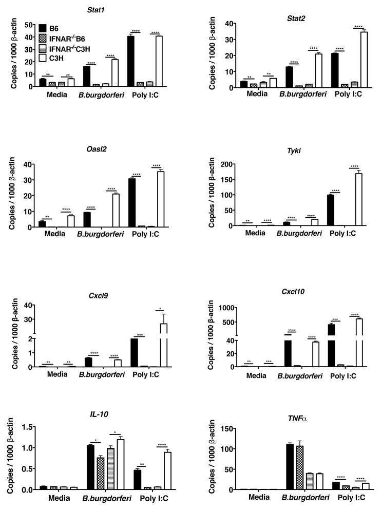
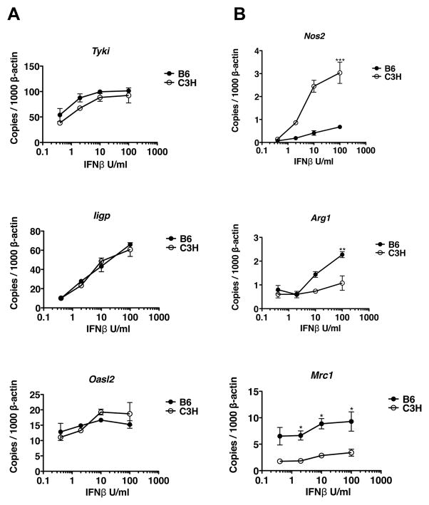
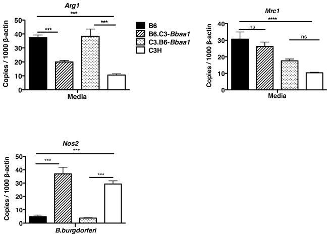
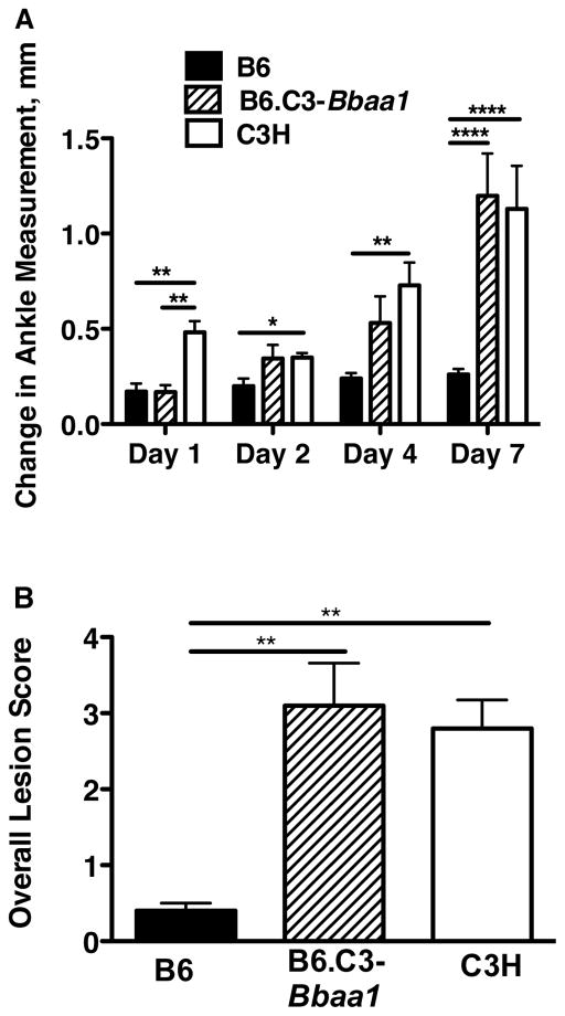
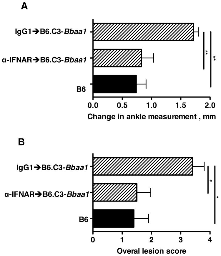
Localized upregulation of type I IFN was previously implicated in development of Borrelia burgdorferi-induced arthritis in C3H mice, and was remarkable due to its absence in the mildly arthritic C57BL/6 (B6) mice. Independently, forward genetics analysis identified a quantitative trait locus on Chr4, termed B. burgdorferi-associated locus 1 (Bbaa1), that regulates Lyme arthritis severity and includes the 15 type I IFN genes. Involvement of Bbaa1 in arthritis development was confirmed in B6 mice congenic for the C3H allele of Bbaa1 (B6.C3-Bbaa1), which developed more severe Lyme arthritis and K/B×N model of rheumatoid arthritis (RA) than did parental B6 mice. Administration of a type I IFN receptor blocking mAb reduced the severity of both Lyme arthritis and RA in B6.C3-Bbaa1 mice, formally linking genetic elements within Bbaa1 to pathological production of type I IFN. Bone marrow-derived macrophages from Bbaa1 congenic mice implicated this locus as a regulator of type I IFN induction and downstream target gene expression. Bbaa1-mediated regulation of IFN-inducible genes was upstream of IFN receptor-dependent amplification; however, the overall magnitude of the response was dependent on autocrine/paracrine responses to IFN-β. In addition, the Bbaa1 locus modulated the functional phenotype ascribed to bone marrow-derived macrophages: the B6 allele promoted expression of M2 markers, whereas the C3H allele promoted induction of M1 responses. This report identifies a genetic locus physically and functionally linked to type I IFN that contributes to the pathogenesis of both Lyme and RA.
Copyright © 2014 by The American Association of Immunologists, Inc.
Conflict of interest statement
Disclosure:
The authors have no financial conflicts.
Figures
Similar articles
-
Genetic Control of Lyme Arthritis by Borrelia burgdorferi Arthritis-Associated Locus 1 Is Dependent on Localized Differential Production of IFN-β and Requires Upregulation of Myostatin.J Immunol. 2017 Nov 15;199(10):3525-3534. doi: 10.4049/jimmunol.1701011. Epub 2017 Oct 6. J Immunol. 2017. PMID: 28986440 Free PMC article.
-
The Cdkn2a gene product p19 alternative reading frame (p19ARF) is a critical regulator of IFNβ-mediated Lyme arthritis.PLoS Pathog. 2022 Mar 24;18(3):e1010365. doi: 10.1371/journal.ppat.1010365. eCollection 2022 Mar. PLoS Pathog. 2022. PMID: 35324997 Free PMC article.
-
MicroRNA-146a provides feedback regulation of lyme arthritis but not carditis during infection with Borrelia burgdorferi.PLoS Pathog. 2014 Jun 26;10(6):e1004212. doi: 10.1371/journal.ppat.1004212. eCollection 2014 Jun. PLoS Pathog. 2014. PMID: 24967703 Free PMC article.
-
Gene expression profiling provides insights into the pathways involved in inflammatory arthritis development: murine model of Lyme disease.Exp Mol Pathol. 2008 Aug;85(1):20-7. doi: 10.1016/j.yexmp.2008.03.004. Epub 2008 Apr 8. Exp Mol Pathol. 2008. PMID: 18462718 Free PMC article. Review.
-
Hamster and murine models of severe destructive Lyme arthritis.Clin Dev Immunol. 2012;2012:504215. doi: 10.1155/2012/504215. Epub 2012 Feb 22. Clin Dev Immunol. 2012. PMID: 22461836 Free PMC article. Review.
Cited by
-
Immune Response to Borrelia: Lessons from Lyme Disease Spirochetes.Curr Issues Mol Biol. 2021;42:145-190. doi: 10.21775/cimb.042.145. Epub 2020 Dec 8. Curr Issues Mol Biol. 2021. PMID: 33289684 Free PMC article.
-
Hitchhiker's Guide to Borrelia burgdorferi.J Bacteriol. 2024 Sep 19;206(9):e0011624. doi: 10.1128/jb.00116-24. Epub 2024 Aug 14. J Bacteriol. 2024. PMID: 39140751 Free PMC article. Review.
-
Lyme arthritis: linking infection, inflammation and autoimmunity.Nat Rev Rheumatol. 2021 Aug;17(8):449-461. doi: 10.1038/s41584-021-00648-5. Epub 2021 Jul 5. Nat Rev Rheumatol. 2021. PMID: 34226730 Free PMC article. Review.
-
Antagonistic Interplay between MicroRNA-155 and IL-10 during Lyme Carditis and Arthritis.PLoS One. 2015 Aug 7;10(8):e0135142. doi: 10.1371/journal.pone.0135142. eCollection 2015. PLoS One. 2015. PMID: 26252010 Free PMC article.
-
Aspects of the Immunopathogenesis of Lyme Arthritis.Microorganisms. 2025 Jul 7;13(7):1602. doi: 10.3390/microorganisms13071602. Microorganisms. 2025. PMID: 40732111 Free PMC article. Review.
References
-
- Steere AC, Schoen RT, Taylor E. The clinical evolution of Lyme arthritis. Ann Intern Med. 1987;107:725–731. - PubMed
-
- Steere AC, Glickstein L. Elucidation of Lyme arthritis. Nat Rev Immunol. 2004;4:143–152. - PubMed
-
- Wormser GP, Dattwyler RJ, Shapiro ED, Halperin JJ, Steere AC, Klempner MS, Krause PJ, Bakken JS, Strle F, Stanek G, Bockenstedt L, Fish D, Dumler JS, Nadelman RB. The clinical assessment, treatment, and prevention of lyme disease, human granulocytic anaplasmosis, and babesiosis: clinical practice guidelines by the Infectious Diseases Society of America. Clin Infect Dis. 2006;43:1089–1134. - PubMed
-
- CDC. Lyme Disease Data US, 1996–2010. 2010 http://www.cdc.gov/lyme/stats/index.html.
Publication types
MeSH terms
Substances
Grants and funding
LinkOut - more resources
Full Text Sources
Other Literature Sources
Medical
Molecular Biology Databases
Miscellaneous
