. 2014 Nov 7;346(6210):748-51.
doi: 10.1126/science.1257522.

Targeting and plasticity of mitochondrial proteins revealed by proximity-specific ribosome profiling

Affiliations

Targeting and plasticity of mitochondrial proteins revealed by proximity-specific ribosome profiling

Christopher C Williams et al. Science. .

Abstract

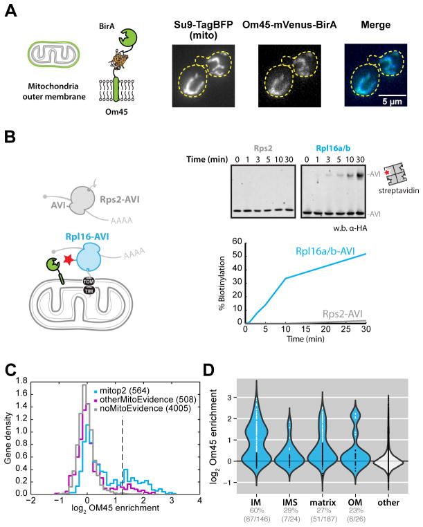
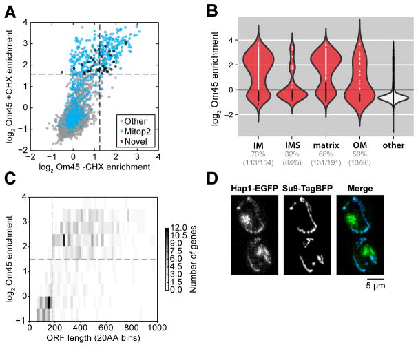
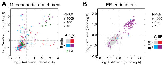
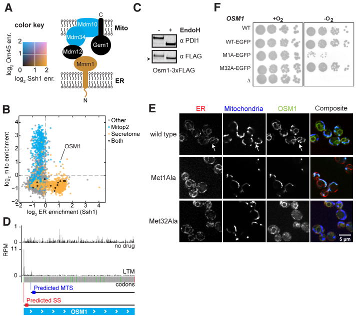
Nearly all mitochondrial proteins are nuclear-encoded and are targeted to their mitochondrial destination from the cytosol. Here, we used proximity-specific ribosome profiling to comprehensively measure translation at the mitochondrial surface in yeast. Most inner-membrane proteins were cotranslationally targeted to mitochondria, reminiscent of proteins entering the endoplasmic reticulum (ER). Comparison between mitochondrial and ER localization demonstrated that the vast majority of proteins were targeted to a specific organelle. A prominent exception was the fumarate reductase Osm1, known to reside in mitochondria. We identified a conserved ER isoform of Osm1, which contributes to the oxidative protein-folding capacity of the organelle. This dual localization was enabled by alternative translation initiation sites encoding distinct targeting signals. These findings highlight the exquisite in vivo specificity of organellar targeting mechanisms.


Figures

Fig. 1
Fig. 1. Cotranslational mitochondrial protein targeting as revealed by proximity-specific ribosome profiling
(A) Localization of biotin ligase to the outer membrane. Schematic of the Om45 fusion protein and confocal fluorescence images of Om45-mVenus-BirA and Su9-TagBFP are shown. (B) Assessment of ribosome orientation at the mitochondrial outer membrane. The kinetics of Avi-tagged Rps2 (gray) and Rpl16a/b (blue) biotinylation by Om45-mVenus-BirA were determined by western blot analysis of streptavidin-shift gels. (C) Histogram of log2 ratios for biotinylated footprints/input footprints in the absence of CHX. Genes annotated in the mitop2 reference set are shown in blue and enrichment threshold is shown as a dashed vertical line. Genes with mitochondrial GO cellular component terms not in mitop2 are shown in magenta. (D) Violin plot showing log2 gene enrichments for proteins grouped by their annotated location within mitochondria in the absence of CHX arrest. Gene enrichments are overlaid as points, with white and black colors indicating enrichment or disenrichment, respectively, as determined by sub-compartment-specific Gaussian mixture modeling (fig. S2A) and quantified below the plot.
Fig. 2
Fig. 2. Comprehensive characterization of RNC targeting to the mitochondria
(A) Scatter plot of log2 gene enrichments for footprints from ribosomes biotinylated by Om45-mVenus-BirA in the presence or absence of CHX. Novel genes represent those that were significantly enriched but had no known mitochondrial annotations. (B) Violin plot showing log2 gene enrichments for proteins grouped by their location within mitochondria in the presence of CHX arrest (as in Fig. 1D). Gene enrichments were determined by sub-compartment-specific Gaussian mixture modeling (fig. S2B). (C) Two-dimensional histogram of genes binned by open reading frame length compared to their log2 gene enrichment in the presence of CHX. (D) Epifluorescence microscopy of Hap1-EGFP and Su9-TagBFP.
Fig. 3
Fig. 3. Conservation of paralogous protein localization
(A) Scatter plot of mitochondrial log2 gene enrichments for ohnolog pairs, whose x- and y-axis assignment is arbitrary. Color codes indicate the expected ohnolog localizations based on existing evidence consolidated from mitop2, GO, and GFP annotations. Point size represents minimum expression level between paralogs. (B) As in (A), for ER enrichments.
Fig. 4
Fig. 4. Dual localization of proteins to the mitochondria and ER
(A) The color-mapped log2 enrichments for ribosome labeling by Om45 (blue) and Ssh1 (orange) are overlaid for members of the ERMES complex. (B) Scatter plot of ER enrichments compared to mitochondrial enrichments in the presence of CHX. Points are colored by their annotated localization. (C) Western blot analysis of N-linked glycans. Extracts from cells expressing Osm1-3xFLAG were blotted for PDI1 or FLAG with or without EndoH treatment. Arrowhead indicates an Osm1 band migrating below the size of the deglycosylated protein. (D) Ribosome occupancy of Osm1 with or without LTM treatment. In-frame AUG codons are shown in green; predicted localization of proteins made from the indicated start codon are shown. (E) Confocal fluorescence microscopy of Osm1-EGFP localization compared to ER (Sec63-mCherry) or mitochondria (Su9-BFP). Wild type, Met1Ala, and Met32Ala variants of Osm1 are shown. (F) Analysis of signal requirements for Osm1 to support anaerobic growth as the sole fumarate reductase gene.

Comment in

References

    1. Elstner M, Andreoli C, Klopstock T, Meitinger T, Prokisch H. The mitochondrial proteome database: MitoP2. Methods Enzymol. 2009;457:3–20. - PubMed
    1. Fox TD. Mitochondrial Protein Synthesis, Import, and Assembly. Genetics. 2012;192:1203–1234. - PMC - PubMed
    1. Kellems RE, Allison VF, Butow RA. Cytoplasmic type 80S ribosomes associated with yeast mitochondria. IV. Attachment of ribosomes to the outer membrane of isolated mitochondria. J Cell Biol. 1975;65:1–14. - PMC - PubMed
    1. Suissa M, Schatz G. Import of proteins into mitochondria. Translatable mRNAs for imported mitochondrial proteins are present in free as well as mitochondria-bound cytoplasmic polysomes. J Biol Chem. 1982;257:13048–13055. - PubMed
    1. Marc P, et al. Genome-wide analysis of mRNAs targeted to yeast mitochondria. EMBO Rep. 2002;3:159–164. - PMC - PubMed

Publication types

MeSH terms

Substances

Associated data